Bone Pathology: Osteonecrosis and Osteomyelitis
Study these flashcardsFree · spaced repetition · iPhone, Android, Mac & web
All cards
1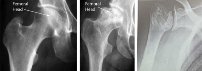
OSTEONECROSIS
Presence of avascular necrosis of the femoral head
Presence of avascular necrosis of the femoral head
2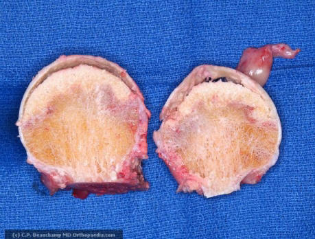
OSTEONECROSIS
Subchondral Collapse.
Subchondral Collapse.
3
[A] Slightly depressed articular surface of the collapse
[B] Wedge-shaped infarct (area of necrosis)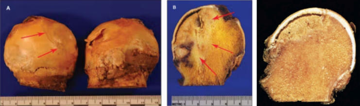
[B] Wedge-shaped infarct (area of necrosis)
Osteonecrosis of femoral head
4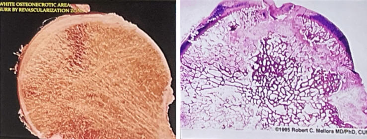
Osteonecrotic head
White osteonecrotic area surrounded by revascularization zone
White osteonecrotic area surrounded by revascularization zone
5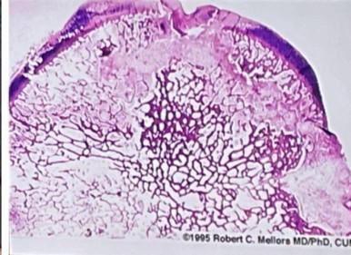
OSTEONECROSIS
Microscopic image of the femoral head
Microscopic image of the femoral head
6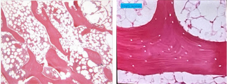
Osteonecrosis, showing an empty lacunae
7
Histologic Findings of osteonecrosis
Dead Bone
Creeping Substitution
Slow Pace of Substitution
Creeping Substitution
Slow Pace of Substitution
8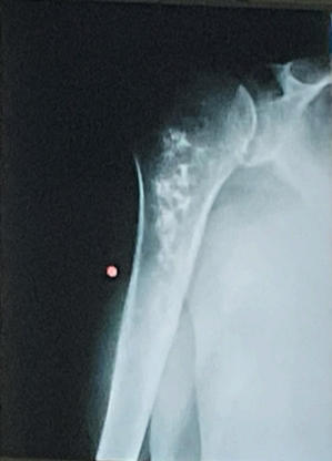
MEDULLARY INFARCTS
Involve cancellous bone and marrow
Cortex unaffected
Involve cancellous bone and marrow
Cortex unaffected
9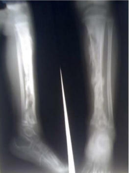
PYOGENIC OSTEOMYELITIS
lytic (black) focus of bone destruction surrounded by zone of sclerosis
lytic (black) focus of bone destruction surrounded by zone of sclerosis
10
[L] Presence of acute suppuration at the metaphyseal region
[R] Inltration of polymorphonuclear leukocytes and periosteal involvement
[R] Abscesses can form in subperiosteal region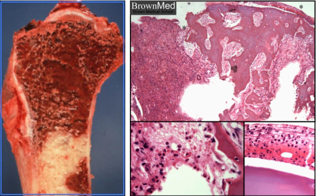
[R] Inltration of polymorphonuclear leukocytes and periosteal involvement
[R] Abscesses can form in subperiosteal region
FIRST 48 HOURS of PYOGENIC OSTEOMYELITIS
11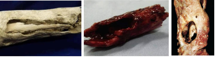
Sequestrum of a dead bone
12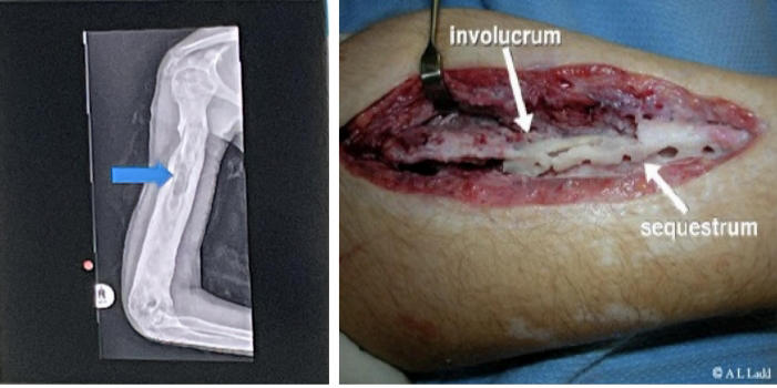
involucrum
13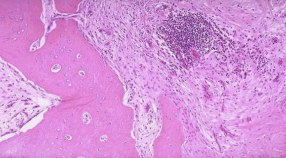
chronic osteomyelitis
Mainly osteoclasts, osteoblasts, broblasts and other bony cells leading to necrosis of the bone marrow
Mainly osteoclasts, osteoblasts, broblasts and other bony cells leading to necrosis of the bone marrow
14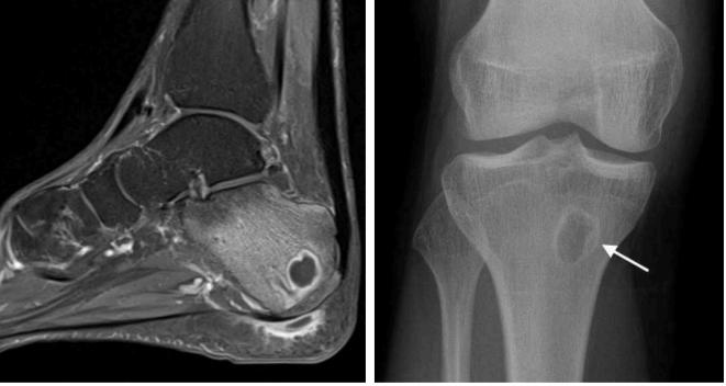
Brodie abscess
15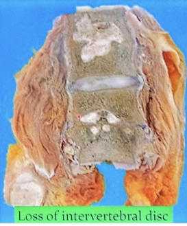
Pott's disease
16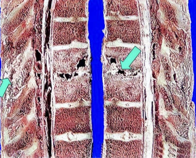
Pott's disease
17
Presence of Langhans-type giant cell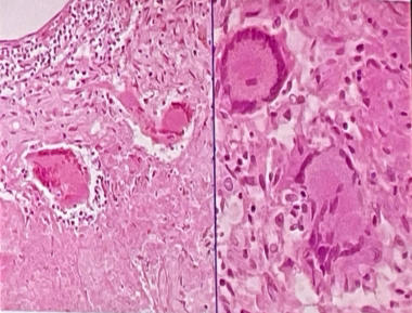
tuberculous myelitis
18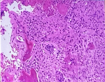
tuberculous myelitis
Presence of caseation necrosis
Presence of caseation necrosis
More Medicine & health flashcards
Hemolytic AnemiasMedicine & health · Englishan OutLearn studentHypoglycemia, Nociception, and AntiemeticsMedicine & health · EnglishKaliLiver and Kidney: Lab Values and DamageMedicine & health · EnglishMatthewBeginning Medical Terminology Chapter 2Medicine & health · EnglishJulieBone Histology (Lab 4)Medicine & health · EnglishKrisMedCardsThyroid Gland Medicine & health · EnglishfaithHead, Face, Neck, Lymphatics HA Exam #2Medicine & health · EnglishMadiruth2CNS Drugs: Generic and Brand NamesMedicine & health · EnglishebradstreetSalon CeresMedicine & health · FrenchJR-DKMastication and FaceMedicine & health · Englishjocyt9COVID 19Medicine & health · EnglishcaePsuedomonas Infection (wound infections)Medicine & health · EnglishKatlynKE
Study these decks in the app
Free on iPhone, Android, Mac and the web. Add any deck to your account in one tap.