Regenerative Anemia and Hemorrhage
Study these flashcardsFree · spaced repetition · iPhone, Android, Mac & web
All cards
1
What are the two major causes of regenerative anemia?
Blood loss and RBC destruction (hemolysis).
2
How long does it take to see initial evidence of regeneration?
About 2–4 days; robust regeneration may take about 7 days.
3
Why can a newly anemic animal initially appear non-regenerative?
The bone marrow has not had enough time to release increased reticulocytes.
4
How can total protein help distinguish blood loss from hemolysis?
Blood loss decreases protein; hemolysis usually leaves protein within the reference interval.
5
What findings support hemolysis rather than blood loss? (4)
↑Bilirubin, bilirubinuria, hemoglobinemia, and/or hemoglobinuria.
6
What is hemorrhage?
Loss of whole blood, including RBCs and plasma/proteins, from the vascular system.
7
How does acute vs. chronic hemorrhage affect regeneration?
Acute is regenerative; chronic may become non-regenerative from iron deficiency.
8
Why may acute hemorrhage not cause obvious anemia immediately?
RBCs and plasma are initially lost proportionally.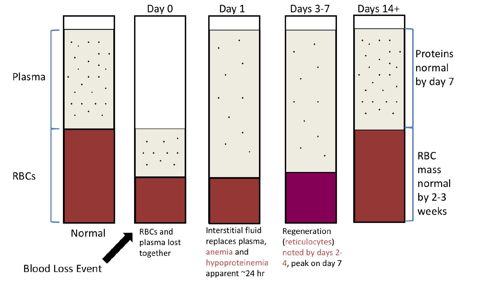
9
When do anemia and hypoproteinemia become apparent after acute blood loss?
Around 24 hours as interstitial fluid replaces plasma volume.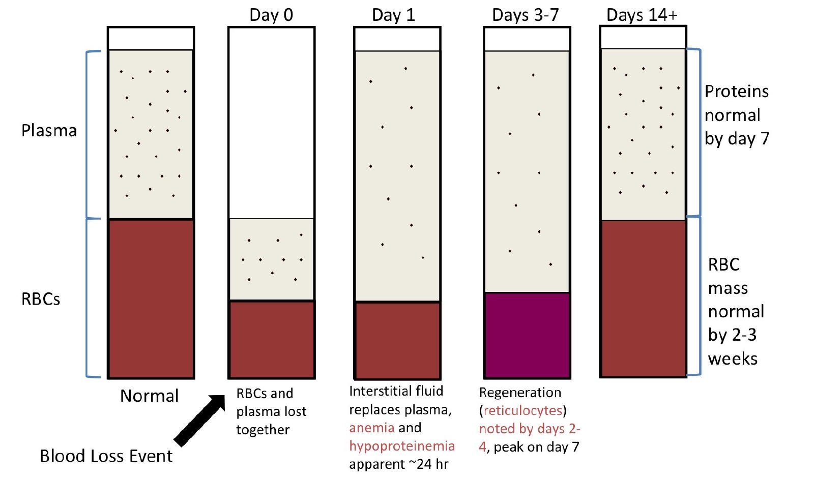
10
When does regeneration occur after acute blood loss?
Reticulocytes appear days 2–4 and peak around day 7.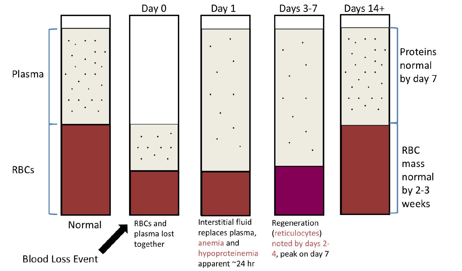
11
When do proteins and RBC mass recover after acute blood loss?
Proteins by ~day 7; RBC mass by ~2–3 weeks.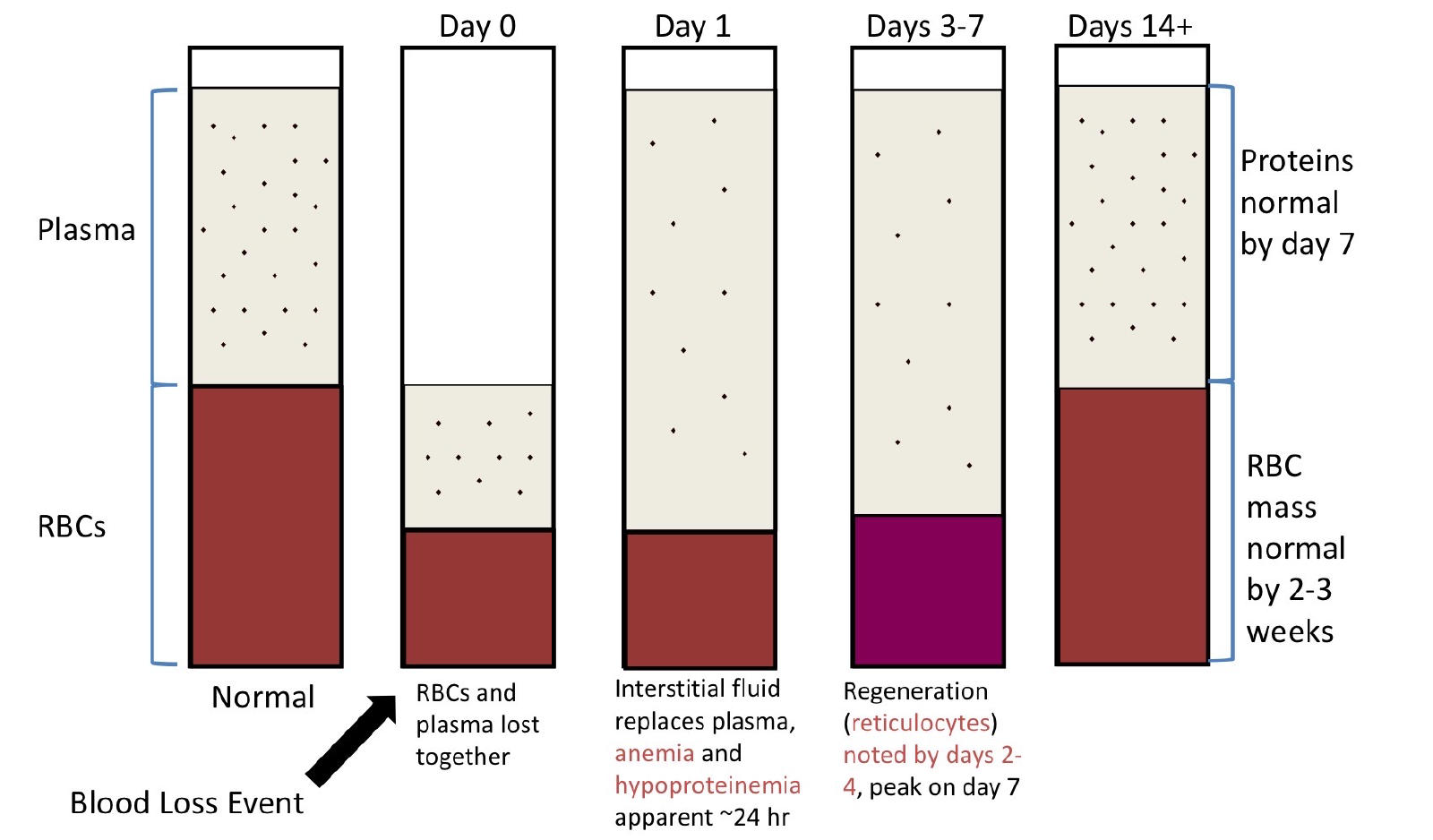
12
What are major causes of hemorrhagic anemia?
Trauma, alimentary blood loss (GI), and hemostatic abnormalities.
13
What happens to iron during internal blood loss?
Macrophages phagocytose RBCs and recycle the iron.
14
Why can chronic external blood loss cause iron deficiency?
Iron is repeatedly lost from the body with the blood.
15
What classic anemia develops with chronic iron deficiency?
Microcytic-hypochromic anemia.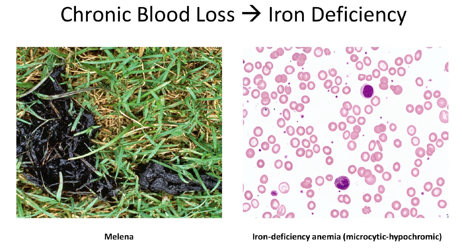
16
What is the laboratory hallmark of hemorrhage? (2)
Anemia + hypoproteinemia.
17
What tests can help investigate hemorrhage? (4)
Fecal occult blood, coagulation panel, effusion analysis, and urinalysis.
18
RBC Destruction Hemolytic Anemias (3)
1. Immune Mediated
2. Oxidative
3. Mechanical
2. Oxidative
3. Mechanical
19
What is hemolysis?
Accelerated destruction of RBCs.
20
What is intravascular vs. extravascular hemolysis?
Intravascular = RBC rupture in blood; extravascular = macrophage phagocytosis of RBCs.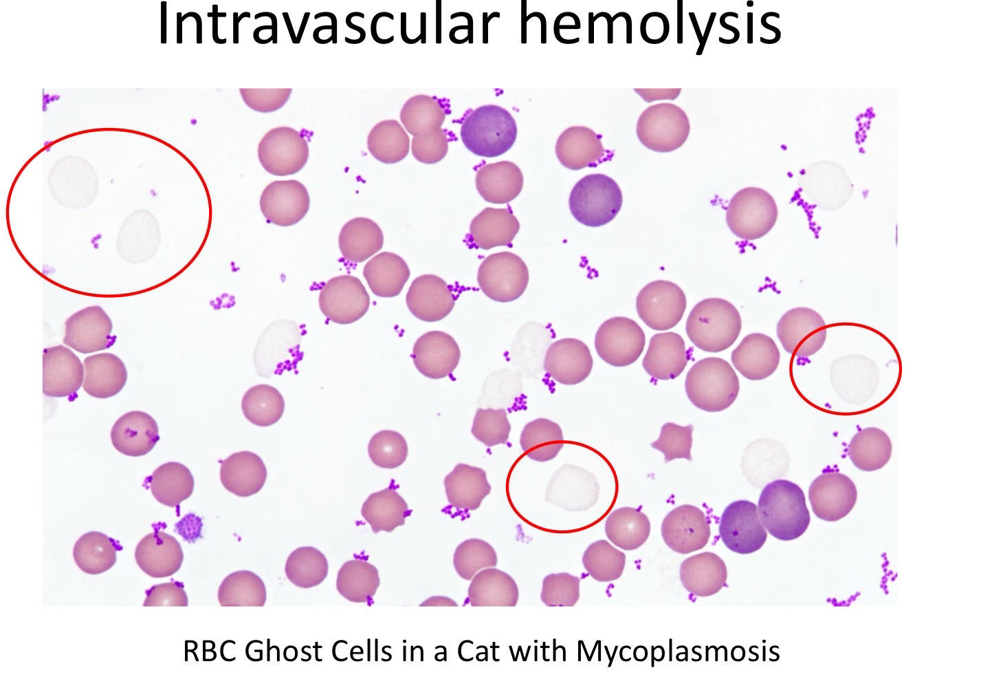
21
What response is usually seen with hemolytic anemia?
Moderate-to-severe regenerative anemia with reticulocytosis.
22
Which type of hemolysis is generally more common?
Extravascular hemolysis.
23
What happens to free Hgb during intravascular hemolysis?
It binds haptoglobin and is taken up by macrophages.
24
What happens when free Hgb overwhelms haptoglobin?
Hemoglobinemia in plasma develops, followed by hemoglobinuria (bloody urine)
25
What findings suggest extravascular hemolysis?
Hyperbilirubinemia, icterus, icteric plasma, and bilirubinuria.
26
What RBC findings support immune-mediated hemolytic anemia (IMHA)?
- autoantibodies macrophages destroy RBCs
- Spherocytes and agglutination.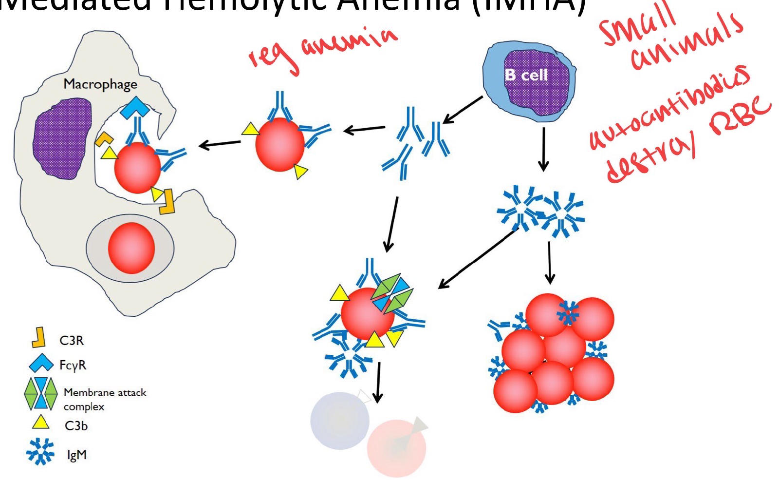
- Spherocytes and agglutination.
27
What RBC finding suggests oxidative hemolysis?
Heinz bodies, best detected with new methylene blue.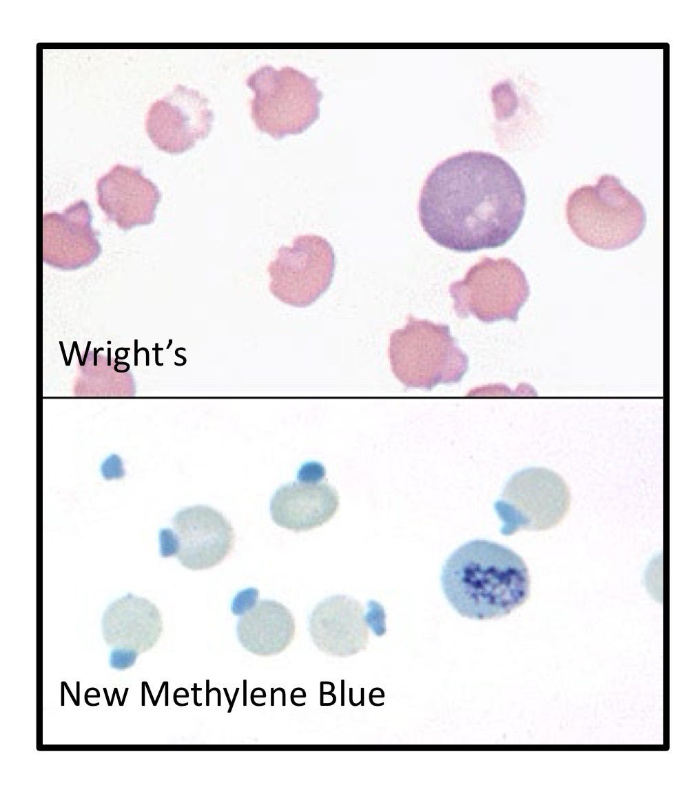
28
What RBC finding can occur with severe intravascular or oxidative hemolysis?
Ghost cells.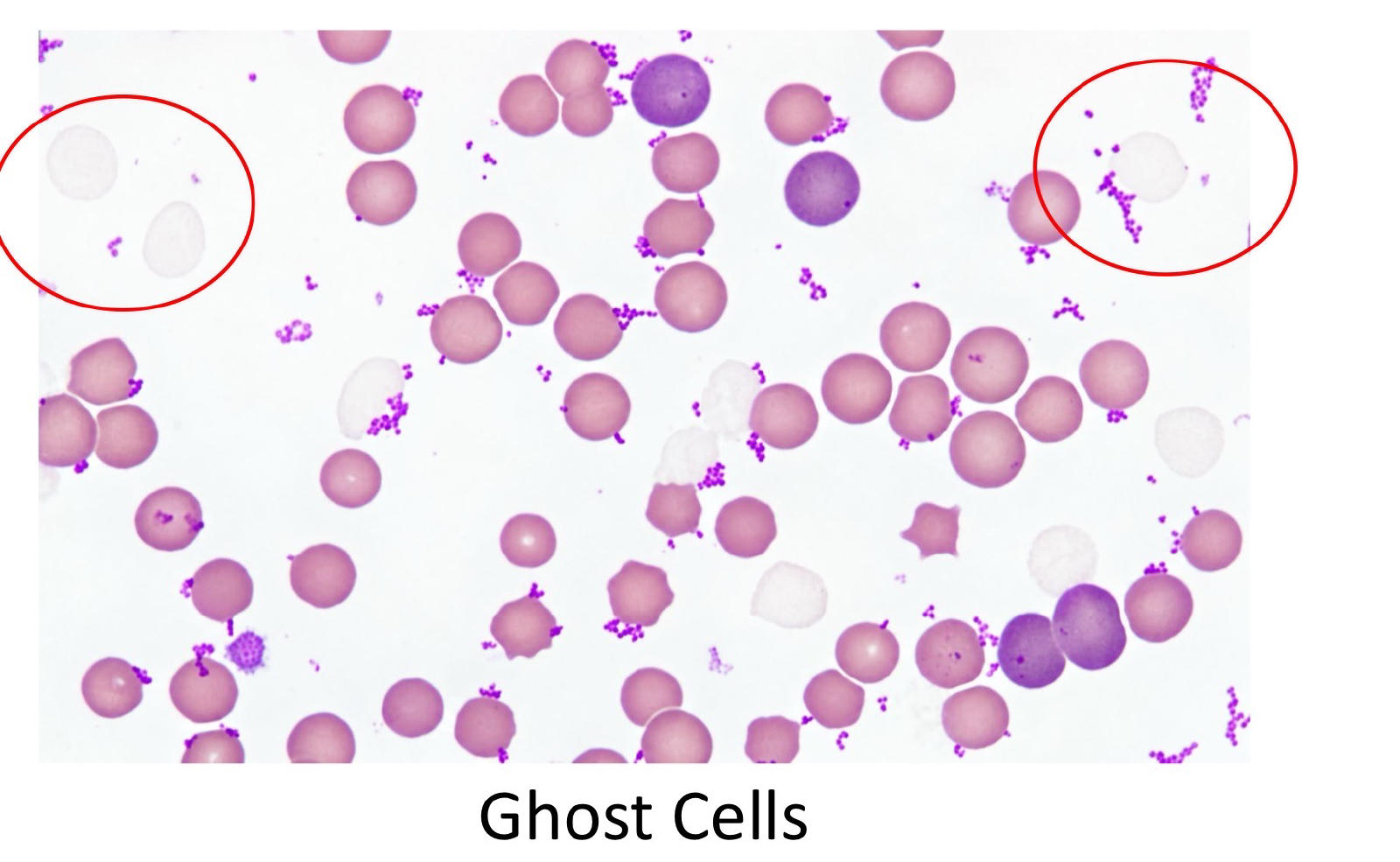
29
What RBC shapes suggest mechanical lysis?
Keratocytes and schistocytes.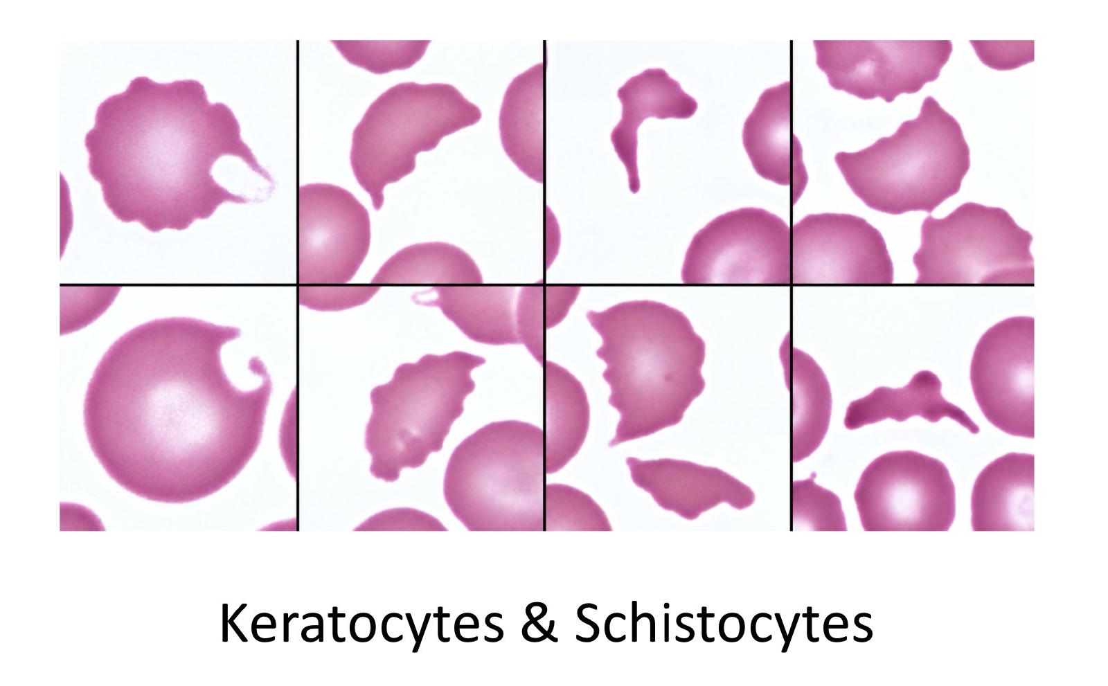
30
What is microangiopathic hemolytic anemia?
Mechanical RBC fragmentation as cells encounter abnormal small vessels or fibrin.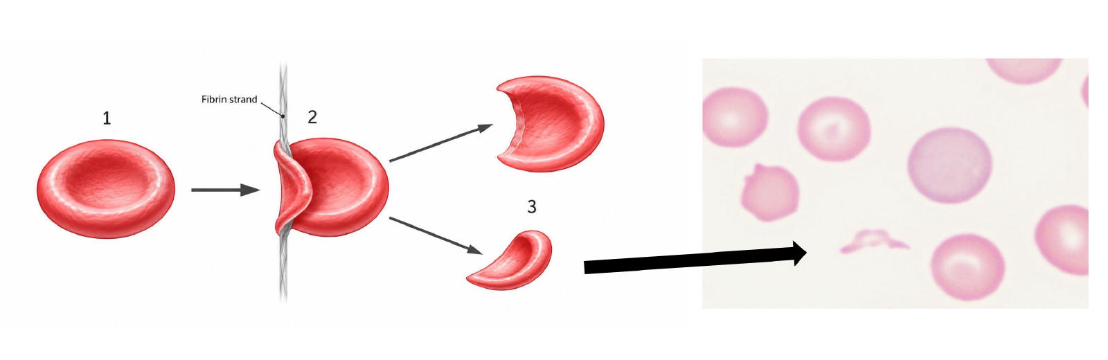
31
What is the laboratory hallmark of hemolysis?
Regenerative anemia with normal plasma protein.
32
What additional tests can investigate hemolysis?
Coombs’ test for immune-mediated anemia, Heinz body stain, PCR for organisms, and specialized RBC testing.
33
What are the two broad mechanisms of non-regenerative anemia?
Decreased erythropoiesis and ineffective erythropoiesis.
34
What are major causes of decreased RBC production?
Inflammation, CKD (chronic kidney disease), iron deficiency, and primary bone marrow disease.
35
What findings characterize non-regenerative anemia? (3)
Minimal polychromasia, few/no nRBCs, and an inadequate reticulocyte response.
36
What RBC changes can occur with iron deficiency?
Hypochromasia, target cells, schistocytes, and keratocytes.
37
What RBC changes can occur with bone marrow disease?
Elliptocytes, dacrocytes, schistocytes, and keratocytes.
38
What is the most common cause of non-regenerative anemia?
Anemia of inflammation.
39
How does hepcidin cause anemia of inflammation?
It traps iron in macrophages and decreases intestinal iron absorption.
40
How else does inflammation decrease RBC mass?
↓EPO production/ response and shortened RBC lifespan.
41
What is the typical anemia of inflammation in lab findings?
Mild normocytic-normochromic non-regenerative anemia with increased marrow iron.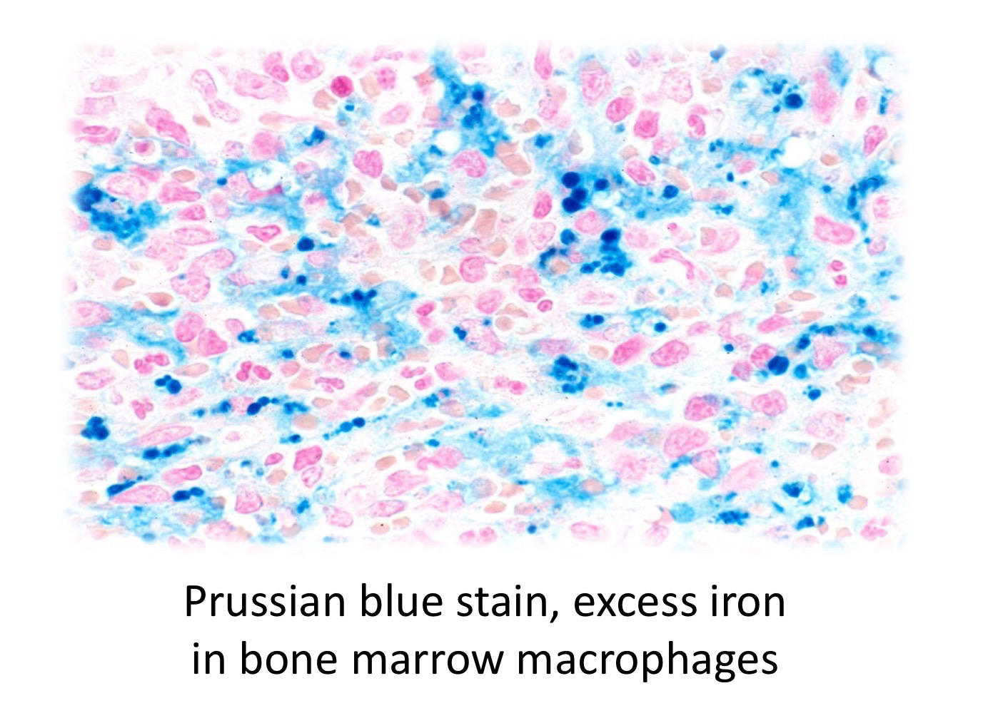
42
What 3 conditions can cause anemia of inflammation?
Infection, sterile inflammation, and immune-mediated disease.
43
What is the major cause of anemia in chronic kidney disease? What other mechanisms contribute to CKD anemia? (5)
Decreased EPO production.
- Iron restriction, uremic toxins, shortened RBC lifespan, inflammation, and GI hemorrhage
- Iron restriction, uremic toxins, shortened RBC lifespan, inflammation, and GI hemorrhage
44
What laboratory pattern supports CKD anemia?
↑BUN/creatinine with moderate-to-severe normocytic-normochromic anemia.
45
What RBC morphology can occur with uremia?
Echinocytosis.
46
What is the hallmark of iron deficiency anemia?
Microcytic-hypochromic RBCs.
47
What are major causes of iron deficiency?
Insufficient intake (neonates) or chronic external blood loss, especially GI bleeding.
48
Why does iron deficiency cause microcytosis?
Low Hgb synthesis causes erythroid precursors to divide extra times, lowering MCV.
49
Why does iron deficiency cause hypochromasia?
Insufficient Hgb production decreases MCHC.
50
How does iron deficiency progress over time?
Regenerative → microcytic-normochromic → microcytic-hypochromic.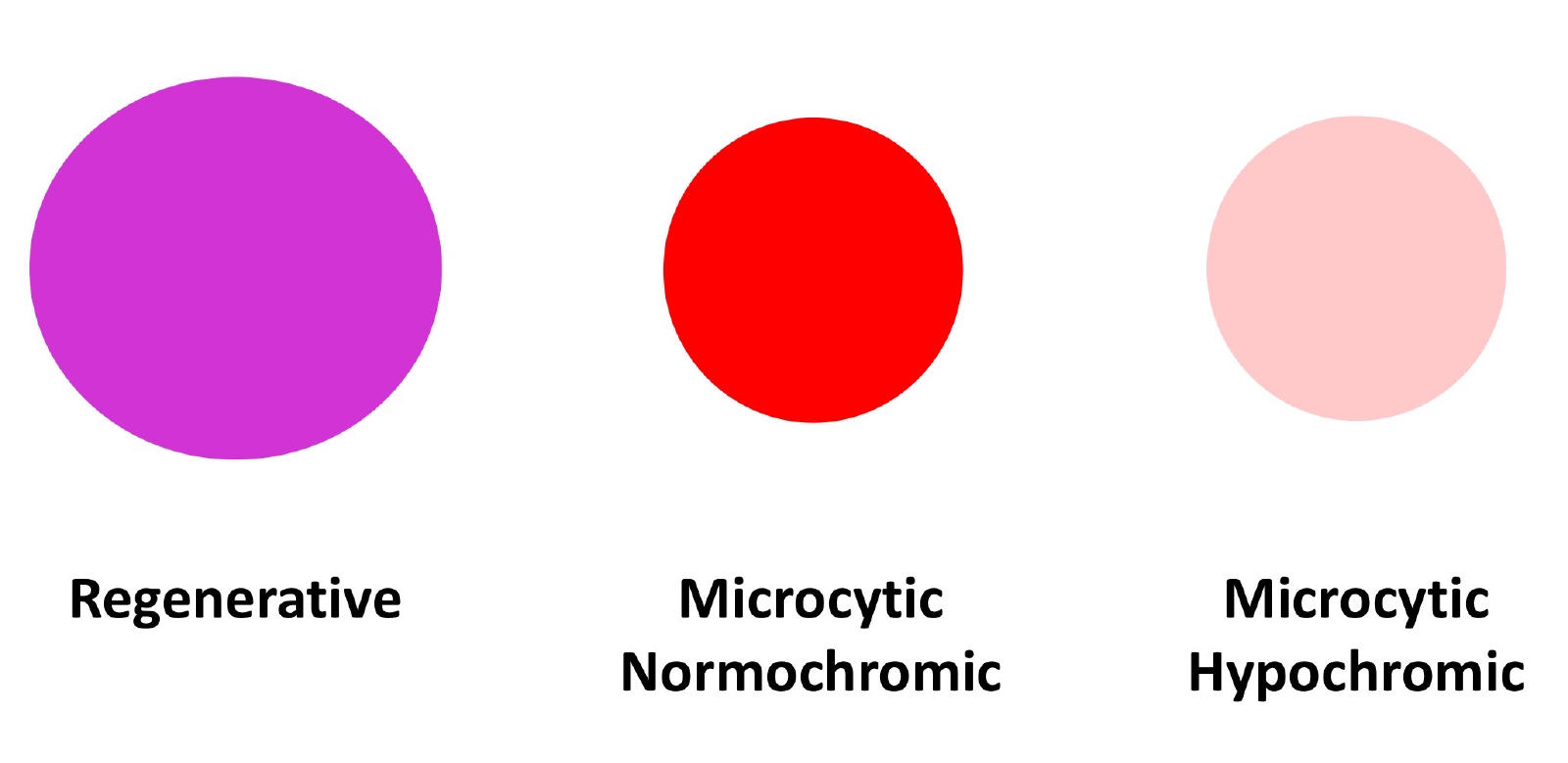
51
What smear findings support iron deficiency?
Microcytes, target cells, hypochromasia, schistocytes, and keratocytes (RBC fragmentation)
52
What platelet change often accompanies iron deficiency?
Thrombocytosis (increases platelets)
53
What findings raise suspicion for primary bone marrow disease?
Moderate-to-severe anemia with concurrent cytopenias, such as pancytopenia.
54
What mechanisms can cause bone marrow disease?
Myelophthisis, altered microenvironment, toxins, immune-mediated disease, and myelofibrosis.
55
What is the usual pattern of decreased/ineffective erythropoiesis?
Normocytic-normochromic-nonregenerative; iron deficiency is the major exception.
56
What is erythrocytosis?
An increased hematocrit.
57
What causes relative (common) erythrocytosis?
Dehydration or splenic contraction.
58
How can you recognize dehydration-associated erythrocytosis?
Both HCT and plasma proteins increase.
59
What is absolute erythrocytosis?
A true increase in RBC mass.
60
What causes primary vs. secondary absolute erythrocytosis?
Primary = RBC neoplasia, polycythemia vera; secondary = increased EPO from hypoxia or rare tumors.
More Medicine & health flashcards
Oncología: Cáncer de PáncreasMedicine & health · SpanishFERNANDACardiovascular Pathology and Muscle PhysiologyMedicine & health · EnglishTerrenceNeurodegeneration and Movement ControlMedicine & health · EnglishCorypares craneales Medicine & health · SpanishAshleyArrhythmiasMedicine & health · Englishcathsimsmišice nogeMedicine & health · LithuanianemAntimicrobial Drugs Part 1Medicine & health · EnglishKatlynKEChirurgie Plastique et Reconstructrice Post-CancerMedicine & health · FrenchClaraPre arrival task: Anatomical MovementsMedicine & health · EnglishLeneGeneticsMedicine & health · EnglishMadiShock/ CompensationMedicine & health · EnglishBrittanyInfectionMedicine & health · Indonesiancgilnursing
Study these decks in the app
Free on iPhone, Android, Mac and the web. Add any deck to your account in one tap.