Neurodegeneration and Movement Control
Study these flashcardsFree · spaced repetition · iPhone, Android, Mac & web
All cards
1
Causes of Cellular Injury in Neurodegeneration
• Oxidative Stress
• Mitochondrial Dysfunction
• Excitotoxicity and calcium
• Neurotrophic factors
• Microglia and cytokines
• Mitochondrial Dysfunction
• Excitotoxicity and calcium
• Neurotrophic factors
• Microglia and cytokines
2
Necrosis
– Ion fluxes across the plasma membrane
– Activation of Ca2+-dependent proteases
– Mitochondrial disruption
– Loss of ATP
– Cellular swelling
– Preservation of nuclear DNA
– Activation of Ca2+-dependent proteases
– Mitochondrial disruption
– Loss of ATP
– Cellular swelling
– Preservation of nuclear DNA
3
Apoptosis
– Cell shrinkage
– Maintenance of the plasma membrane
– Cytoskeletal depolymerization
– Fragmentation of nuclear DNA
– Requires protein synthesis
– Maintenance of the plasma membrane
– Cytoskeletal depolymerization
– Fragmentation of nuclear DNA
– Requires protein synthesis
4
Causes of cell death
– Depletion of energy stores
– Failure of ion pumps
– Neuronal depolarization
– Increases in intracellular calcium
– Free radical formation
– Acidosis
– Protein aggregation
– ER Stress Response
– Autophagy
– Failure of ion pumps
– Neuronal depolarization
– Increases in intracellular calcium
– Free radical formation
– Acidosis
– Protein aggregation
– ER Stress Response
– Autophagy
5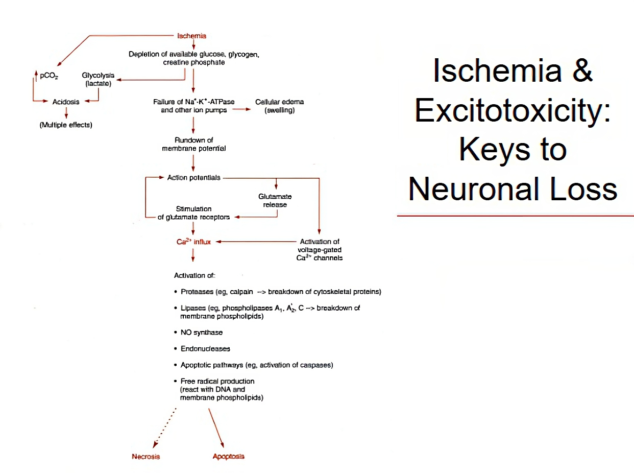
Image occlusion: name the hidden labels
6
Image occlusion: name the hidden labels
7
Image occlusion: name the hidden labels
8
Image occlusion: name the hidden labels
9
Image occlusion: name the hidden labels
10
Image occlusion: name the hidden labels
11
Image occlusion: name the hidden labels
12
Image occlusion: name the hidden labels
13
Image occlusion: name the hidden labels
14
Common Factors in Neurodegenerative Disease
• Oxidative stress
• Axonal Injury
• Accumulation of Cytoskeletal Proteins
• Pathology presents as a result of cell death
• Age-related onset
• Disaster theory???
• Axonal Injury
• Accumulation of Cytoskeletal Proteins
• Pathology presents as a result of cell death
• Age-related onset
• Disaster theory???
15
Protein Aggregation: Cell Death
• Proteasome proteolyses ubiquitinated substrates
• It is an endopeptidase complex that can be compared to a ‘trash can’
• Undegradable protein aggregates play a role in a number of neurodegenerative diseases
– Polyglutamine repeats
– Parkin
• Uncleavable aggregates lead to cellular dysfunction and death
• It is an endopeptidase complex that can be compared to a ‘trash can’
• Undegradable protein aggregates play a role in a number of neurodegenerative diseases
– Polyglutamine repeats
– Parkin
• Uncleavable aggregates lead to cellular dysfunction and death
16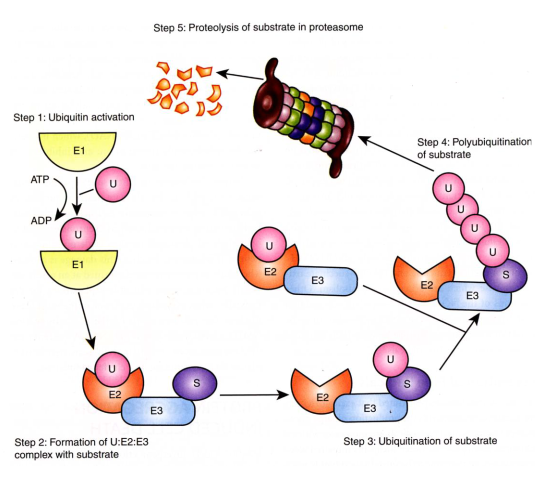
Image occlusion: name the hidden labels
17
Image occlusion: name the hidden labels
18
Image occlusion: name the hidden labels
19
Image occlusion: name the hidden labels
20
Brain Anatomy and Movement Control
• Movement control operates primarily through the basal ganglia
• Anatomy and physiology of basal ganglia are extremely complicated
• Nobody really knows how it all works
• Underlying hypothesis do exist and are testable
• Much of what we do know comes from the study of disease
• Anatomy and physiology of basal ganglia are extremely complicated
• Nobody really knows how it all works
• Underlying hypothesis do exist and are testable
• Much of what we do know comes from the study of disease
21
Basal Ganglia Structures
• Receives input from the Cerebral cortex
• Striatum (including caudate nucleus and putamen)
• Global Pallidus (lateral and medial)
• Subthalamic nucleus
• Substantia nigra (pars compacta and pars reticulata)
• Output goes from the thalamus back to the cerebral cortex
• Striatum (including caudate nucleus and putamen)
• Global Pallidus (lateral and medial)
• Subthalamic nucleus
• Substantia nigra (pars compacta and pars reticulata)
• Output goes from the thalamus back to the cerebral cortex
22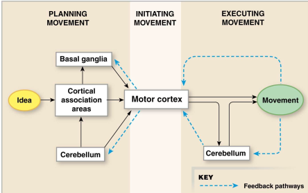
Image occlusion: name the hidden labels
23
Image occlusion: name the hidden labels
24
Image occlusion: name the hidden labels
25
Main Neurotransmitters of the Basal Ganglia
• γ-amino butyric acid (GABA)
• Acetylcholine (Ach)
• Dopamine (Da)
• Glutamate (Glu)
• Adenosine (Ad)
• Neurokinins (Substance P, SP)
• Opiod Peptides (endorphins/met-enkephalin, Enk)
• Acetylcholine (Ach)
• Dopamine (Da)
• Glutamate (Glu)
• Adenosine (Ad)
• Neurokinins (Substance P, SP)
• Opiod Peptides (endorphins/met-enkephalin, Enk)
26
Basal Ganglia Connections
• SNC - Substantia Nigra Compacta
• STN – Subthalamic nucelus
• LGP – Lateral Global Pallidus
• SNR – Substantia Nigra Reticulata
• MGP Medial Global Pallidus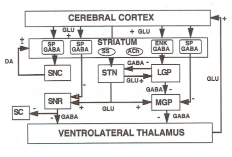
• STN – Subthalamic nucelus
• LGP – Lateral Global Pallidus
• SNR – Substantia Nigra Reticulata
• MGP Medial Global Pallidus
27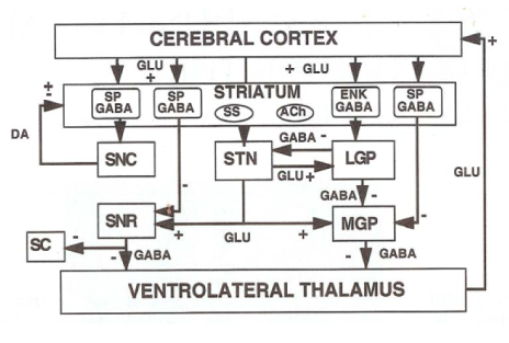
Image occlusion: name the hidden labels
28
Image occlusion: name the hidden labels
29
Image occlusion: name the hidden labels
30
Image occlusion: name the hidden labels
31
Image occlusion: name the hidden labels
32
Image occlusion: name the hidden labels
33
Image occlusion: name the hidden labels
34
Direct Movement Control
• Direct movement control is the act of higher function to exact a positive change in activity (Start/Stop)
• Filled arrows are increased during movement control
• Open arrows are reduced during movement control
• Increased Glutamate outflow from Ventrolateral Thalamus leads to reinforcement of the decision to move.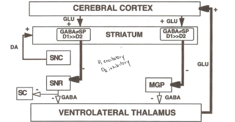
• Filled arrows are increased during movement control
• Open arrows are reduced during movement control
• Increased Glutamate outflow from Ventrolateral Thalamus leads to reinforcement of the decision to move.
35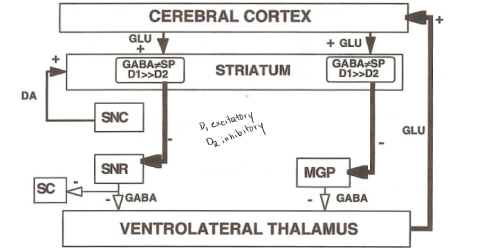
Image occlusion: name the hidden labels
36
Image occlusion: name the hidden labels
37
Image occlusion: name the hidden labels
38
Image occlusion: name the hidden labels
39
Image occlusion: name the hidden labels
40
Indirect Movement Control
• Indirect movement control is best thought of as the inhibition of non-required movement
• Thick with closed arrow (increased), Thin with open arrow (decreased)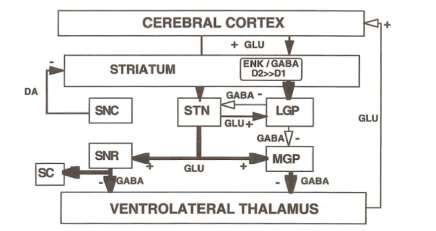
• Thick with closed arrow (increased), Thin with open arrow (decreased)
41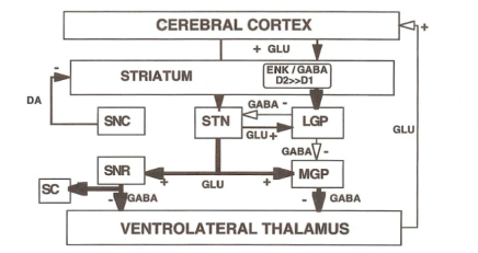
Image occlusion: name the hidden labels
42
Image occlusion: name the hidden labels
43
Image occlusion: name the hidden labels
44
Image occlusion: name the hidden labels
45
Dopaminergic Transmission
• Dopamine Transporter
• Vesicular monoamine transporter
• Dopamine receptors
– Two families
– Five types
– D1 & 5 vs D2,3,4
• Monoamine oxidase (MAO)
• Catechol-O-Methyl Transferase (COMT)
• Vesicular monoamine transporter
• Dopamine receptors
– Two families
– Five types
– D1 & 5 vs D2,3,4
• Monoamine oxidase (MAO)
• Catechol-O-Methyl Transferase (COMT)
46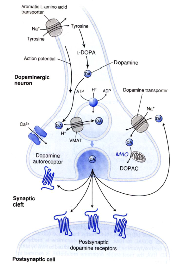
Image occlusion: name the hidden labels
47
Image occlusion: name the hidden labels
48
Image occlusion: name the hidden labels
49
Image occlusion: name the hidden labels
50
Image occlusion: name the hidden labels
51
Dopamine Receptors
• All types metabotropic receptors
• Two-main families
– D1-like - link to $G_\alpha s$- increase adenylate cyclase activity - indirect excitatory effect - D1 & D5
– D2-like - link to $G_\alpha i$ - increase phosphodiesterase activity - indirect inhibitory effect (induce IPSP) - D2, D3, & D4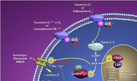
• Two-main families
– D1-like - link to $G_\alpha s$- increase adenylate cyclase activity - indirect excitatory effect - D1 & D5
– D2-like - link to $G_\alpha i$ - increase phosphodiesterase activity - indirect inhibitory effect (induce IPSP) - D2, D3, & D4
52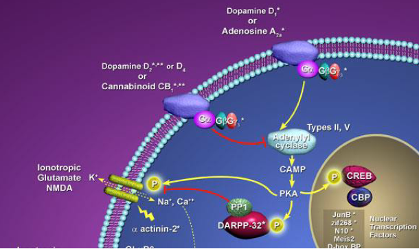
Image occlusion: name the hidden labels
53
Image occlusion: name the hidden labels
54
Image occlusion: name the hidden labels
55
Pharmacology of D-Receptors
• Pharmacological targeting of D-receptors forms the basis of many CNS drugs
• Anti-psychotics
– Typical (Haloperidol) and Atypical (Aripiprazole)
• Anti-emetics - metoclopramide
• Anti-arrythmics - procainamide
• Antidepressants - imipramine
• Anti-Parkinsonians - pramipexole
• Anti-psychotics
– Typical (Haloperidol) and Atypical (Aripiprazole)
• Anti-emetics - metoclopramide
• Anti-arrythmics - procainamide
• Antidepressants - imipramine
• Anti-Parkinsonians - pramipexole
56
Movement Disorders
• Basal ganglia
• Movement control and Neurotransmission
• Dopaminergic Signaling
– Pathway
– Receptors
– Functions
• Hypo and Hyperkinetic Diseases
– Pathology
– Dopaminergics
– Other therapies
• Movement control and Neurotransmission
• Dopaminergic Signaling
– Pathway
– Receptors
– Functions
• Hypo and Hyperkinetic Diseases
– Pathology
– Dopaminergics
– Other therapies
57
Hypokinesia (dyskinesia)
abnormally decreased mobility or motor function (both start and stop)
58
Hyperkinesia
abnormally increased muscular activity
59
Bradykinesia
slowed ability to start or continue movement
60
Chorea
irregular, unpredictable, and involuntary muscle jerks (distinct form Tics which are coordinated abnormal movements)
61
Athetosis
slow and writhing abnormal movements
62
Dystonia
abnormal posture resulting form tonic contractions
63
Parkinson’s Disease
• Classic Hypokineticdisorder
• Associated dystonia
• Essential Tremor
• Young and Older onset forms of disease
• Five stages
• Associated dystonia
• Essential Tremor
• Young and Older onset forms of disease
• Five stages
64
Parkinson’s Disease (Causes)
• Environmental factors
– Exogenous toxins (MPTP)
– Trauma
• Genetic factors
– Low concordance rates
– Multiple potential targets
• Parkin, UCHL1, $\alpha$-synuclein, Leucine repeat rich kinase-2,
PTEN-induced kinase 1
• Mitochondrial DNA
• Italian kindred
– α-synuclein
– Exogenous toxins (MPTP)
– Trauma
• Genetic factors
– Low concordance rates
– Multiple potential targets
• Parkin, UCHL1, $\alpha$-synuclein, Leucine repeat rich kinase-2,
PTEN-induced kinase 1
• Mitochondrial DNA
• Italian kindred
– α-synuclein
65
Genetics of Parkinson’s Disease
• Parkin - E3 ubiquitin ligase
• UCHL1 – a deubiquitinating enzyme
• $\alpha$-synuclein – SNARE chaperone interacts with tubulin synaptic rearrangement
• Tau – microtubule associated protein
• Dardarin (LRRK2) – signaling protein that operates like Ras to control autophagy and induce apoptosis
• PTEN-induced kinase 1(PINK1) –serine/threonine kinase that is thought to protect cells from mitochondrial stress
• UCHL1 – a deubiquitinating enzyme
• $\alpha$-synuclein – SNARE chaperone interacts with tubulin synaptic rearrangement
• Tau – microtubule associated protein
• Dardarin (LRRK2) – signaling protein that operates like Ras to control autophagy and induce apoptosis
• PTEN-induced kinase 1(PINK1) –serine/threonine kinase that is thought to protect cells from mitochondrial stress
66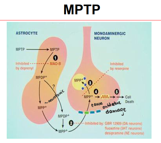
Image occlusion: name the hidden labels
67
Image occlusion: name the hidden labels
68
Image occlusion: name the hidden labels
69
Image occlusion: name the hidden labels
70
Image occlusion: name the hidden labels
71
Image occlusion: name the hidden labels
72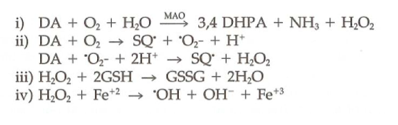
Dopamine Oxidation and Oxidative Stress
73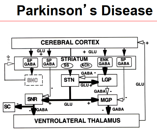
Image occlusion: name the hidden labels
74
Image occlusion: name the hidden labels
75
Image occlusion: name the hidden labels
76
Image occlusion: name the hidden labels
77
Image occlusion: name the hidden labels
78
Dopamine Therapy
• Carlsson (1957) showed DA in the striatum
• Reserpine in animals induced PD by striatal loss of DA
• Ehringer & Hornykielwicz (1960) DA i.v reduced akinesis of reserpine
• Cotzias (1967) found high dose oral levodopa effective in idiopathic PD.
• Reserpine in animals induced PD by striatal loss of DA
• Ehringer & Hornykielwicz (1960) DA i.v reduced akinesis of reserpine
• Cotzias (1967) found high dose oral levodopa effective in idiopathic PD.
79
Levodopa Metabolism
• Peripheral vs central conversion
• Carbidopa
– Inhibitor of AAAD
– Unable to cross Blood Brain Barrier
– Decreases peripheral conversion
– Increases central effect and reduces peripheral side effects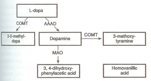
• Carbidopa
– Inhibitor of AAAD
– Unable to cross Blood Brain Barrier
– Decreases peripheral conversion
– Increases central effect and reduces peripheral side effects
80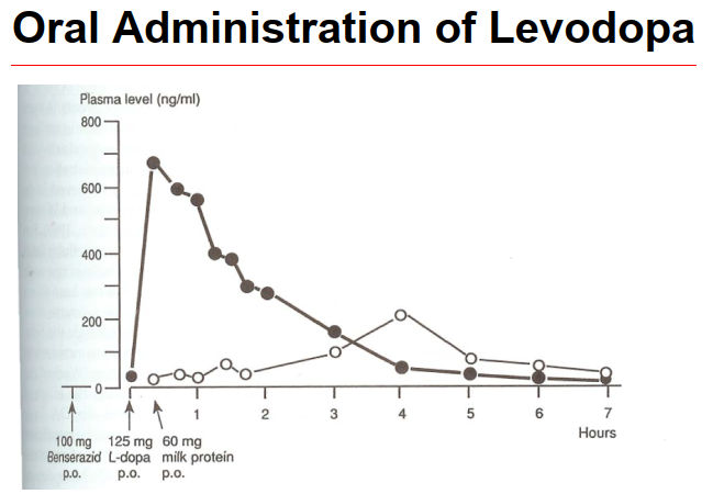
Image occlusion: name the hidden labels
81
Huntington’s Disease
• Choreas have been described for over 300 years
• Formally described in 1872 by George Huntington
• Occurs in 5 to 10 per 100,000 people
• Inherited as an autosomal dominant disorder
• Caused by trinucletoide repeats in gene IT15
• Disease usually starts in mid-thirties to mid-forties
• Personality change and adventitious movement develops into chorea, cognitive dysfunction and memory loss
• Death usually occurs as a result of secondary factors within 10 to 30 years
• Formally described in 1872 by George Huntington
• Occurs in 5 to 10 per 100,000 people
• Inherited as an autosomal dominant disorder
• Caused by trinucletoide repeats in gene IT15
• Disease usually starts in mid-thirties to mid-forties
• Personality change and adventitious movement develops into chorea, cognitive dysfunction and memory loss
• Death usually occurs as a result of secondary factors within 10 to 30 years
82
Excitotoxicity and Huntington’s
• Patients are often thin
– High metabolic rate??
• Striatal injection of NMDA agonists produces symptoms
• 3-nitropropionic acid (3-NP) complex II inhibitor produces striatal lesions
– High metabolic rate??
• Striatal injection of NMDA agonists produces symptoms
• 3-nitropropionic acid (3-NP) complex II inhibitor produces striatal lesions
83
Huntingtin (IT15 gene)
• Expressed in cytoplasm
• No clear function
• Binds to Huntingtin associated protein 1 (HAP1)
• Most common in NO synthesizing neurons
• Mutant form contains polyglutamine repeats (CAG)
• More repeats more severe disease
• Maybe caspase substrate
• Binds to Glyceraldehyde-3-phosphates dehydrogenase
• Polyglutamines bind to sugars causing aggregation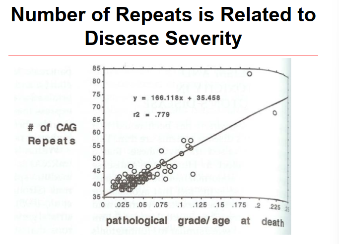
• No clear function
• Binds to Huntingtin associated protein 1 (HAP1)
• Most common in NO synthesizing neurons
• Mutant form contains polyglutamine repeats (CAG)
• More repeats more severe disease
• Maybe caspase substrate
• Binds to Glyceraldehyde-3-phosphates dehydrogenase
• Polyglutamines bind to sugars causing aggregation
84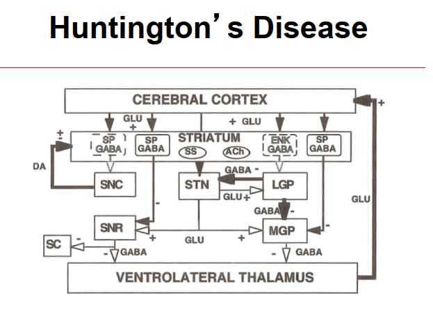
Image occlusion: name the hidden labels
85
Image occlusion: name the hidden labels
86
Image occlusion: name the hidden labels
87
Pharmacology for Huntington's
• NMDA antagonists
• Antioxidants (Omega-3 Fatty acids)
• Minocycline (apoptosis inhibitor)
• Neurotrophic growth factors (GDNF)
• Antidopaminergic (Tetrabenazine TBZ VMAT inhibitor)
• Inhibition of aggregation
– Disaccharides bind polyQ
– Trehalose
• Antipsychotics (Fluphenazine, D2 effect)
• Rapamycin (inhibit the kinase mTOR)
• Antioxidants (Omega-3 Fatty acids)
• Minocycline (apoptosis inhibitor)
• Neurotrophic growth factors (GDNF)
• Antidopaminergic (Tetrabenazine TBZ VMAT inhibitor)
• Inhibition of aggregation
– Disaccharides bind polyQ
– Trehalose
• Antipsychotics (Fluphenazine, D2 effect)
• Rapamycin (inhibit the kinase mTOR)
88
Symptomatic Pharmacology
• Early in disease depressed, irritable, anxious, or psychotic
– Fluoxetine
– Carbamazepine
– Caution can impair mobility
• Large-amplitude chorea
– Reserpine
– Benzodiazepines
• Juvenile onset and rigidity
– DA agonists
– Fluoxetine
– Carbamazepine
– Caution can impair mobility
• Large-amplitude chorea
– Reserpine
– Benzodiazepines
• Juvenile onset and rigidity
– DA agonists
89
Amyotrophic Lateral Sclerosis
• Disease onset usually occurs in mid-thirties to mid-fifties
• Presents as peripheral weakness and/or loss of function (heterogeneous presentation makes diagnosis difficulty)
• Leads to paralysis and death
• Disease progression is usually 2-3 years
• No known effective therapy
• Presents as peripheral weakness and/or loss of function (heterogeneous presentation makes diagnosis difficulty)
• Leads to paralysis and death
• Disease progression is usually 2-3 years
• No known effective therapy
90
Guamanian ALS-PDC
Guamanian ALS-PDC is a neurological disease that is characterized by a combination of symptoms of amyotrophic lateral sclerosis (ALS) and Parkinson's disease.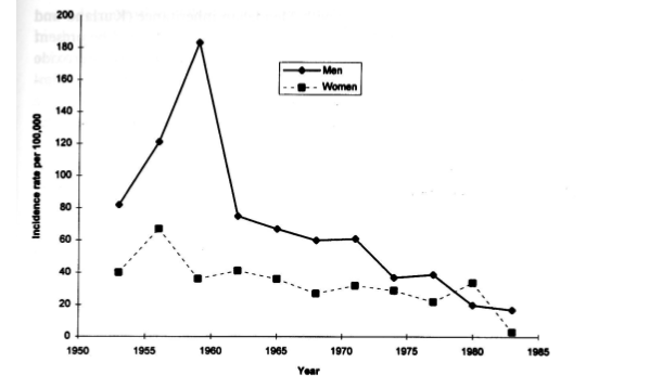
91
Age-dependency to ALS Survival
The older you are when diagnosed, the shorter you live afterward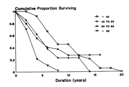
92
Gender Bias in ALS
More men are affected by ALS than women
93
SOD Mutation and ALS
It is believed that mutations in the SOD1 gene lead to the accumulation of toxic SOD1 protein aggregates in motor neurons, which ultimately results in the death of these neurons. This, in turn, leads to the progressive loss of muscle function and eventual paralysis, which are hallmark symptoms of ALS.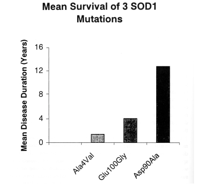
94
Image occlusion: name the hidden labels
95
Image occlusion: name the hidden labels
96
Image occlusion: name the hidden labels
97
Image occlusion: name the hidden labels
98
Image occlusion: name the hidden labels
99
Image occlusion: name the hidden labels
100
Measures of ALS Progression
• ALS-FRS
– Functional rating scale
– Considers upper and lower motor neuron loss and specific functional effects
– Disease progression related to phenotypic presentation
• Ultrasound
– Can determine loss of motor unit density
• Electromyography
– Adverse events, fasiculations, bloackage events
– Disorder recruitment
• Pre-FALS Study
– Identify biomarkers
– Functional rating scale
– Considers upper and lower motor neuron loss and specific functional effects
– Disease progression related to phenotypic presentation
• Ultrasound
– Can determine loss of motor unit density
• Electromyography
– Adverse events, fasiculations, bloackage events
– Disorder recruitment
• Pre-FALS Study
– Identify biomarkers
101
ALS Therapeutic Strategies
• Symptomatic
– BiPAP
– Mucolytic agents
– Antidepressants
– Mild Exercise
• Neuroprotection/Antioxidants
– Vitamin E
– Selegiline
– NAC
• Increased Mitochondrial Function
– Dexpramipexole
• Neurotrophic factors
– IGF-1
– Myotrophin
• Reduced Excitation
– Riluzole
• Increases ventilator free life
– Lamotrigine
• Voltage gated sodium channel blocker
– Ceftriaxone
• Increased glutamate uptake by astrocytic cells EAAT2 expression
– Retigabine
• Stimulate Potassium channel opening (hyperpolarize)
• Anti-inflammatory
– Mastinib
• CD117 tyrosine kinase inhibitor (myeloid cells)
– BiPAP
– Mucolytic agents
– Antidepressants
– Mild Exercise
• Neuroprotection/Antioxidants
– Vitamin E
– Selegiline
– NAC
• Increased Mitochondrial Function
– Dexpramipexole
• Neurotrophic factors
– IGF-1
– Myotrophin
• Reduced Excitation
– Riluzole
• Increases ventilator free life
– Lamotrigine
• Voltage gated sodium channel blocker
– Ceftriaxone
• Increased glutamate uptake by astrocytic cells EAAT2 expression
– Retigabine
• Stimulate Potassium channel opening (hyperpolarize)
• Anti-inflammatory
– Mastinib
• CD117 tyrosine kinase inhibitor (myeloid cells)
102
Riluzole
• Sodium channel blocker
• High voltage calcium channel blocker
• NMDA receptor antagonist
• Antagonized by pertussis toxin
– Potential GPCR
• Metabolized in liver by P450 enzymes
– Potential hepatotoxicity
• Slows down progression of disease ?
• High voltage calcium channel blocker
• NMDA receptor antagonist
• Antagonized by pertussis toxin
– Potential GPCR
• Metabolized in liver by P450 enzymes
– Potential hepatotoxicity
• Slows down progression of disease ?
103
Other Potential Therapies for ALS
• Tau-tubulin kinase inhibitors (TDP-43)
• Antisense oligos (C9orf72)
• Antisense FUS
• RAN antibodies
• Antisense oligos (C9orf72)
• Antisense FUS
• RAN antibodies