Eosinophil Biology and Maturation
Study these flashcardsFree · spaced repetition · iPhone, Android, Mac & web
All cards
1
What gives eosinophils their strong affinity for eosin?
Highly basic (cationic) proteins in the secondary granules, mainly Major Basic Protein
2
From which maturation stage can eosinophils first be recognized?
The promyelocyte/myelocyte stage
3
Which progenitor gives rise to eosinophils in the marrow?
How long does eosinophil development in the marrow take?
Which cytokines are the major players in eosinophil production?
How long does eosinophil development in the marrow take?
Which cytokines are the major players in eosinophil production?
1. CFU-Eo
2. About 2-6 days
3. IL-3, GM-CSF and IL-5
2. About 2-6 days
3. IL-3, GM-CSF and IL-5
4
Do eosinophils have a marrow maturation/storage pool?
Yes, and its size is species specific
5
What is the eosinophil blood half-life (T1/2) in dogs?
Does a marginal pool exist for eosinophils?
Does a marginal pool exist for eosinophils?
1. Less than 1 hour
2. Yes
2. Yes
6
How does eosinophil tissue lifespan compare to neutrophils?
Longer (6+ days), and cytokines can extend it
7
Do eosinophils recirculate after leaving the blood?
No, just like neutrophils
8
Where do eosinophils preferentially reside?
What is the approximate blood:tissue eosinophil ratio? EXAM
What is the approximate blood:tissue eosinophil ratio? EXAM
Skin, GI tract and lung
About 1:200-300
About 1:200-300
9
Which chemokine is a chemoattractant for eosinophils?
Eotaxin
10
Which mast cell-derived tetrapeptides attract eosinophils?
ECF-A (eosinophil chemotactic factor-A)
11
Which chemotactic factors do eosinophils share with neutrophils?
LTB4, C5a, C567 and PAF
12
Which mediators from parasites or mast cells attract eosinophils?
Histamine, antigen/antibody complexes, and parasite or damaged tissue products
13
What is the major function of eosinophils against helminths?
Helminth killing (with antibody and complement as opsonization)
14
What is the regulatory role of eosinophils in type I hypersensitivity? How important is eosinophil phagocytosis of bacteria?
They phagocytose immune complexes and neutralize mast cell products
Very minor
Very minor
15
What condition is seen in a transtracheal wash with many eosinophils from a horse with Dictyocaulus arnfieldi?
Eosinophilic inflammation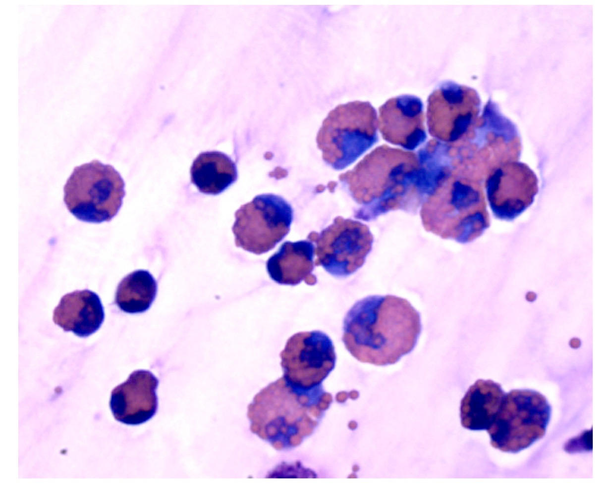
16
What causes eosinopenia? What effect can ____ have on eosinophilia from other causes?
Corticosteroids, as part of the stress leukogram
They can attenuate or dampen it
They can attenuate or dampen it
17
What are the main causes of eosinophilia?
Parasitism, allergies/hypersensitivities (skin, gut, lung) and sometimes fungal disease (esp. horses)
18
How does IgE/IgG-driven hypersensitivity cause eosinophilia?
Mast cell granule release is chemotactic to eosinophils
19
What happens to eosinophilia on second exposure to an antigen?
Sensitized T cells produce a more marked eosinophilia
20
Which neoplasms can cause paraneoplastic eosinophilia?
Mast cell tumors and lymphoma (especially T cell)
21
Is blood eosinophilia consistent with eosinophilic tissue inflammation? EXAM
No, and it is not proportional to severity of tissue disease
22
Why do basophils stain as they do?
Highly acidic (anionic) granule proteins with a strong affinity for basic stains
23
Which mediators do basophil granules contain?
Histamine, heparin and sulfated mucopolysaccharides
24
How do basophil granules appear in the dog and cat?
Less numerous in the dog and pale lavender in the cat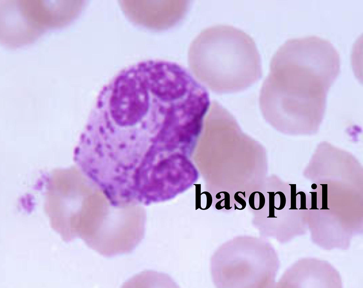
25
Which cells’ maturation and nuclear morphology do basophils parallel?
Neutrophils
26
Which progenitor gives rise to basophils?
CFU-Baso
27
Which cytokines drive basophil production? How large is the basophil marrow storage pool?
The same as eosinophils: IL-3, GM-CSF and IL-5
Minimal
Minimal
28
What is the approximate basophil blood half-life?
About 6 hours
29
What relationship exists between blood basophils and tissue mast cells?
An inverse relationship (e.g., cats vs. rabbits)
- a species with lots of basophils in the blood tends to have few mast cells in its tissues, and the reverse
- Rabbits have relatively high blood basophil numbers and few tissue mast cells.
• Cats have very few blood basophils and abundant tissue mast cells.
- a species with lots of basophils in the blood tends to have few mast cells in its tissues, and the reverse
- Rabbits have relatively high blood basophil numbers and few tissue mast cells.
• Cats have very few blood basophils and abundant tissue mast cells.
30
What triggers basophil degranulation?
Antigen binding and crosslinking of surface Ig (release of inflammatory mediators)
- An antigen (allergen) binds and links together two antibodies (IgE) already stuck to the basophil's surface, and this crosslinking triggers degranulation
- An antigen (allergen) binds and links together two antibodies (IgE) already stuck to the basophil's surface, and this crosslinking triggers degranulation
31
What is the only known source of heparin and activators of lipoprotein lipase?
Basophils
32
Why is basopenia not clinically recognized?
So few basophils are present normally
33
What conditions are associated with basophilia?
What does basophilia often accompany?
What does basophilia often accompany?
Lipid disorders, feline heartworm disease and myeloid neoplasms
Eosinophilia
Eosinophilia
34
Which are the largest cells in a blood smear?
Monocytes
35
How is monocyte nuclear and cytoplasmic morphology described?
Ameboid nuclei with abundant, blue-gray, often vacuolated cytoplasm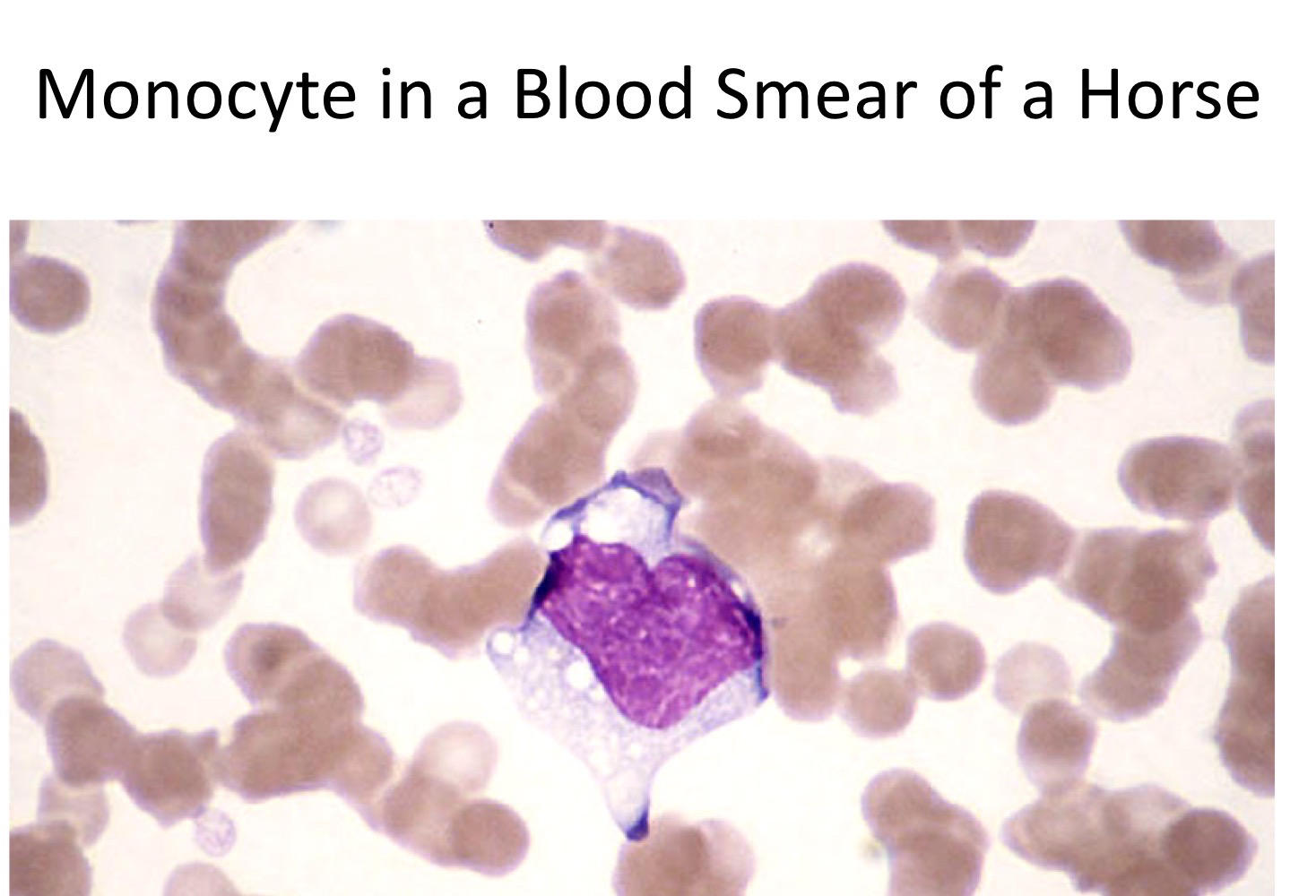
36
What can increased monocyte vacuolation indicate?
Activation, but rule out artefact from long EDTA storage
37
Which progenitor gives rise to monocytes?
The bi-potential CFU-GM
38
Which cytokines drive monocyte production?
IL-3, GM-CSF and M-CSF
39
Do monocytes have a marrow storage pool?
No, the blood is the storage pool for tissue macrophages
40
How long is monocyte blood circulation time?
About 24 hours
41
Do monocytes recirculate after entering tissue?
No, they become tissue macrophages
42
Which species have a monocyte marginal pool?
Human, mouse, dog and rabbit
43
Which functions do macrophages perform? 4
1. Phagocytosis (fungi, foreign material)
2. antigen presentation
3. cytokine production
4. removal of effete cells
5. catabolism of plasma proteins
2. antigen presentation
3. cytokine production
4. removal of effete cells
5. catabolism of plasma proteins
44
How well does blood monocytosis correlate with tissue macrophage accumulation?
Poorly, but it often occurs with inflammation
45
When can monocytosis occur during acute neutrophilic inflammation?
if there is associated tissue destruction and chronic inflammation
46
Which species consistently show steroid-induced monocytosis?
Dogs
47
Which persistent stimuli cause monocytosis?
Viral, fungal and atypical bacterial infections
48
Which diseases increase tissue demand for macrophages and cause monocytosis? 5
Which diseases increase tissue demand for macrophages and cause monocytosis? 5
1. Immune mediated disease
2. necrosis
3. malignancy
4. hemolysis
5. pyogranulomatous disease
2. necrosis
3. malignancy
4. hemolysis
5. pyogranulomatous disease
49
What is the clinical significance of monocytopenia?
None
50
Which fungal organisms were seen inside macrophages from a cat skin lesion?
Sporothrix, causing sporotrichosis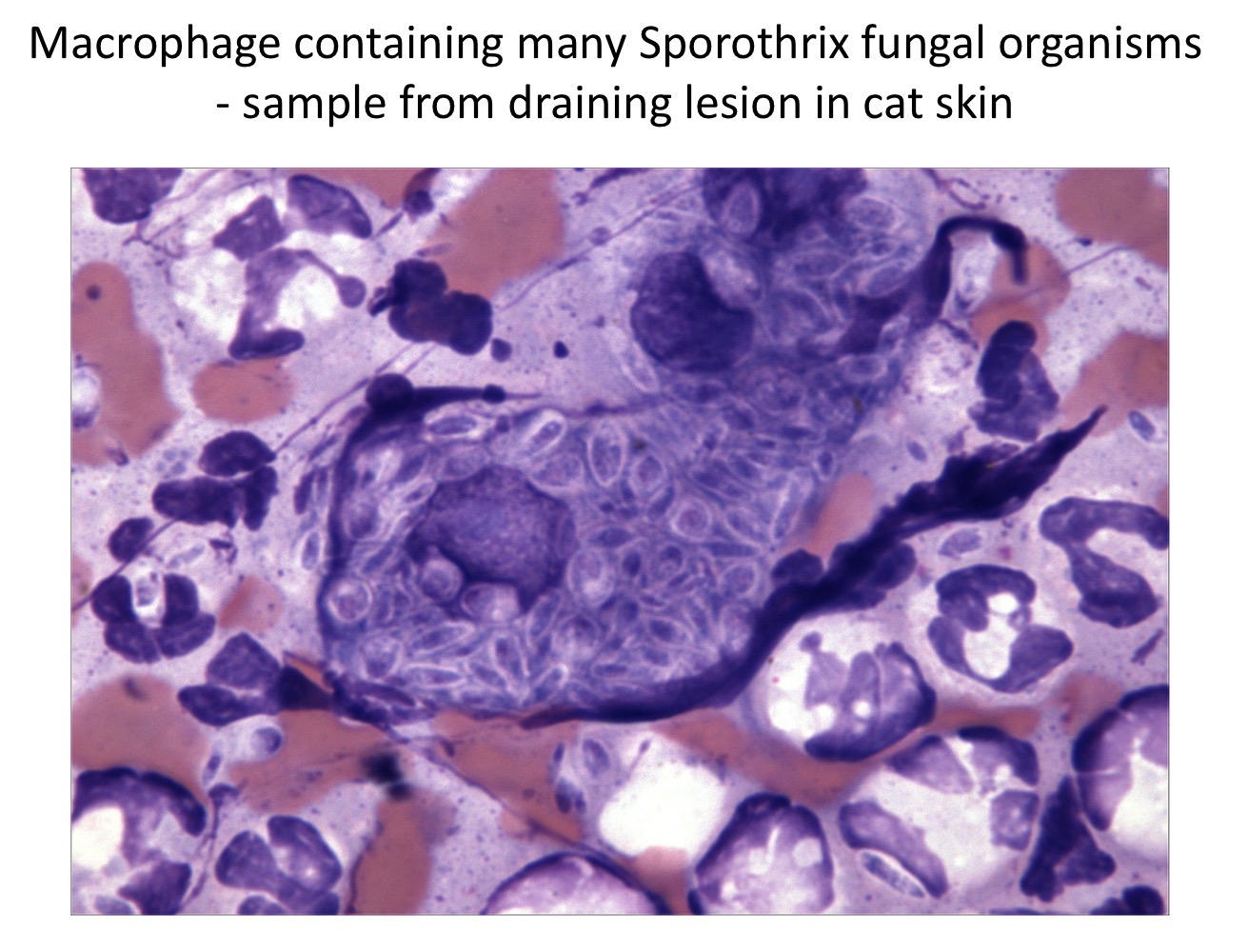
More Medicine & health flashcards
EPS⚕️Medicine & health · SpanishGonzálezFizyoloji: Sindirim ve PankreasMedicine & health · TurkishikranurAnxiety, OCD, and NeurotransmittersMedicine & health · EnglishTashaHematopoiesis and ErythropoiesisMedicine & health · EnglishelleezeeeeSerous Fluid and MembranesMedicine & health · EnglishkarebearTendon Disorders ExaminationMedicine & health · EnglishEshaHis Lab 6 - Nervous System Medicine & health · EnglishKrisMedCardsProduction RBC,WBCMedicine & health · EnglishBrittanyAcute GUMedicine & health · EnglishMadiType II DM MedicationsMedicine & health · EnglishcathsimsRETROPERITONEAL CAVITYMedicine & health · EnglishKrisMedCardsAcute GI d/oMedicine & health · EnglishMadi
Study these decks in the app
Free on iPhone, Android, Mac and the web. Add any deck to your account in one tap.