Veterinary Hematology: Anemia and Reticulocytes
Study these flashcardsFree · spaced repetition · iPhone, Android, Mac & web
All cards
1
What is anemia?
A decrease in red blood cell mass.
2
How does the body normally respond to anemia?
Hypoxia stimulates erythropoietin (EPO), causing increased erythropoiesis and reticulocyte release.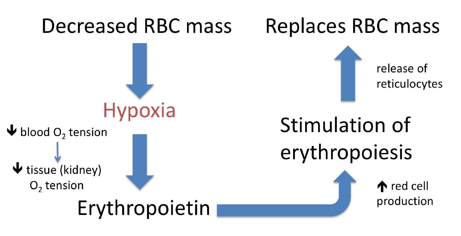
3
What bone marrow change occurs during a regenerative response to anemia?
Erythroid hyperplasia.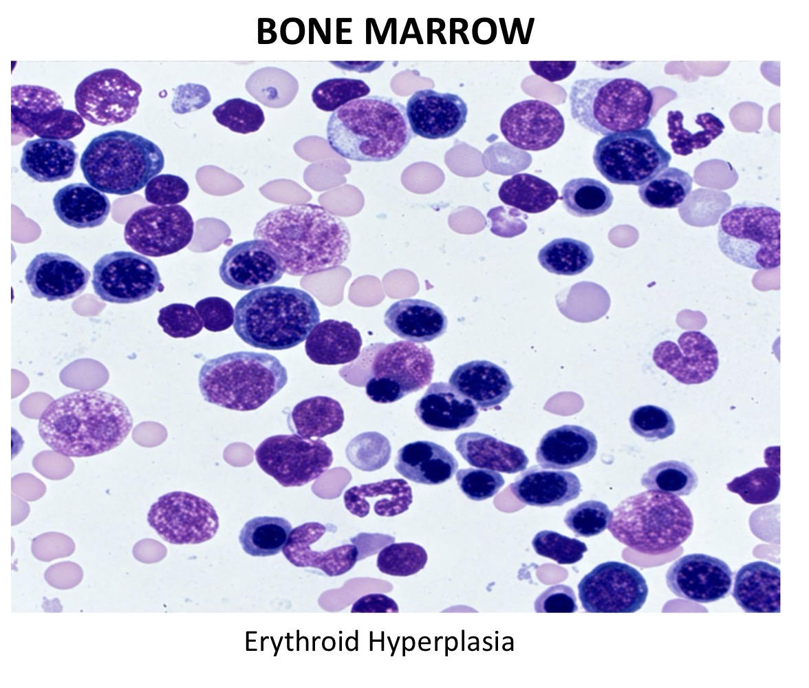
4
What are polychromatophilic RBCs (polychromatophils)?
Reticulocytes visible on a routine blood smear.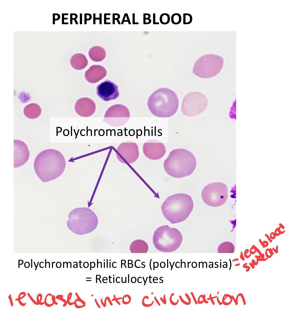
5
How do mammalian reticulocytes differ from mature RBCs?
They are larger and bluer/purpler because they contain residual RNA and less hemoglobin.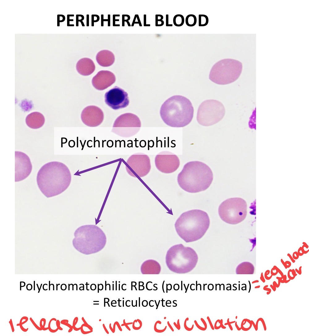
6
What is “reticulin” in a reticulocyte?
Residual RNA, ribosomes, and organelles that become visible with new methylene blue stain.
7
Which major veterinary species does NOT release reticulocytes into peripheral blood?
Horses.
8
What are the two types of feline reticulocytes?
Aggregate and punctate reticulocytes.
9
Which feline reticulocytes indicate current active regeneration and are counted?
Aggregate reticulocytes.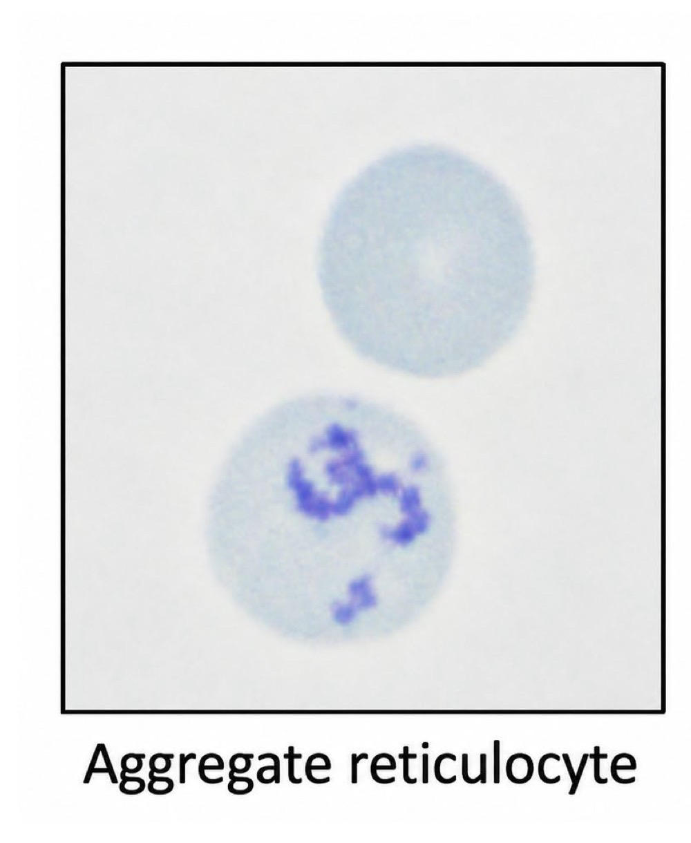
10
What are punctate reticulocytes in cats?
Older RBCs released about 2–3 weeks earlier that are not counted when assessing current regeneration.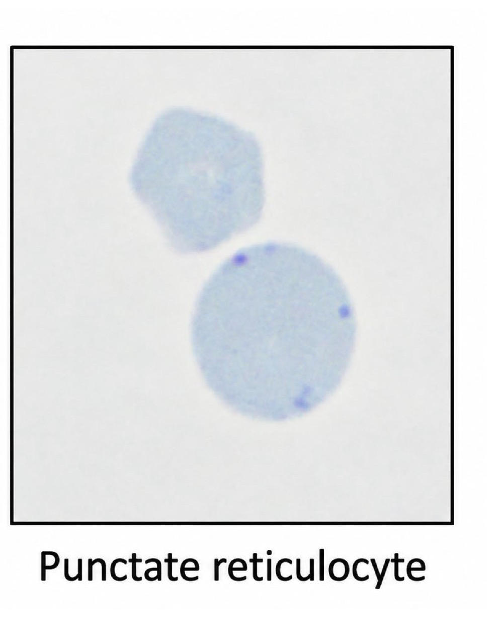
11
What is basophilic stippling?
Residual ribosomal complexes visible in RBCs with routine hematology stains.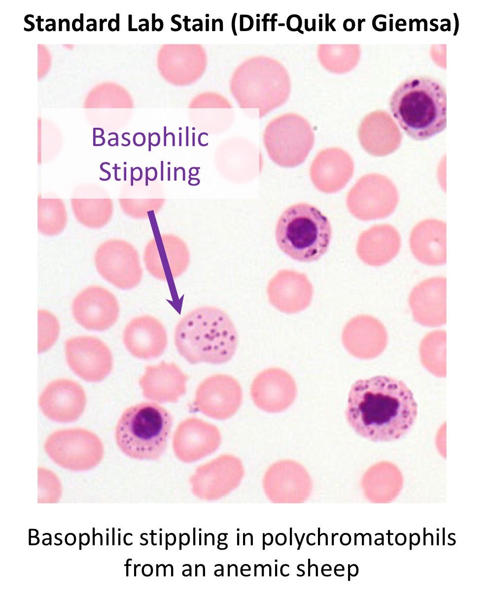
12
Besides regeneration, what important disease can cause basophilic stippling?
Lead toxicosis.
13
How do avian polychromatophils differ from mammalian reticulocytes in size?
Avian polychromatophils are smaller than mature RBCs.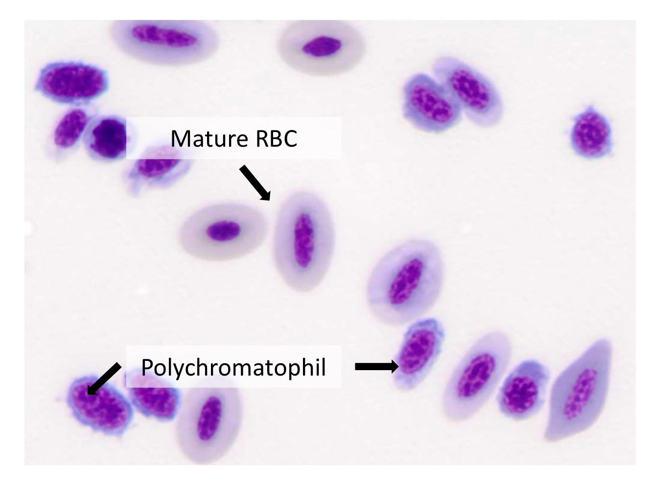
14
What factors determine the degree of reticulocytosis?
Severity and duration of anemia and the bone marrow’s ability to increase erythropoiesis.
15
How does severe anemia affect the regenerative response?
Lower HCT causes greater hypoxia and generally a stronger regenerative response.
16
How can marked reticulocytosis affect RBC indices?
It can increase MCV and decrease MCH/MCHC, producing a macrocytic, hypochromic pattern.
17
What 6 areas should be evaluated on an erythrogram?
RBC mass, regenerative response, plasma proteins, RBC indices, RBC morphology, and plasma color.
18
What is the best metric of RBC mass and easiest metric for identifying anemia?
Hematocrit (HCT/PCV).
19
Regenerative response in erythorgram ?
reticulocytes and NRBCs
20
What is the difference between HCT and PCV?
HCT is calculated from RBCC count × MCV; PCV is physically measured.
21
What does blood hemoglobin concentration best estimate?
The oxygen-carrying capacity of blood.
22
What is the normal approximate relationship between HCT and Hgb?
About 3:1; for example, HCT 30% ≈ Hgb 10 g/dL.
23
Why can Hgb be misleading during intravascular hemolysis? LOOK UP INFO
Free Hgb from lysed RBCs is still measured, altering the normal HCT:Hgb relationship.
24
What does MCV (mean corpuscular volume) measure? What is it helpful for classifying? (3)
Average RBC volume or size.
1. regeneration
2. nutritional/mineral associated anemia
3. FELV associated anemia
1. regeneration
2. nutritional/mineral associated anemia
3. FELV associated anemia
25
How are RBCs classified using MCV?
- Normal MCV = normocytic
- increased = macrocytic
- decreased = microcytic.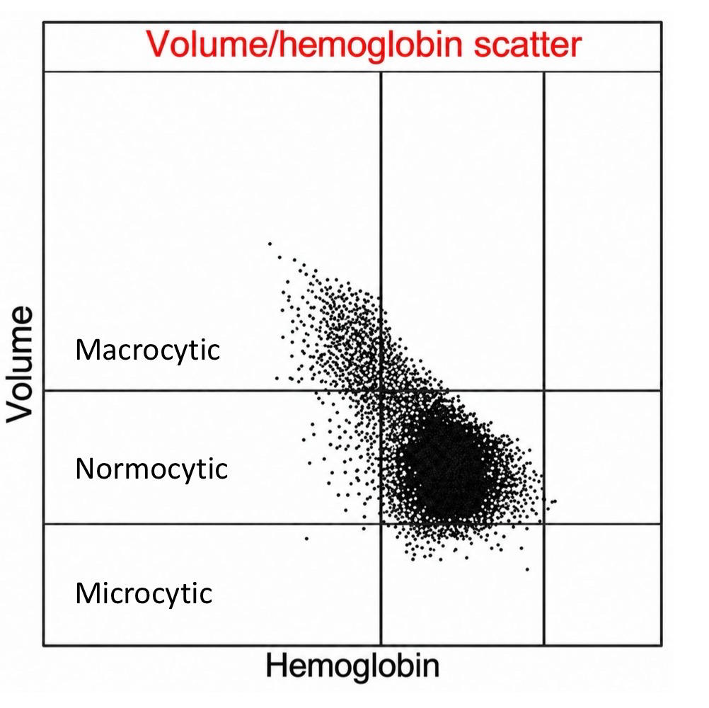
- increased = macrocytic
- decreased = microcytic.
26
What does MCHC (mean corpuscular hemoglobin concentration) measure? What is it helpful classifying?
Average hemoglobin concentration within RBCs.
1. Regeneration
2. Nutritional associated anemia
3. Hemolysis
1. Regeneration
2. Nutritional associated anemia
3. Hemolysis
27
How are RBCs classified using MCHC?
- Normal = normochromic
- Decreased = hypochromic.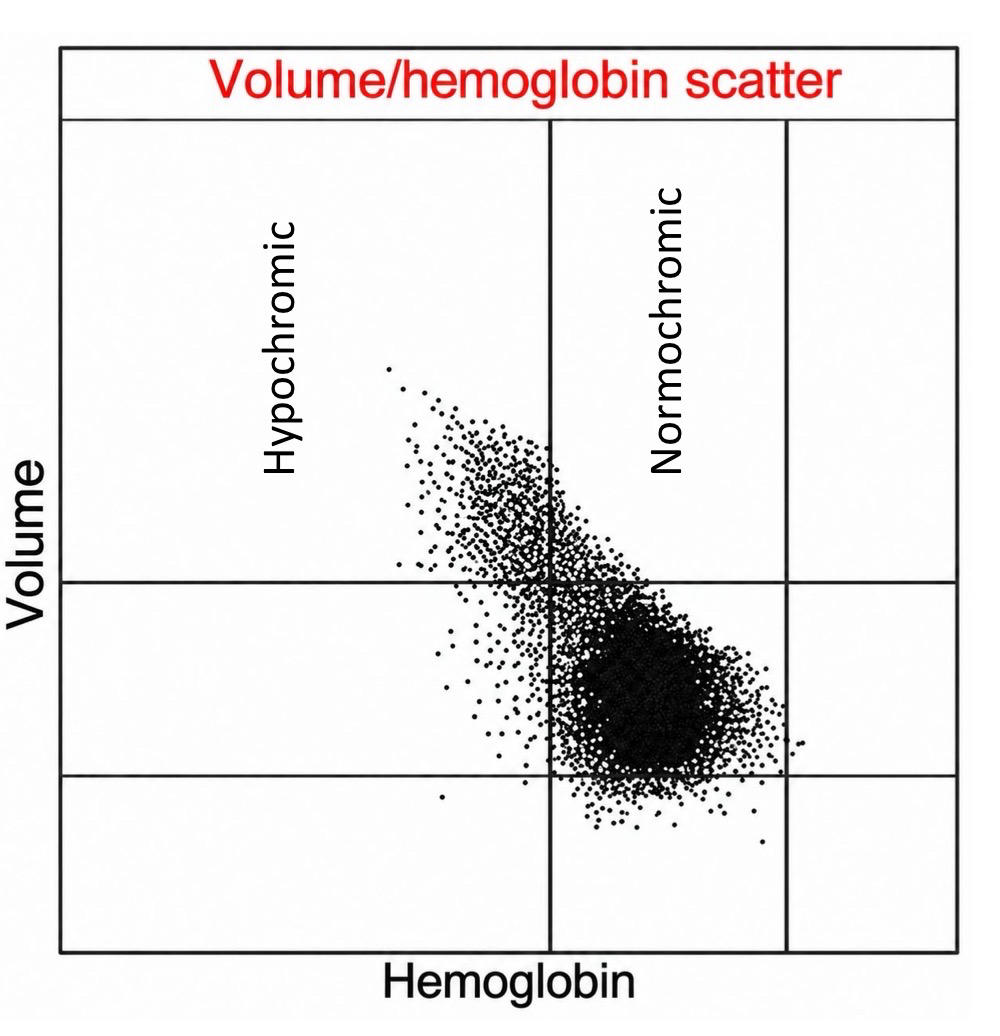
- Decreased = hypochromic.
28
What does an increased MCHC indicate?
It is not a true hyperchromic change and suggests hemolysis.
29
What does RDW (red cell distribution width) measure?
Variation in RBC size; it is the coefficient of variation of MCV.
30
What does increased RDW suggest when evaluating anemia?
Increased size variation (anisocytosis), which can support regeneration.
31
What is the BEST metric for assessing RBC regeneration?
Absolute reticulocyte count.
32
Why can an anemia initially appear non-regenerative? What is it called?
- It takes about 2–4 days after the stimulus for increased reticulocytes to appear in blood.
- “pre-regenerative” phase
- “pre-regenerative” phase
33
How long do circulating reticulocytes take to fully mature?
About 24–48 hours.
34
Which method of reticulocyte counting is preferred?
- Automated optical analysis
- Manual new methylene blue counting is more labor-intensive and less accurate.
- Manual new methylene blue counting is more labor-intensive and less accurate.
35
What should be evaluated on a blood smear besides regeneration?
1. RBC arrangement
2. Regeneration evidence
3. Morphology
4. Microorganisms.
2. Regeneration evidence
3. Morphology
4. Microorganisms.
36
Where should you look for large cells, platelet clumps, and microfilariae on a blood smear?
The feathered edge
37
Where are erythrocyte morphology and leukocyte differentials best evaluated?
body and monolayer of blood smear
38
What is the difference between rouleaux and agglutination?
Rouleaux is orderly stacking of RBCs; agglutination is irregular RBC clumping.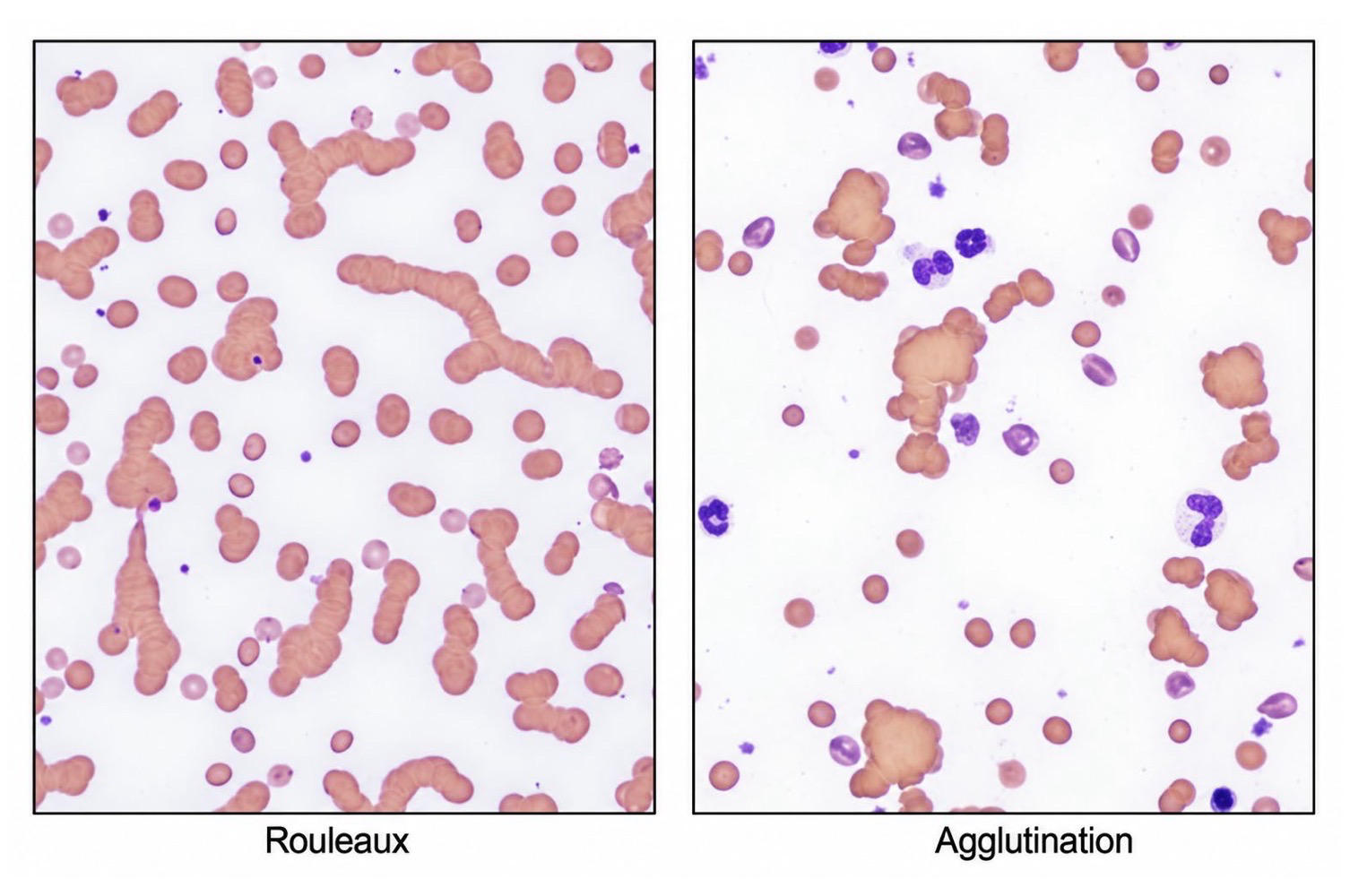
39
How does saline help distinguish rouleaux from agglutination?
Rouleaux disperses with saline; true agglutination persists.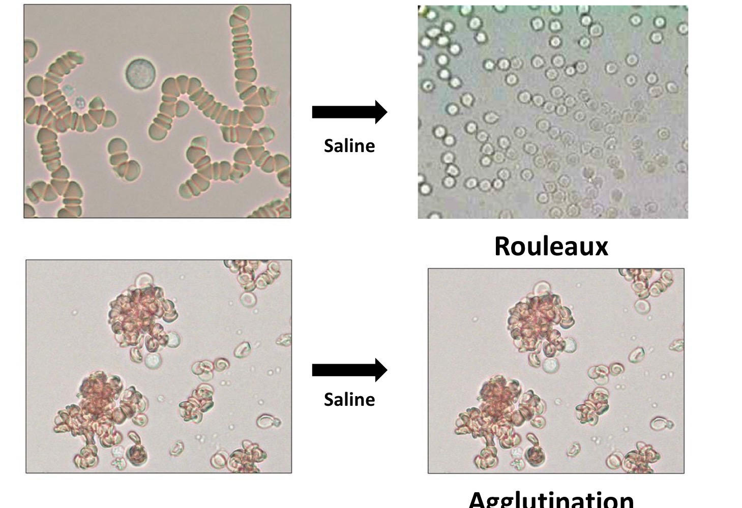
40
What blood smear findings support regeneration? (4)
Anisocytosis, polychromasia, nucleated RBCs, and sometimes basophilic stippling.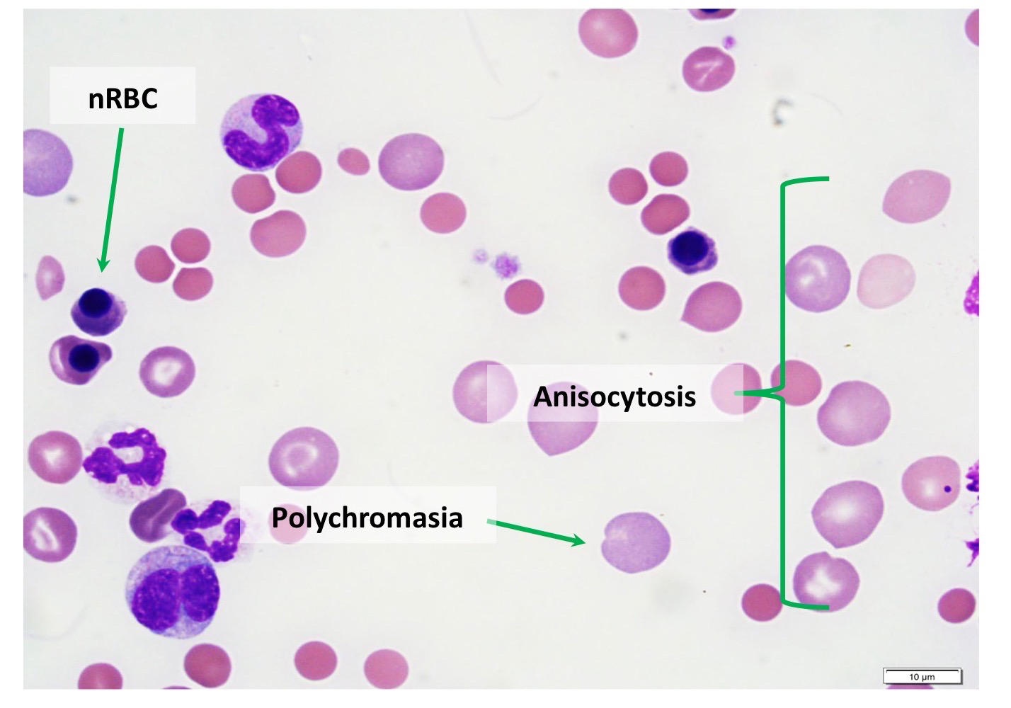
41
What causes appropriate rubricytosis?
A regenerative erythroid response.
42
What can cause inappropriate nucleated RBCs without regeneration? (4)
Glucocorticoids, splenic disease, heat stroke, or lead toxicosis.
43
Most common hemoparasites
Hemotrophic mycoplasma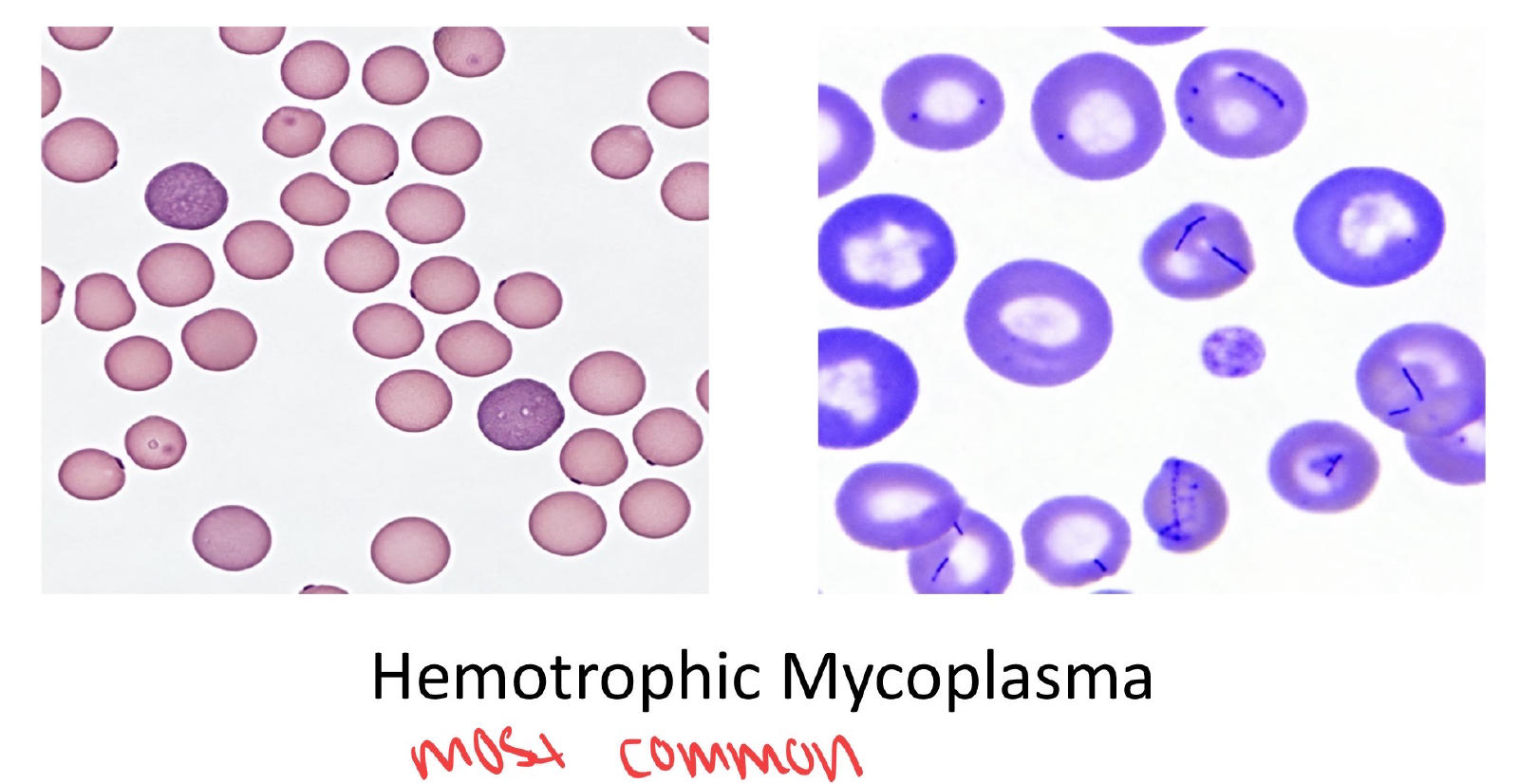
44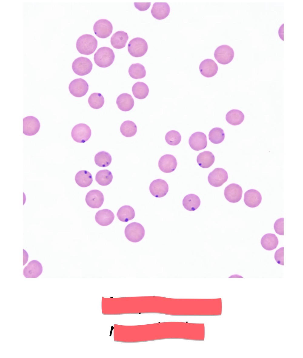
Anaplasma marginale
45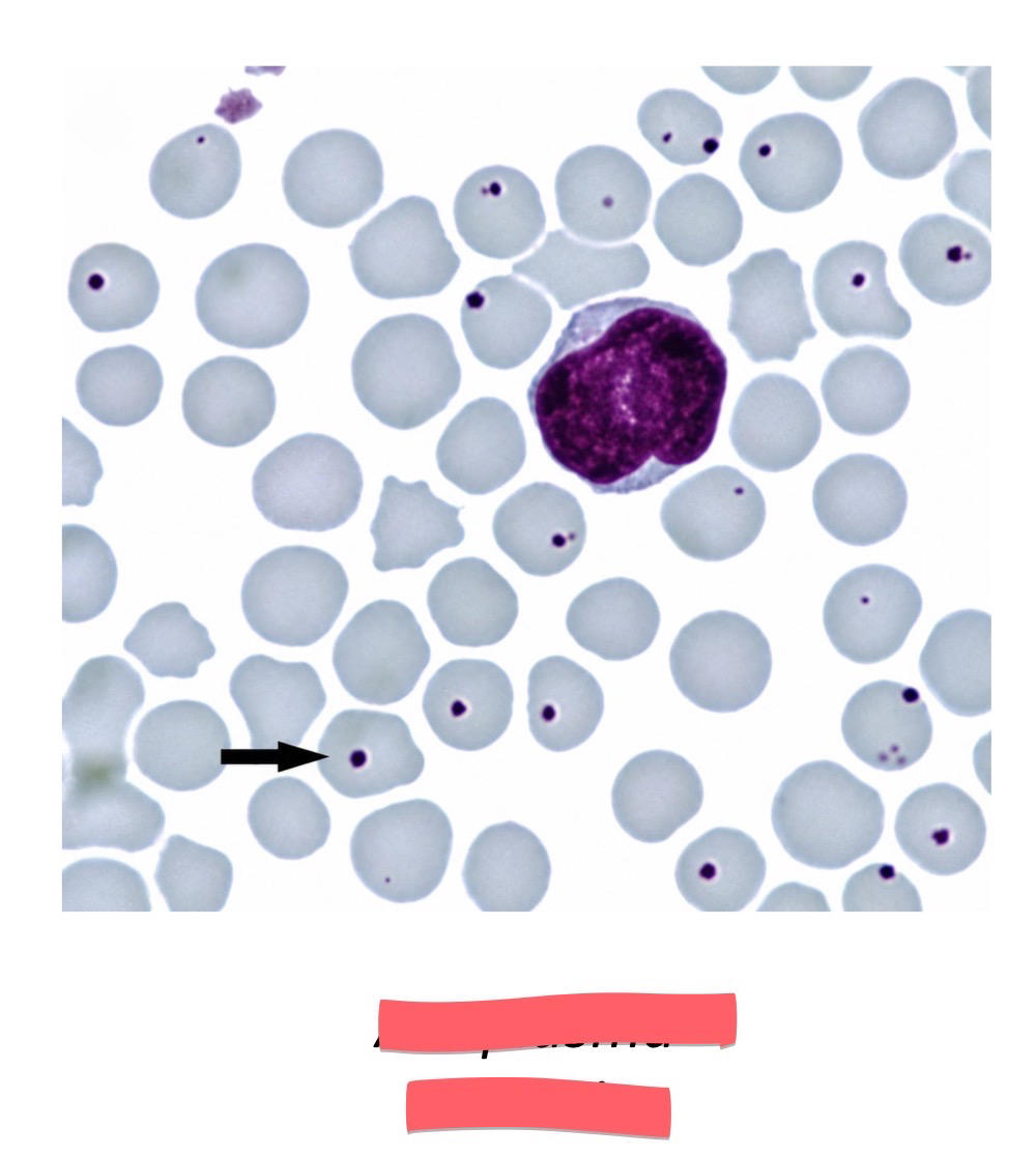
Anaplasma centrale
46
Piroplasms (2)
1. Babesia
2. Theileria species
2. Theileria species
47
What RBC inclusion can mimic Anaplasma organisms?
Howell-Jolly bodies.
48
What should be your basic diagnostic pathway after identifying anemia?
Determine regenerative vs. non-regenerative → assess MCV/MCHC pattern → evaluate RBC morphology.
49
What are the major causes of regenerative vs. non-regenerative anemia?
Regenerative = blood loss or RBC destruction; non-regenerative = bone marrow disease or systemic disease.
50
An anemic dog has increased MCV and decreased MCHC. What does this pattern suggest?
Regenerative anemia.
51
An anemic animal has normal MCV and normal MCHC. What broad pattern does this suggest?
Non-regenerative anemia.
52
What can cause increased MCV with normal MCHC?
Regeneration, Poodle breed effect, or FeLV-associated anemia.
53
What can cause decreased MCV with normal MCHC?
Iron deficiency, portosystemic shunts, or breed-associated microcytosis.
54
Which dog breeds can have breed-associated microcytosis?
Akita, Basenji, Shiba Inu, Jindo, and Chow Chow.
55
What does decreased MCV with decreased MCHC strongly suggest?
Iron deficiency.
More Medicine & health flashcards
Coagulation and HemostasisMedicine & health · EnglishMontanaHypersensitivityMedicine & health · EnglishKimLiver DiseaseMedicine & health · EnglishfaithLecture 7 - Autoimmunity, Autoimmune DiseasesMedicine & health · EnglishKimTD chimiothérapieMedicine & health · FrenchClaraAdvanced Anatomy & InjuriesMedicine & health · EnglishAristotelisNeurotransmittersMedicine & health · EnglishAdUvertMedicine typesMedicine & health · EnglishNovaUnit 1 - Circulatory SystemMedicine & health · EnglishAnantMedical Terminology J-RMedicine & health · EnglishKayleeAnatomy and Physiology QuestionsMedicine & health · EnglishBaoNeuroscience: The SynapseMedicine & health · EnglishLaura
Study these decks in the app
Free on iPhone, Android, Mac and the web. Add any deck to your account in one tap.