Lecture 7 - Autoimmunity, Autoimmune Diseases
Study these flashcardsFree · spaced repetition · iPhone, Android, Mac & web
All cards
1
What are autoimmune diseases?
• Disorders in which immune responses are targeted toward self-antigens and result in damage to organs and tissues in the body
• Could be T cell mediated or due to Abs to host Ags
• More than 100 autoimmune diseases have been discovered, and these can involve various organ system
• Autoimmune diseases are a leading cause of chronic illness and death, affecting about 5% of the world’s population, including more than 20 million people in the United States alone
• Could be T cell mediated or due to Abs to host Ags
• More than 100 autoimmune diseases have been discovered, and these can involve various organ system
• Autoimmune diseases are a leading cause of chronic illness and death, affecting about 5% of the world’s population, including more than 20 million people in the United States alone
2
How do autoimmune diseases develop?
1. Self-tolerance
- central (thymus or bone marrow)
- peripheral (lymph nodes, spleen)
2. Genetics
3. Hormonal influence
4. Exposure of cryptic antigens due to tissue damage following, e.g., infections, toxins, injury
5. Microbial infections: the main mechanism is molecular mimicry
- central (thymus or bone marrow)
- peripheral (lymph nodes, spleen)
2. Genetics
3. Hormonal influence
4. Exposure of cryptic antigens due to tissue damage following, e.g., infections, toxins, injury
5. Microbial infections: the main mechanism is molecular mimicry
3
What is self-tolerance?
- ability of the immune system to discriminate “self” from “non-self” or “foreign”, unresponsiveness to self Ags
- Lymphocytes must be “educated” at two levels (layers of protection) during development or immune response in order for self-tolerance to develop:
- Lymphocytes must be “educated” at two levels (layers of protection) during development or immune response in order for self-tolerance to develop:
4
What occurs in central self-tolerance?
- Central: which occurs in thymus for T cells and in BM for B cells. CD4+ T cells or B cells having receptors reactive to self Ags will be removed, some self-reactive CD4+ T cells are not deleted but released into circulation to differentiate into Treg cells.
• Some self-reactive B cells also are released to modify their receptors, so they become non-reactive to Ags (receptor editing).
• If some B cells have BCRs that weakly react with self Ags, they will be induced to become anergic (downregulate their receptor)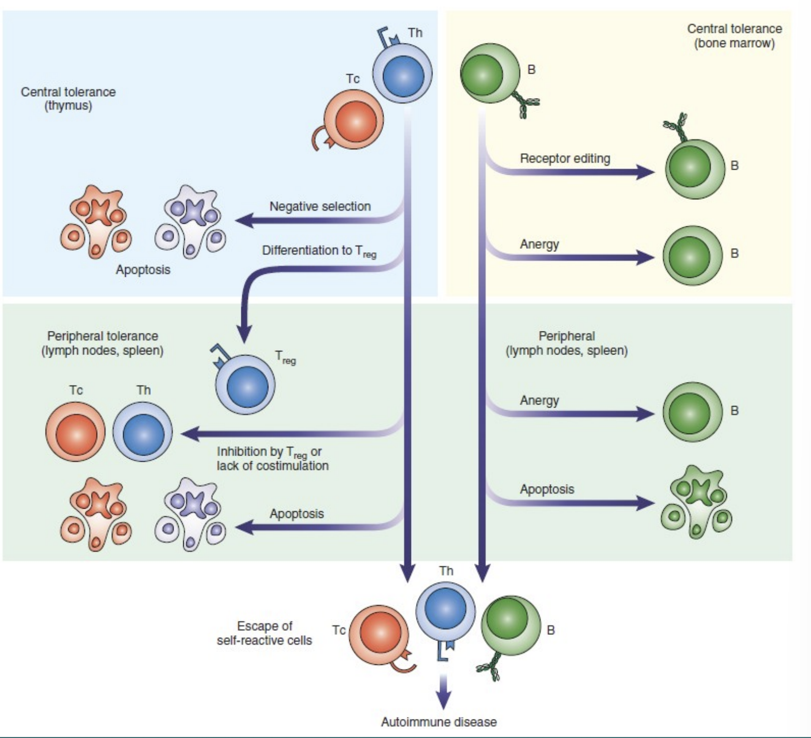
• Some self-reactive B cells also are released to modify their receptors, so they become non-reactive to Ags (receptor editing).
• If some B cells have BCRs that weakly react with self Ags, they will be induced to become anergic (downregulate their receptor)
5
What occurs in peripheral self-tolerance?
Peripheral: occurs in spleen or lymph nodes
• T cells reactive to self Ags will be removed either through apoptosis, inhibition by Treg cells, by losing a co-stimulatory signal from APCs, or through binding of CTLA-4 (inhibits T cells)
• B cells reactive to self Ags will either be removed by apoptosis, become anergic after repetitive exposure to self Ags, or become inactivated through activation of CD22 receptor
• T cells reactive to self Ags will be removed either through apoptosis, inhibition by Treg cells, by losing a co-stimulatory signal from APCs, or through binding of CTLA-4 (inhibits T cells)
• B cells reactive to self Ags will either be removed by apoptosis, become anergic after repetitive exposure to self Ags, or become inactivated through activation of CD22 receptor
6
What can happen if after self-tolerance fails?
In some individuals, self-tolerance can fail even after this second layer of protection; if this happens, autoimmunity can arise.
7
What are autoimmune disease thought to be caused by?
The development of autoimmune disease is thought to be caused by complex interactions between genetics, exposure to environmental factors, and defects in immune regulation.
8
Why would genetics play a factor in autoimmune diseases developing?
• Important factor (autoimmune diseases more prevalent among family members, or more prevalent among monozygotic (genetically identical) twins than dizygotic (non-identical) twins or siblings
• Most studies in humans on MHC genes
• Polymorphism in Non-MHC genes can also be involved
• Most studies in humans on MHC genes
• Polymorphism in Non-MHC genes can also be involved
9
What is an example of a certain inherited HLA gene that may cause an autoimmune disease?
- people inheriting HLA-B27 allele are at 100 times higher risk of developing ankylosing spondylitis
10
What examples of polymorphism in non-MHC genes can be involved in autoimmune disease?
- IL2RA gene, which is involved in T-cell activation and maintenance of Tregs
- CTLA4 gene, which has an inhibitory effect on T-cell activation
- BLK gene, which is involved in B-cell activation and development
- CTLA4 gene, which has an inhibitory effect on T-cell activation
- BLK gene, which is involved in B-cell activation and development
11
Why are women are at higher risk of developing autoimmune disease
than men?
than men?
Estrogens promote type 2 helper cell (Th2) response, resulting in more B cell activation and antibody production, whereas androgens promote type 1 helper cell (Th1) response with activation of CD8+ T cells
12
How can exposure of cryptic antigens due to tissue damage following, e.g., infections, toxins, injury cause autoimmune disease?
- autoantibodies to the lens of the eye following an ocular injury
- autoantibodies to sperm after a vasectomy
- autoantibodies to DNA following damage to skin cells by overexposure to UV rays from the sun
- autoantibodies to sperm after a vasectomy
- autoantibodies to DNA following damage to skin cells by overexposure to UV rays from the sun
13
How can microbial infections cause autoimmune disease?
• The main mechanism is molecular mimicry:
• Bacterial or viral agents contain antigens that closely resemble the structure or amino acid sequence of self-antigens. Exposure to such foreign antigens may trigger immune responses that cross-react with similar self-antigens
• Bacterial or viral agents contain antigens that closely resemble the structure or amino acid sequence of self-antigens. Exposure to such foreign antigens may trigger immune responses that cross-react with similar self-antigens
14
What is an example of a microbial infection causing autoimmune disease?
- association between Streptococcus pyogenes and rheumatic
fever
- an autoimmune disorder that primarily affects the joints and the heart. Patients develop antibodies to the M protein and N-acetyl glucosamine components of the bacteria, which cross-react with cardiac myosin, causing damage to the heart
fever
- an autoimmune disorder that primarily affects the joints and the heart. Patients develop antibodies to the M protein and N-acetyl glucosamine components of the bacteria, which cross-react with cardiac myosin, causing damage to the heart
15
What is the etiology of autoimmune disease?
Although the precise etiology of autoimmunity is unknown, there is much evidence that suggests that this heterogeneous disease entity is caused by complex interactions between genetic and environmental factors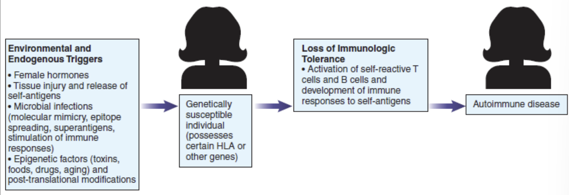
16
What is systemic lupus erythematosus (SLE)?
• Chronic inflammatory disease
• Females are more likely to develop SLE than males, by a ratio of 9:1
• Females are more likely to develop SLE than males, by a ratio of 9:1
17
What is the immunopathology of SLE?
• More than 100 autoantibodies identified
• Abs against: dsDNA, histones, as well as autoantibodies to lymphocytes, erythrocytes, platelets, phospholipids, ribosomal components, and endothelium. The typical patient has several circulating autoantibodies.
• Anti-dsDNA complexed with complement circulate (cannot be cleared from circulation) and deposit on glomerular basement membrane (GBM) of the kidneys
• Joint, skin, renal involvement
• Lupus anticoagulants
• Abs against: dsDNA, histones, as well as autoantibodies to lymphocytes, erythrocytes, platelets, phospholipids, ribosomal components, and endothelium. The typical patient has several circulating autoantibodies.
• Anti-dsDNA complexed with complement circulate (cannot be cleared from circulation) and deposit on glomerular basement membrane (GBM) of the kidneys
• Joint, skin, renal involvement
• Lupus anticoagulants
18
What is the initial evaluation of SLE?
- CBC, platelet count, creatinine levels, and urinalysis to
assess renal function
- it may show leukopenia, possible thrombocytopenia and anemia, ESR may also be high, but CRP may be low or normal
assess renal function
- it may show leukopenia, possible thrombocytopenia and anemia, ESR may also be high, but CRP may be low or normal
19
If SLE is suspected, what tests are done?
- (anti-nuclear antibodies) ANA will be done which is the most important screening test
- After ANA screening is positive, further tests should be done to differentiate the type of ANA
- After ANA screening is positive, further tests should be done to differentiate the type of ANA
20
What antibodies are highly specific for SLE?
Antibodies to dsDNA and antibodies to the Sm nuclear antigen are highly specific for SLE
21
What indirect immunofluorescence (IIF) method is used for ANA detection?
Fluorescent anti-nuclear antibody (FANA) testing has been the most widely used and accepted test because it is highly sensitive, detects a wide range of antibodies, and is inexpensive and easy to perform
22
What are the steps for FANA?
• A commercially prepared slide with fixed HEp2 cell line
• Incubation with patient’s serum (serial dilutions, starting with 1:80 or 1:160), ANA will bind to HEp2 nucleus
• Wash, to remove unbound Ab molecules
• Add anti-human Ig labeled with a fluorescent dye and incubate
• Wash again to remove unbound Ab
• Put a cover slip, mount the slide and view it under microscope (400× magnification)
• Titers above 160 are clinically significant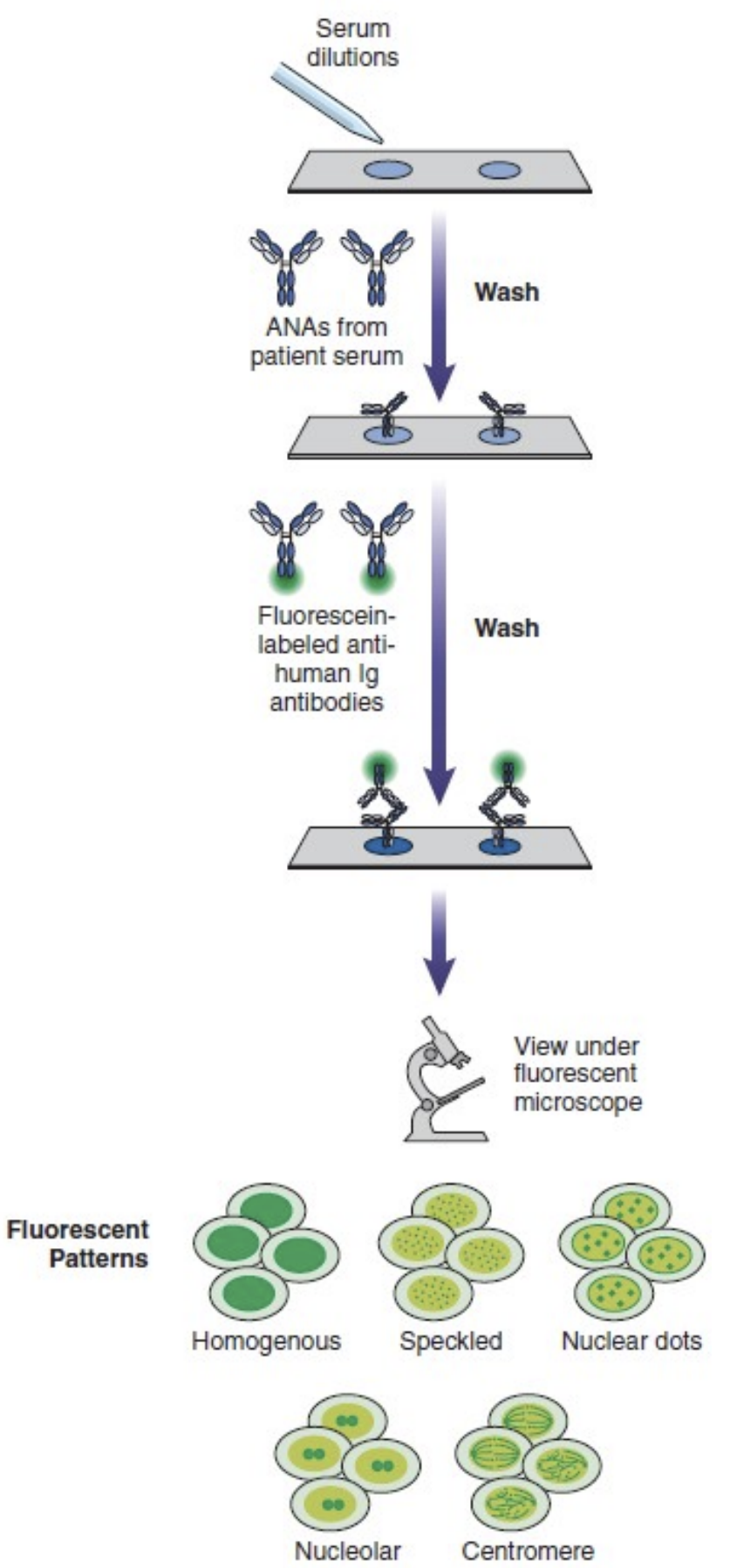
• Incubation with patient’s serum (serial dilutions, starting with 1:80 or 1:160), ANA will bind to HEp2 nucleus
• Wash, to remove unbound Ab molecules
• Add anti-human Ig labeled with a fluorescent dye and incubate
• Wash again to remove unbound Ab
• Put a cover slip, mount the slide and view it under microscope (400× magnification)
• Titers above 160 are clinically significant
23
What are the main fluorscence patterns for ANA reaction?
1. homogenous
2. speckled
3. nucleolar
2. speckled
3. nucleolar
24
Which fluorescence pattern for ANA reaction is this?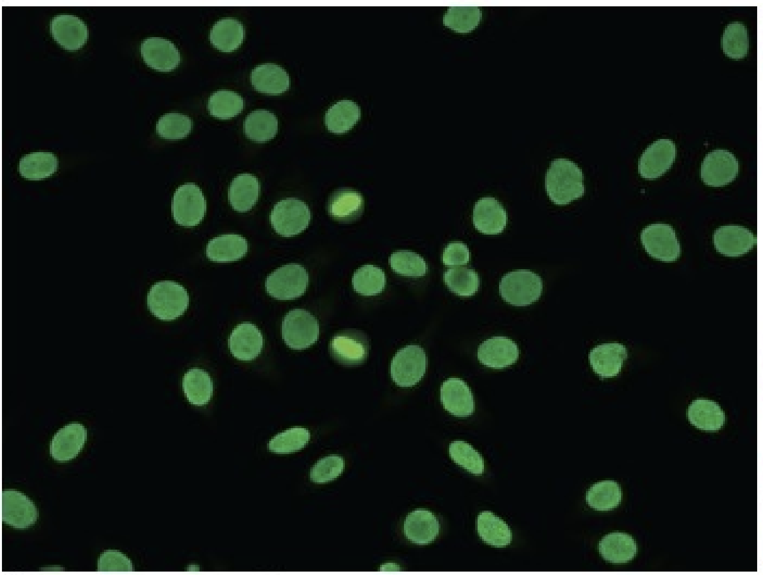
- Homogenous!
- associated with antibodies to dsDNA, histones, and nucleosomes
- found in SLE patients, drug-induced lupus, chronic autoimmune hepatitis (AIH), and juvenile idiopathic arthiritis
- associated with antibodies to dsDNA, histones, and nucleosomes
- found in SLE patients, drug-induced lupus, chronic autoimmune hepatitis (AIH), and juvenile idiopathic arthiritis
25
Which fluorescence pattern for ANA reaction is this?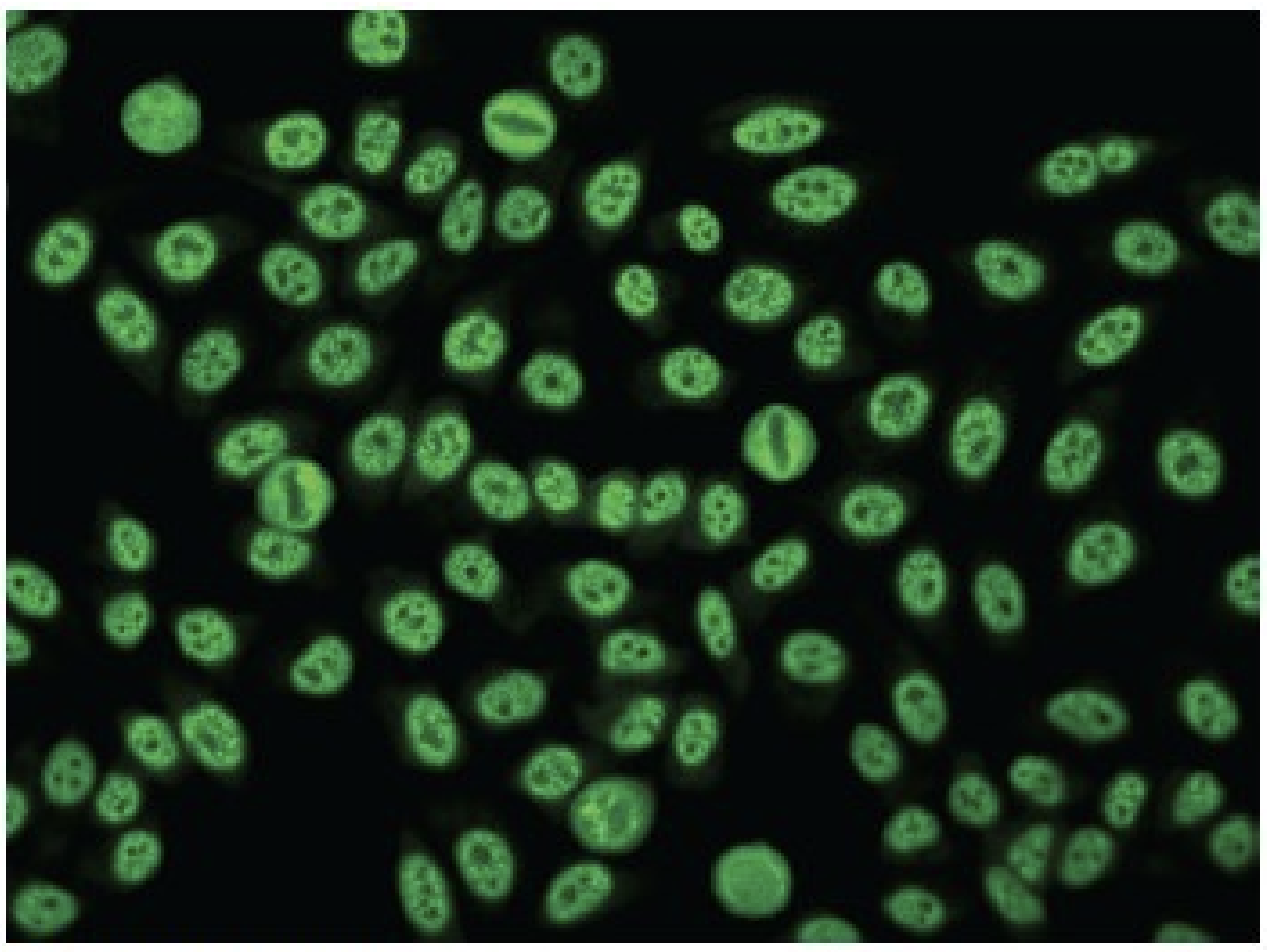
- Speckled!
- associated with antibodies to ENAs
- found in SLE patients, Sjogren's syndrome, systemic sclerosis (SSc), and other SARDs
- associated with antibodies to ENAs
- found in SLE patients, Sjogren's syndrome, systemic sclerosis (SSc), and other SARDs
26
Which fluorescence pattern for ANA reaction is this?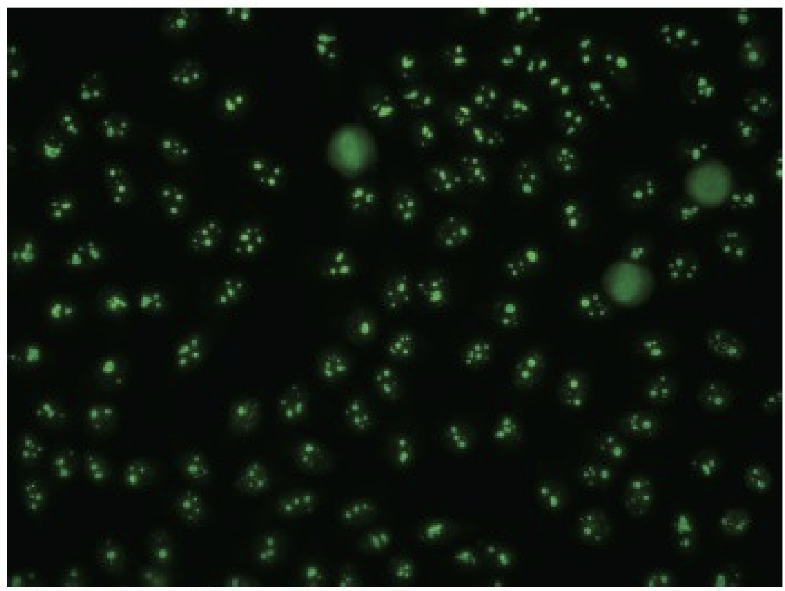
- Nucleolar!
- primarily caused by antibodies to RNA and RNP
- seen in patients with SSc, but can also be seen in other SARDs
- primarily caused by antibodies to RNA and RNP
- seen in patients with SSc, but can also be seen in other SARDs
27
What is rheumatoid arthritis (RA)?
• Caused by an inflammatory process that results in the destruction of bone and cartilage in the joints
• Infiltration of the inflamed synovium with T and B lymphocytes, plasma cells, dendritic cells, mast cells, and granulocytes is evidence of immunologic activity within the joint.
• Infiltration of the inflamed synovium with T and B lymphocytes, plasma cells, dendritic cells, mast cells, and granulocytes is evidence of immunologic activity within the joint.
28
How many genetic regions are associated with rheumatoid arthritis (RA)?
>100 genetic regions
29
What are the strongest associations with rheumatoid arthritis (RA)?
- specific HLA-DRB1 alleles or PTPN22 gene polymorphisms
- these patients are positive for rheumatoid factor (RF) or antibodies to cyclic citrullinated peptides (CCP)
- these patients are positive for rheumatoid factor (RF) or antibodies to cyclic citrullinated peptides (CCP)
30
What are some risk factors for rheumatoid arthritis (RA)?
• Epigenetic factors, such as altered DNA methylation affecting the transcription of critical genes, are also believed to play a role in the pathogenesis of RA.
• The strongest environmental risk factor for RA is believed to be cigarette smoking, which doubles the risk of developing the disease.
• The strongest environmental risk factor for RA is believed to be cigarette smoking, which doubles the risk of developing the disease.
31
What is the immunopathology for rheumatoid arthritis (RA)?
• The balance between proinflammatory and anti-inflammatory cytokines in RA appears to be tipped toward continual inflammation: IL-1, IL-6, IL-17 and TNF-a (TNF-a plays the key role)
• These cytokines trigger the release of matrix metalloproteinases from fibroblasts and macrophages; these enzymes degrade important structural proteins in the cartilage
• Bone lesions due to hyperactivation of osteoclasts.
• TNF-a and RANKL ((receptor activator of nuclear factor kappa-B ligand), inhibit osteoblast differentiation and bone formation, they activate osteoclasts
• Two key Abs
• These cytokines trigger the release of matrix metalloproteinases from fibroblasts and macrophages; these enzymes degrade important structural proteins in the cartilage
• Bone lesions due to hyperactivation of osteoclasts.
• TNF-a and RANKL ((receptor activator of nuclear factor kappa-B ligand), inhibit osteoblast differentiation and bone formation, they activate osteoclasts
• Two key Abs
32
What are the two key antibodies associated with Rheumatoid Arthritis (RA)?
1. Rheumatoid factor (RF)
2. anti-CCP or ACPA
2. anti-CCP or ACPA
33
What is Rheumatoid Factor?
an IgM against FC portion of IgG, enhances activity of macrophages and Ag presentation to APCs
34
What is anti-CCP or ACPA?
- Anti-cyclic citrullinated peptide antibody [anti-CCP or ACPA])
- citrullinated proteins released from dead granulocytes or macrophages
- overexpression of these antigens may provoke an immune response in individuals with certain HLA-DRB1 alleles.
- citrullinated proteins released from dead granulocytes or macrophages
- overexpression of these antigens may provoke an immune response in individuals with certain HLA-DRB1 alleles.
35
What is the initial test for Rheumatoid Factor (RF)?
- initial test is RF
- negative RF does not exclude RA,
- positive RF is not specific to RA
- RF is also positive in about 5% of healthy individuals and in 10% to 25% of those over the age of 65. RF can be found in patients with other SARDs, such as SLE, Sjögren’s syndrome, scleroderma, as well as in people with some chronic infections
- negative RF does not exclude RA,
- positive RF is not specific to RA
- RF is also positive in about 5% of healthy individuals and in 10% to 25% of those over the age of 65. RF can be found in patients with other SARDs, such as SLE, Sjögren’s syndrome, scleroderma, as well as in people with some chronic infections
36
How is RF tested?
• RF is done using latex agglutination on a slide, detects only IgM isotype
• ELISA and nephelometry are more sensitive and precise, they detect IgG isotype (more specific)
• ELISA and nephelometry are more sensitive and precise, they detect IgG isotype (more specific)
37
How is anti-CCP tested?
• Anti-CCP is done using ELISA, great test for early diagnosis where RF may be negative
• A large number of RF-negative RA patients are anti-CCP positive
• A large number of RF-negative RA patients are anti-CCP positive