Gynecological Ultrasound and Imaging
Study these flashcardsFree · spaced repetition · iPhone, Android, Mac & web
All cards
1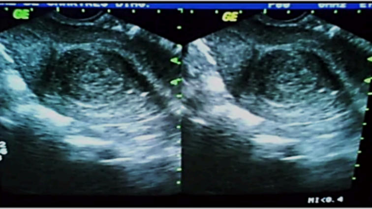
Ultrasound of Submucous Myoma
2
3.5 – 5 MHz sector
TRANSABDOMINAL APPROACH
3
4 – 7 MHz
TRANSVAGINAL APPROACH
4
Can be used when there are cysts or masses in the vaginal
wall or along the labia
wall or along the labia
TRANSLABIAL APPROACH
5
Done in virgins if the uterus and adnexa are not visualized
through the abdominal approach; Useful in patients with polycystic ovarian morphology,
through the abdominal approach; Useful in patients with polycystic ovarian morphology,
TRANSRECTAL APPROACH
6
visualize the endometrial cavity and assess fallopian tube patency
SONOHYSTEROGRAM
7
X-ray Examination of the uterus and fallopian tubes
HYSTEROSALPINGOGRAM
8
timing for sonohysterogram
4-7 days to 10 days from LMP
9
Cannula is inserted through the cervical canal
Leech Wilkenson Cannula
10
Contraindication for donohysterogram
Pelvic Inammatory diseases
11
detect an endometrial polyp
Sonohysterogram
12
To check fallopian tube patency
Hysterosalpingogram
13
No spillage
tubal obstruction
14
WHAT TO ASSESS?
size, shape, contour, orientation, and sppearance of Myometrium, Endometrium, and Cervix
15
Relationship of the uterus to the vagina in a sagital plane
VERSION
16
Relationship of the uterus to the cervix
FLEXION
17
Most common position of the uterus
o Anteverted: Cervix angles forward
o Anteexed: Body is exed forward
o Anteexed: Body is exed forward
18
hugging the urinary bladder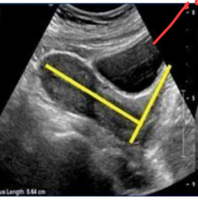
anteverted
19
uterus is pointing backwards towards the rectum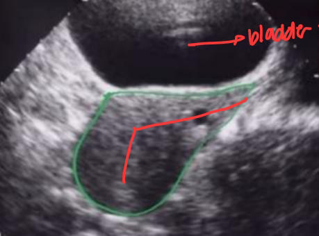
Retroverted Uterus
20
arcuate arteries are calcied
Monckeberg’s Sclerosis
21
medium-level echoes.
➢Granular in texture
➢Granular in texture
MYOMETRIUM
22
Varies in thickness and appearance because of the effects of estrogen and progesterone
ENDOMETRIUM
23
Thin, broken echogenic line (1 – 2 mm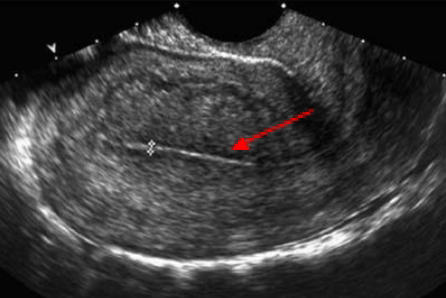
Menstrual phase
24
Hypoechoic thickening (4 – 8 mm)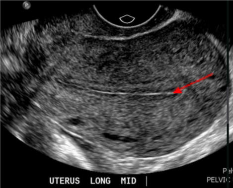
Proliferative phase
25
Triple layer (6 – 10 mm)
Periovulatory phase
26
Hyperechoic thickening (7 – 14 mm)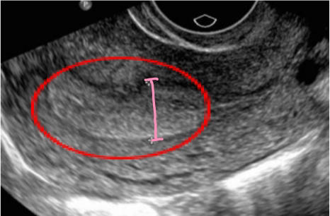
Secretory phase
27
Menstruating Patient: endometrial thickness >14 mm
Endometrial Hyperplasia
28
Postmenopausal: Endometrial thickness >14 mm
endometrial cancer
29
7 – 12 mm in length; Extends laterally from the uterus
FALLOPIAN TUBE
30
Parts of the fallopian tube
○ Intramural
○ Isthmus
○ Ampulla
○ Infundibulum / fimbriae
○ Isthmus
○ Ampulla
○ Infundibulum / fimbriae
31
CAUSES OF ENDOMETRIAL THICKENING
➢ Early intrauterine pregnancy
➢ Incomplete abortion
➢ Ectopic pregnancy (Arias-Stella reaction)
➢ Incomplete abortion
➢ Ectopic pregnancy (Arias-Stella reaction)
32
Hormone-related atypical endometrial change characterized by hypertrophy and vacuolization of glandular epithelial cells, associated with marked nuclear pleomorphism, enlargement, and hyperchromasia.
Arias Stella reaction
33
unopposed stimulation of the endometrium by estrogen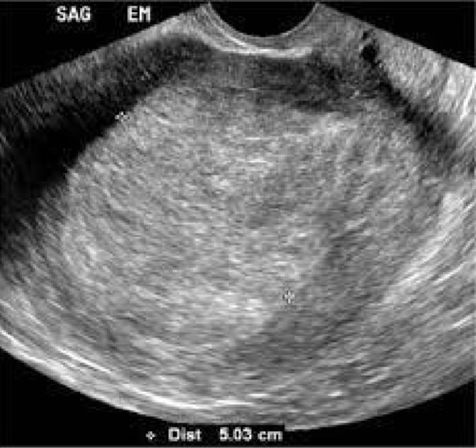
ENDOMETRIAL HYPERPLASIA
34
treatment for endometrial hyperplasia if medications do not work
Curetage
35
not appreciated during the secretory phase 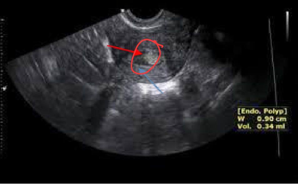
ENDOMETRIAL POLYP
36
If a polyp becomes large
may cause infertility or ectopic pregnancy by tubal blockade
37
Treatment for endometrial polyp
Dilatation and curetage
38
Polypectomy via curetage
if polyp is inside the endometrium
39
Best time to look for polyp
menstrual phase or the early proliferative phase
40
To check for endometrial polyps
sonohysterogram
41
MC gynecologic malignancy; thickened, heterogeneous endometrium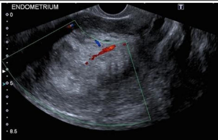
ENDOMETRIAL CARCINOMA
42
endometrium becomes thickened and may present with cystic changes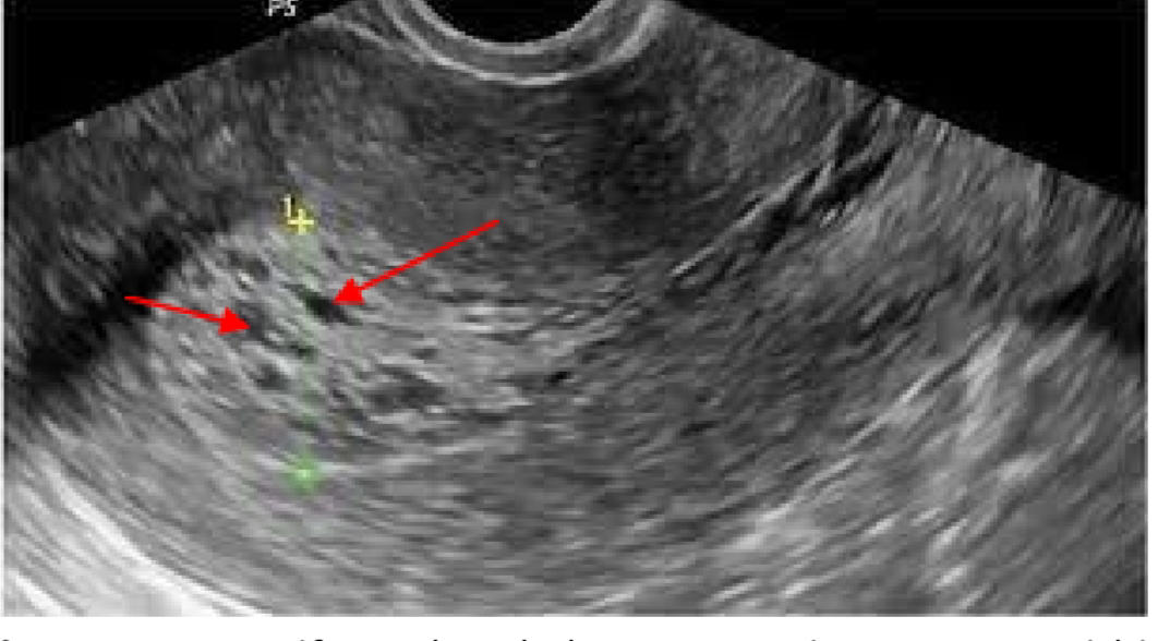
TAMOXIFEN-RELATED ENDOMETRIAL CHANGES
43
Circular and hypoechoic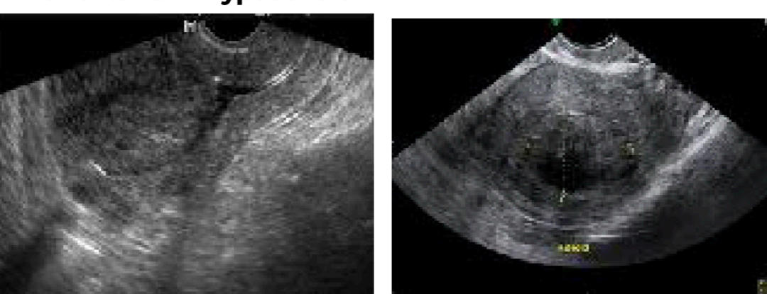
SUBMUCOSAL LEIOMYOMA
44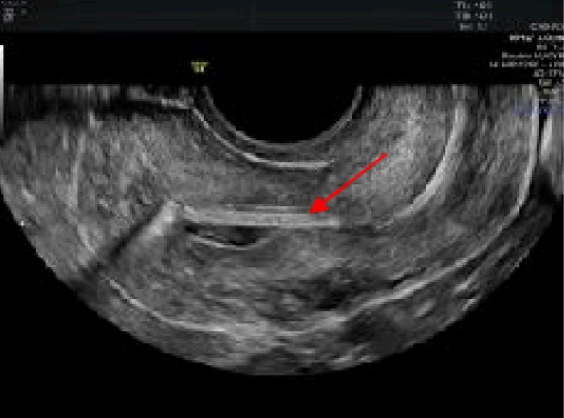
IUD in the uterine cavity
45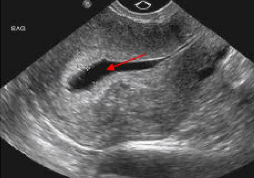
Fluid in the uterine cavity
46
Gas within the endometrial cavity; Most commonly occurs after a septic abortion; Common in induced abortions involving catheters
ENDOMETRITIS
47
air echoes within endometrial cavity (echogenic foci with artifact – white)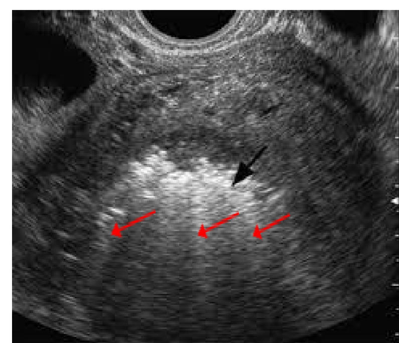
ENDOMETRITIS
48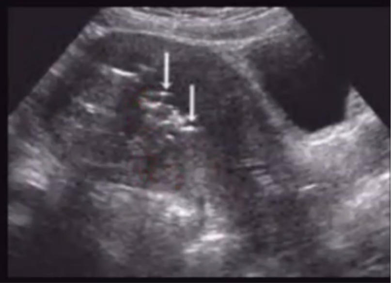
Endometritis with air echoes
49
Due to an Imperforate hymen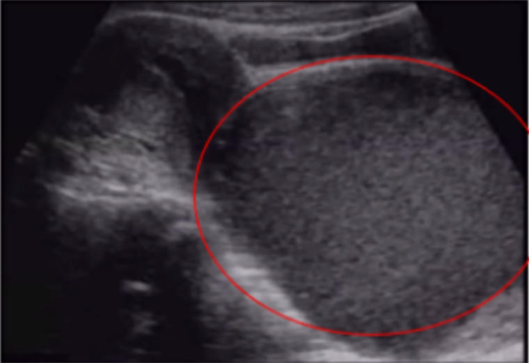
HEMATOCOLPOS / HYDROMETROCOLPOS
50
Accumulation of menstrual blood,within the vagina due to an obstruction of the vaginal outow tract.
Hematocolpos
51
Accumulation of uid in both the uterus (hydrometra) and the vagina (hydrocolpos) due to obstruction of the genital outow tract.
Hydrometrocolpos
52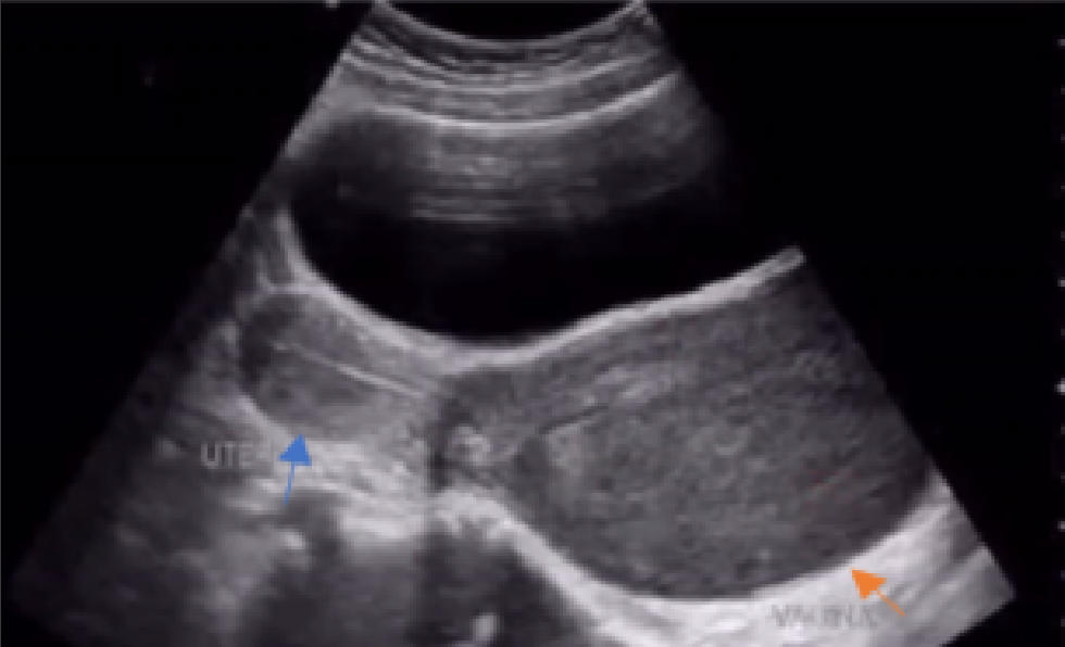
Vagina lled with fluid collection
53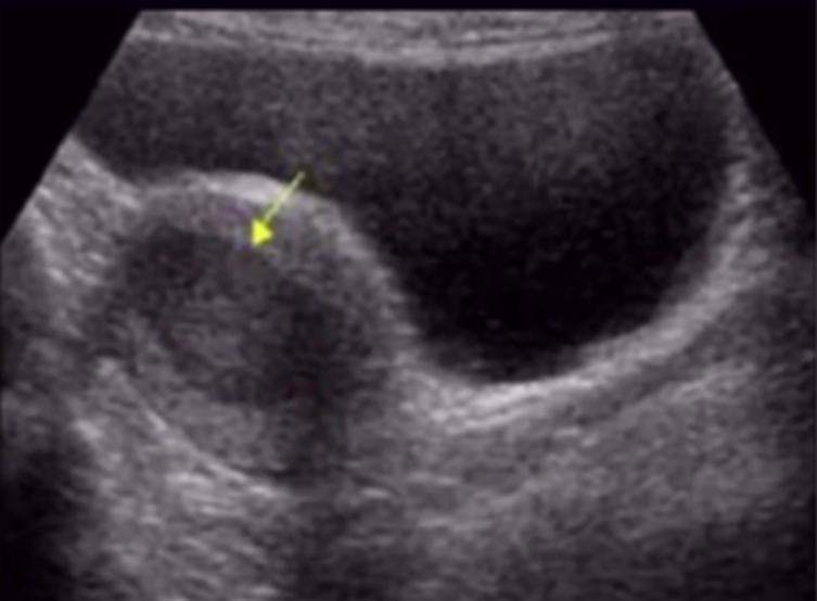
Collection of blood inside the uterus
54
The most common neoplasia in the myometrium; oes not cause dysmenorrhea
LEIOMYOMA
55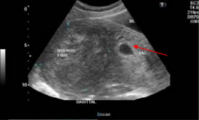
Intramural leiomyoma with gestational sac (red arrow)
56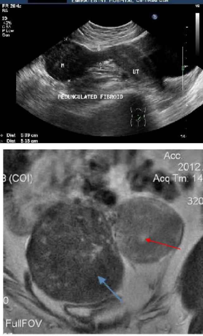
Subserosal fibroid
57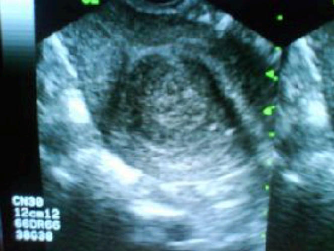
SUBMUCOSAL Projects to the uterine cavity
58
Presence of endometrial tissue in the myometrium; Severe form of dysmenorrhea; Heterogeneous* patern of the myometrium
ADENOMYOSIS
59
Enlarged uterus with normal appearing endometrium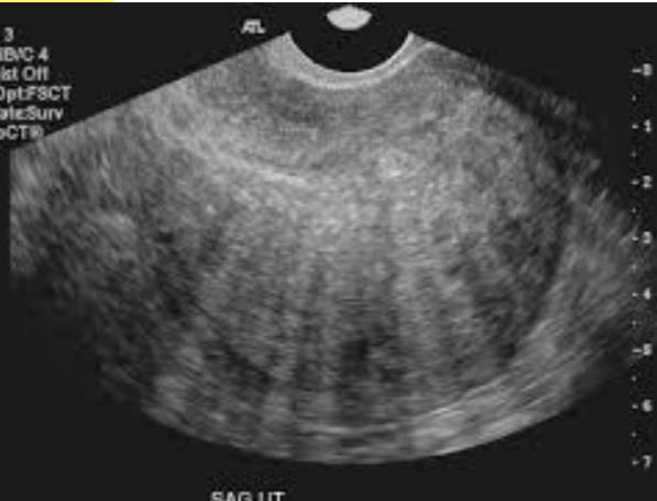
Diffuse adenomyosis
60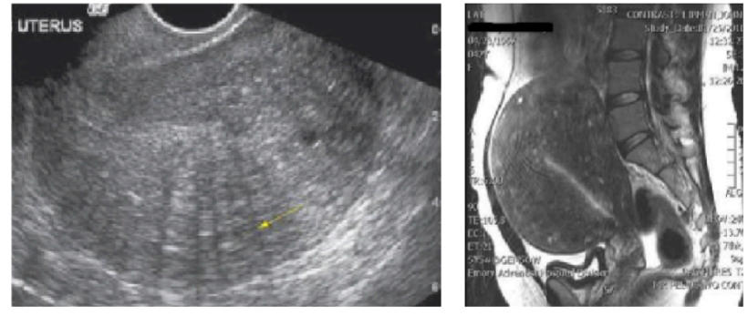
Enlarged uterus with heterogeneous* myometrium
61
Most common (benign)
Nabothian Cyst
62
Absent uterus; failed to form
Uterine Aplasia
63
Result of unilateral developmental arrest
Uterus Unicornis, Unicollis
64
Most common uterine anomaly with renal agenesis; 2 uterus, 2 cervix, 1 vagina.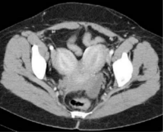
UTERUS BICORNIS, BICOLLIS
65
2 uterus, 1 cervix, 1 vagina
UTERUS BICORNIS, UNICOLLIS
66
Indentation of the uterine fundus
UTERUS ARCUATUS
67
FAILURE OF RESORPTION OF THE MEDIAN SEPTUM
Septate or subseptate uterus
68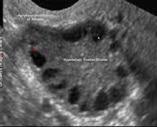
polycystic ovary
69
More than 25 mm; mature follicle fails to involute
Follicular cyst
70
Seen in pregnancy; Regress after 3 menstrual cycle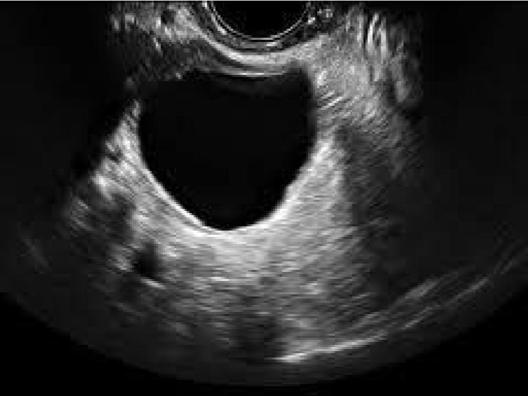
Corpus Luteal Cyst
71
Network of fine interdigitation avascular strands; Fish-net appearance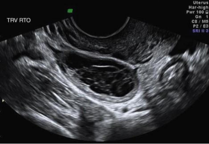
HEMORRHAGIC CYST
72
Occurs in pregnancy (looks like Poly Cystic Ovaries)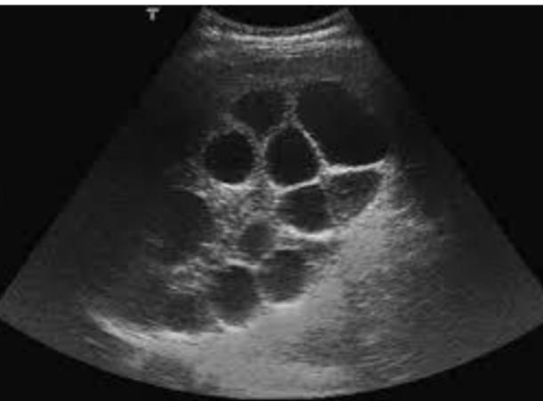
Theca Lutein Cyst
73
Endometrial tissue outside of the uterus; Possibility of infertility; Symptom: Severe dysmenorrhea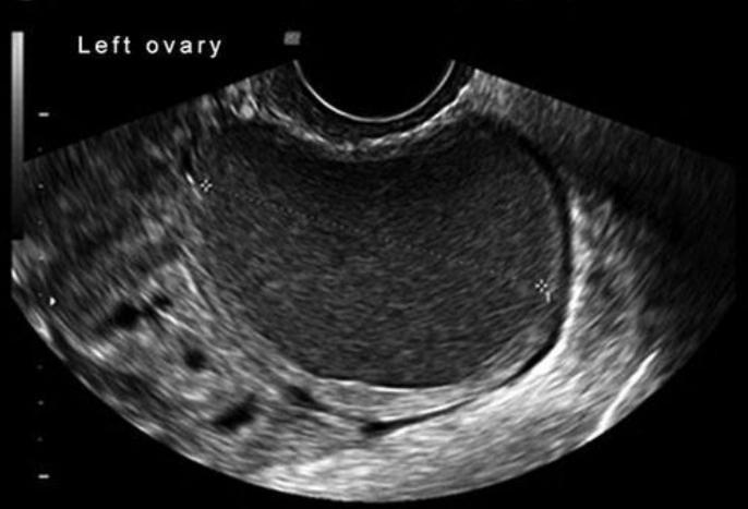
ENDOMETRIOMA / ”CHOCOLATE CYST”
74
Contains fats, bones, parts of the skin, and hair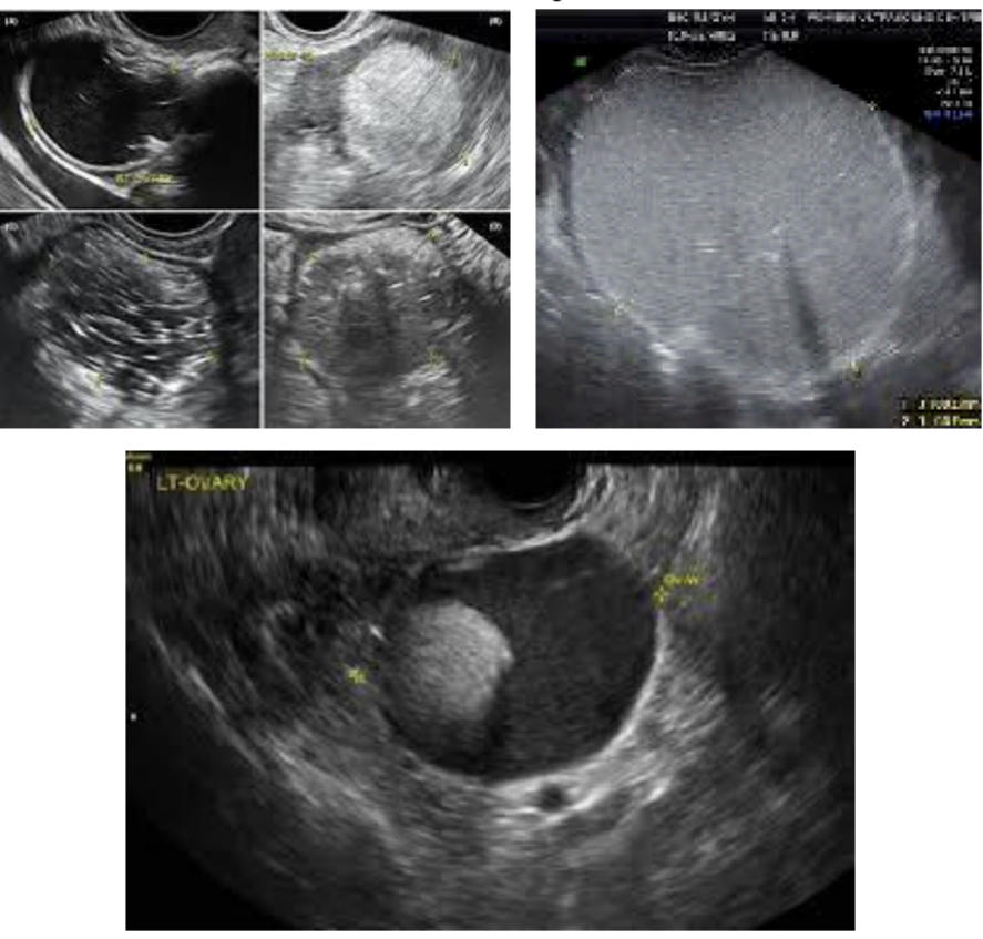
Dermoid cysts
75
Due to chronic anovulation
POLYCYSTIC OVARIAN SYNDROME
76
Polycystic Ovarian Morphology (PCOM) Diagnosis
Follicle Number Per Ovary (FNPO) ≥20 follicles (2 – 9
mm) in at least one ovary; Ovarian volume (OV) ≥10 mL in at least one ovary; Follicle Number Per Section (FNPS) ≥10 follicles in one ovary (one plane or slice)
mm) in at least one ovary; Ovarian volume (OV) ≥10 mL in at least one ovary; Follicle Number Per Section (FNPS) ≥10 follicles in one ovary (one plane or slice)
77
menstruation has been irregular for more than 8 years
diagnose PCOM
78
➢ Irregular thick walls
➢ Solid mural nodules
➢ Increase CA-125
➢ Lesions are greater than 4 cm
➢ Solid mural nodules
➢ Increase CA-125
➢ Lesions are greater than 4 cm
NEOPLASTIC/OVARIAN CANCER
79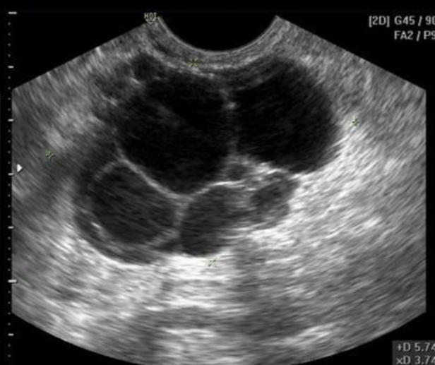
Ovarian Carcinoma
80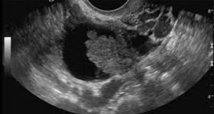
Ovarian cancer
81
The most common type of ovarian tumor; Unilocular, thin septations; Can be bilateral; Suggestive of malignant
Serous Cystadenoma and Cystadenocarcinoma
82
Serous Cystadenoma and Cystadenocarcinoma associated with:
Papillary projections
Echogenic materials
Ascites
Echogenic materials
Ascites
83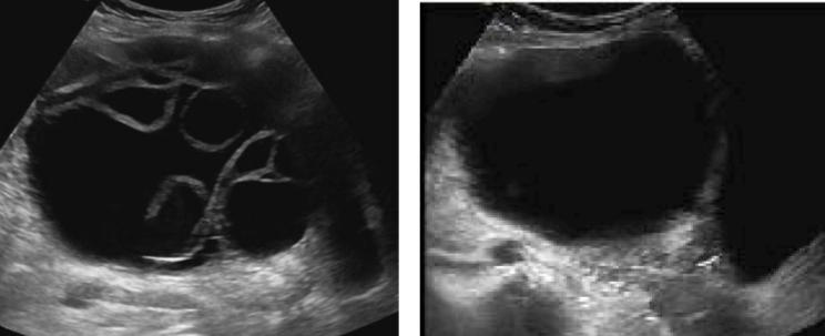
Serous cystadenoma
84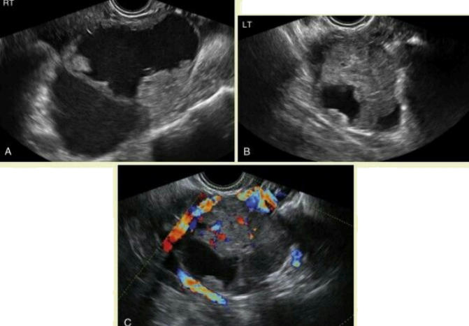
Bilateral papillary serous cystadenocarcinoma
85
2nd most common epithelial tumor; he patient may appear pregnant due to the huge size (15 – 30 cm)
Mucinous Cystadenoma and Cystadenocarcinoma
86
Thin septations
cystadenoma
87
Thickened septations or solid components
cystadenocarcinoma
88
Ruptured Mucinous Cystadenoma/cystadenocarcinoma
pseudomyxoma peritonei
89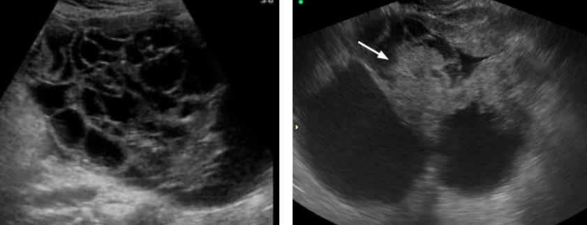
Mucinous Cystadenocarcinoma
90
Nearly all are malignant; Present as a cystic mass with papillary projections; 2nd most common epithelial malignancy
Endometroid Tumor
91
Mullerian duct in origin; Non-specic in appearance; Complex mass that can be cystic or solid
Clear Cell Carcinoma
92
Denitely malignant
➢ Hypoechoic solid masses
➢ May contain calcications
➢ Differentiation: Pathologic
➢ Hypoechoic solid masses
➢ May contain calcications
➢ Differentiation: Pathologic
Brenner Tumor
93
Most radiosensitive type of tumor; Morphologically identical to male testicular seminoma; Occurs in women less than 30 years of age
Dysgerminoma
94
Causes/correlated with increased AFP
Similar appearance to dysgerminoma
Similar appearance to dysgerminoma
Endometrial Sinus Tumor (Yolk Sac)
95
Occur in post-menopausal women
95% unilateral
Most estrogenically active ovarian tumor
95% unilateral
Most estrogenically active ovarian tumor
Granulosa Cell Tumor
96
Occurs in women under 30 years old; 70% occurs in post-menopausal women
Thecoma and Fibroma
97
Triad of benign tumor (broma), ascites, and pleural effusion
Meigs Syndrome
98
Most common origins are the breast and the GIT
METASTATIC TUMORSBaby is radiosensitive
99
Reserved for tumors containing signet ring cells, usually gastric in origin
Krukenberg Tumor
100
From conception to 13th week AOG; Greatest risk for maldevelopment, injury, and death; Baby is radiosensitive
FIRST TRIMESTER
101
Beter for direct visualization of the embryo at 0 – 8 weeks AOG
Transvaginal UTS
102
no embryo is found
anembryonic pregnancy or blighted ovum
103
used to measure cardiac activity (6 wks)
M-mode
104
Used only in >13 wks AOG
Doppler
105
Presence of intradecidual sign
Very Early Pregnancy or ENDOMETRIAL CYST
106
First structure to be visible within the gestational sac
➢ Visible by TVS at the 6th week AOG
➢ Visible by TVS at the 6th week AOG
Secondary Yolk Sac
107
yolk sac exceeding 6 mm
NON- VIABLE PREGNANCY
108
baby gets the nutrients from the yolk sac from
0 to the 8th week AOG
109
takes over after the 8th week.
Placenta
110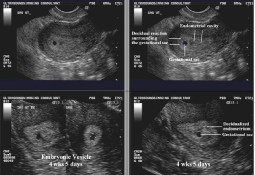
Early pregnancy ultrasound
111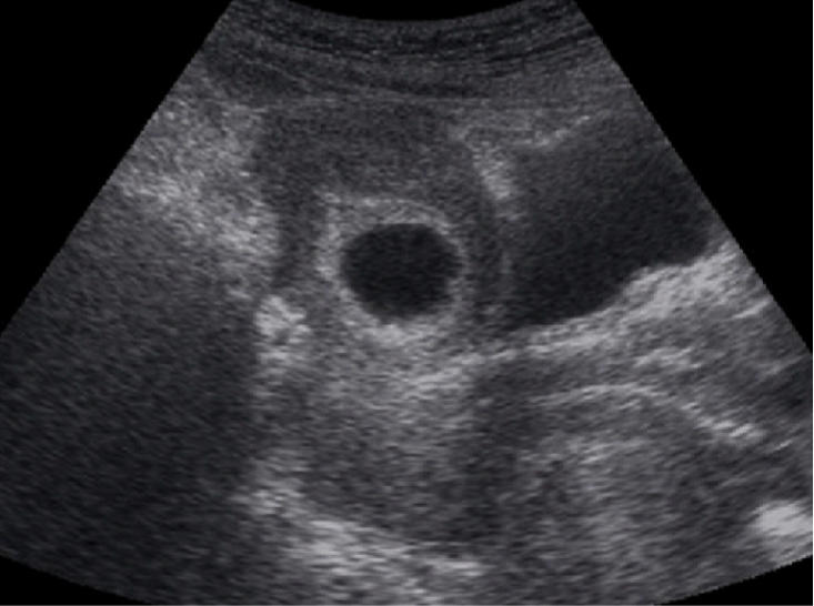
Intradecidual sign of early intrauterine pregnancy
112
Echogenic disc-like structure approximately 2 mm long; Seen at the end of the 5th week AOG
EMBRYO
113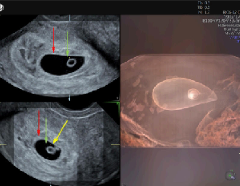
Gestational sac, yolk sac, and the embryo
114
a common early pregnancy complication, may indicate a threatened abortion or a subtle sign of placenta abruptio
subchorionic
hemorrhage
hemorrhage
115
marks early abdominal wall formation
8 weeks AOG.
116
Mean sac diameter >8 mm
“blighted
ovum”
ovum”
117
distorted gestational sac
Abortion in Progress
118
Typically occurs 6 weeks AOG and above
ECTOPIC PREGNANCY
119
Simultaneous intrauterine and extrauterine pregnancy
Heterotopic pregnancy
120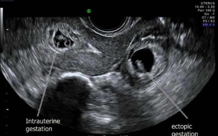
Heterotopic pregnancy
121
Free uid (FF) in the pelvic cavity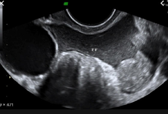
ruptured ectopic pregnancy or hemorrhage
122
fertilization of a 23X sperm
COMPLETE MOLE
123
Snowstorm appearance; Increased β-hcg by 3x
COMPLETE MOLE
124
fertilization of an ovum by 2 sperms
Triploid (69XXX, 69XXY, 69XYY)
Triploid (69XXX, 69XXY, 69XYY)
PARTIAL MOLE
125
SECOND TRIMESTER AND THIRD TRIMESTER Document the following:
1. Fetal life- Heartbeat
2. Number- Single or twins
3. Presentation- Cephalic or Breech
2. Number- Single or twins
3. Presentation- Cephalic or Breech
126
Assessment of the gestational age
Biparietal Diameter (BPD), Femur Length and Abdominal Circumference (AC)
127
MSD
<6 weeks gestational sac
128
CRL of embryo
6-13 weeks
129
BPD, FL, and AC
13 weeks and above
130
Taken at the level of the thalamus
BPD
131
Measured at the axial plane at the,junction of the umbilical vein and the left portal vein
Abdominal circumference (AC)
132
Estimated
Fetal Weight
(EFW)
Fetal Weight
(EFW)
➢ BPD + AC = EFW
➢ AC + FL = EFW
➢ AC + FL = EFW
133
1.st seen at 8 weeks AOG as focal thickening at the periphery of the gestational sac
PLACENTA
134
Causes of thick placenta: >4cm
Diabetes, Fetal hydrops, Anemia, Triploidy
135
Measure the vertical height of the deepest uid pocket in each quadrant of the uterus and sum up the 4 measurements (4 pockets)
AMNIOTIC FLUID INDEX (AFI)
136
AFI <5 cm
Oligohydramnios
137
Single pocket
>8 cm
>8 cm
Polyhydramnios
138
Dichorionic, diamniotic
3 – 4 days
139
Monoamniotic, dichorionic
4 – 7 days
140
Monochorionic,monoamniotic
After 8 days
141
conjoined twins
>13 days
142
Abnormal accumulation of uid in at least 2 body cavities
HYDROPS FETALIS
143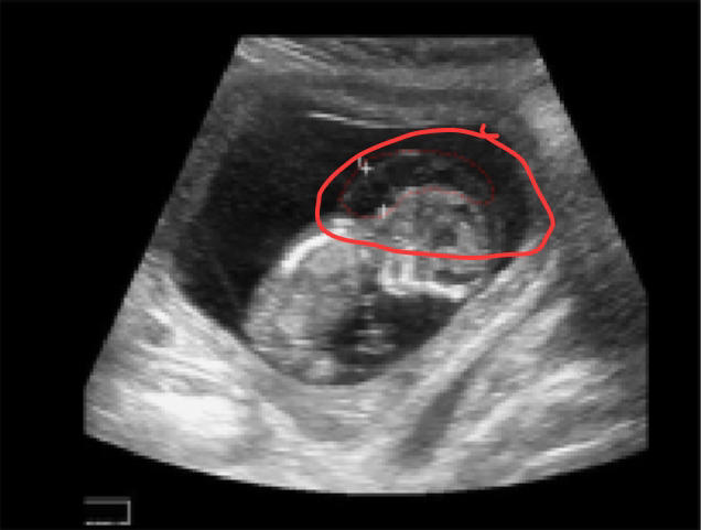
Fluid collection in the fetal nape
144
Most common cause of neural tube defects; Absence of the cranium and cerebral hemispheres;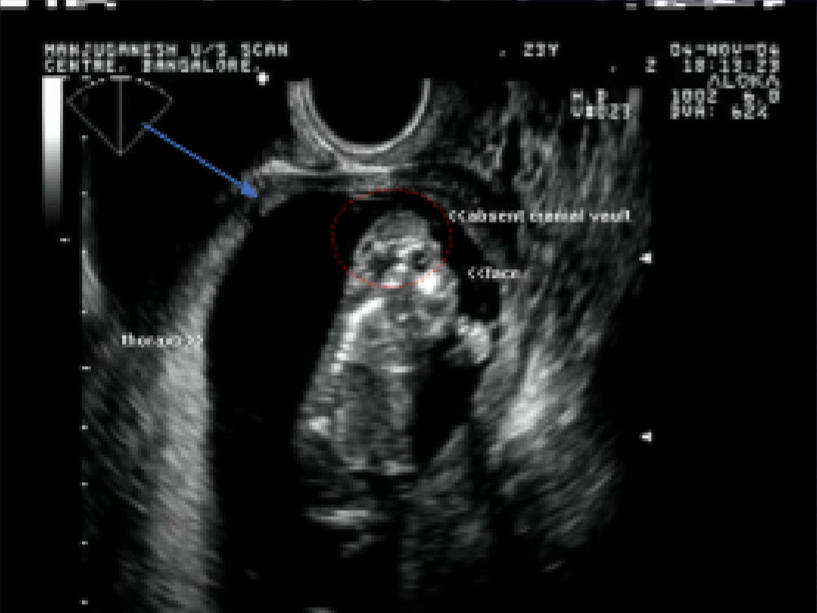
ANENCEPHALY
145
Abnormal uid accumulation in the ventricles; Due to total cerebral infarction in utero caused by bilateral thrombosis of the internal carotid arteries; no cerebral hemispheres.
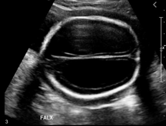
HYDRANENCEPHALY
146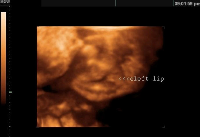
Cleft deformity
147
paramedian abdominal wall defect; Defect in the anterior abdominal wall, usually to the right of the umbilicus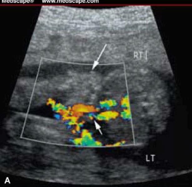
GASTROSCHISIS
148
Midline mass protruding through the abdominal wall
➢ Covering membrane present
➢ Has a beter survival rate than gastroschisis
➢ Covering membrane present
➢ Has a beter survival rate than gastroschisis
OMPHALOCOELE
149
Complex mass occupying the presacral region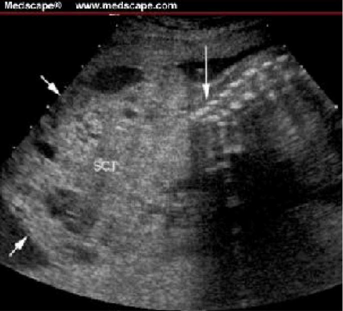
SACROCOCCYGEAL TERATOMA
150
Self-Breast examination
Monthly, 1 week AFTER period
151
Clinical breast examination
EVERY 3 YEARS for women 20 – 39 years old
YEARLY for women 40 years old and above
YEARLY for women 40 years old and above
152
Mammogram; Invented in the year 2015
Start at 35 – 40 years old
153
earliest recorded case of breast cancer
1600 BC Edwin Smith Papyrus
154
Indicated to asymptomatic patients that is >40 years old
Screening Mammogram
155
Indicated to symptomatic patient
Diagnostic Mammogram
156
Indication for patient with injectable implants
BREAST MRI
157
15 – 18 per breast; Milk-producing regions of the breast
lobes
158
○ Extralobular duct + lobule
○ Where cancer can occur
○ Functional unit of breast
○ Where cancer can occur
○ Functional unit of breast
TDLU
159
○ Appear tree-like on the breast imaging
○ Trunk: Mammary (lactiferous) ducts
○ Branches: Secondary ducts leading to the lobules
○ Trunk: Mammary (lactiferous) ducts
○ Branches: Secondary ducts leading to the lobules
Ducts
160
Used for intraductal lesions, bloody nipples
Galactography
161
A persistent, non healing nipple lesion
Paget’s Disease
162
Involvement of the main glandular units and posterior atachments
poor prognosis
163
Lesions conned to the TDLU
DCIS
164
localization of lesions in the horizontal plane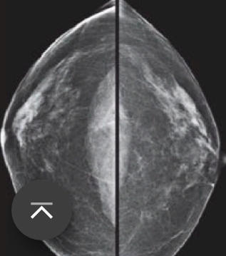
Cranio-Caudal (CC) View
165
Involvement of axilla where the lymph nodes are seen; localization of
lesions in the vertical plane.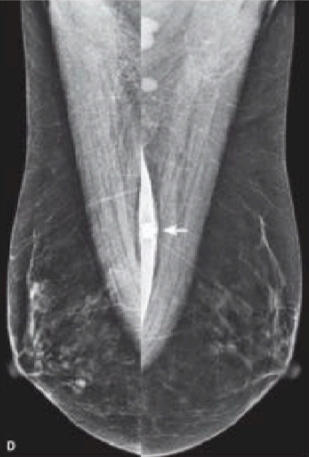
lesions in the vertical plane.
MEDIOLATERAL OBLIQUE VIEW (MLO)
166
Describe the position of the lesion in “O’Clock” position +
ABC zone
ABC zone
TRIANGULATION
167
Key component of triangulation in breast imaging
Posterior Nipple Line
168
Category 0
Needs additional imaging
169
Category 1
Negative in mammogram
170
Category 2
Benign lesion
171
Category 3
Probably Benign
172
Category 4
Suspicious lesions
173
Category 5
Highly Suggestive of Malignancy
174
Category 6
Known Biopsy-Proven Malignancy
175
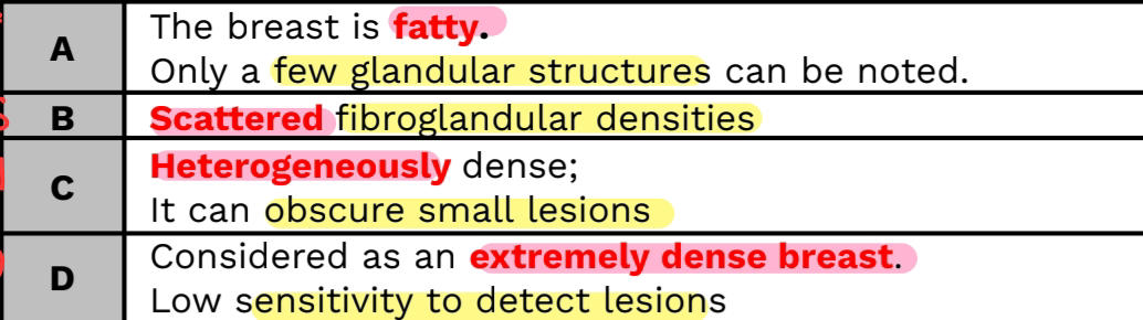
Refers to the amount of broglandular tissue in the breast
relative to the amount of fat.
relative to the amount of fat.
BREAST COMPOSITION
176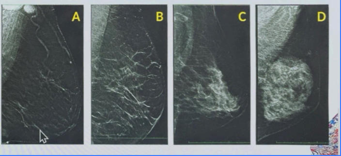
177
irregular lesion
BI-RADS 4 (suspicious) or 5 (highly suggestive of malignancy)
178
Circumscribed margins
cyst broadenomas, high grade intraductal carcinomas, medullary carcinomas
179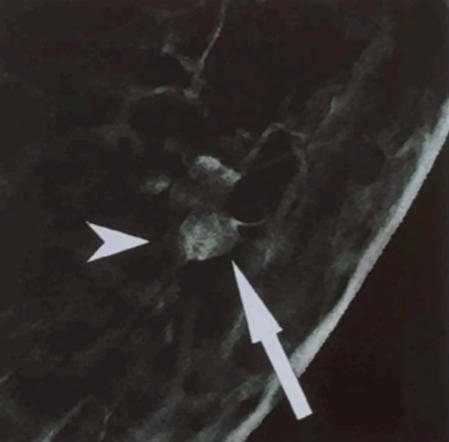
BI RADS score is 5
180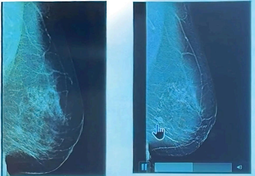
obscured margins,
181
INDISTINCT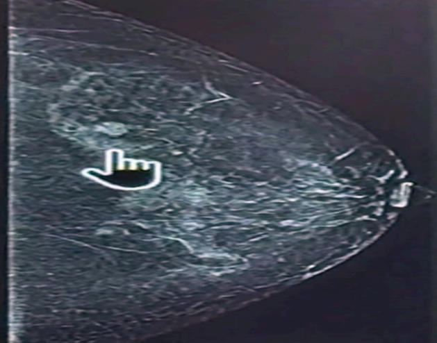
BI RADS 4
182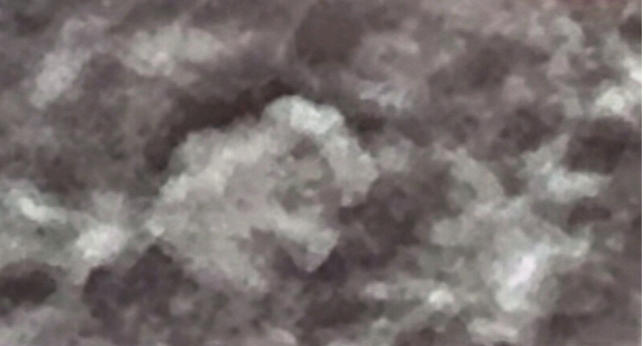
LOBULATED
183
SPICULATED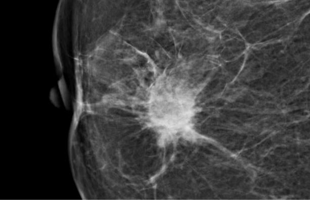
BI-RADS 5
184
Refers to X-ray atenuation relative to fibroglandular tissue
BREAST DENSITY
185
Low density lesions:
lipoma
186
Equal density
benign and malignant
187
High-density
cysts
188
LAYERING in new BI RAD 2025
Milk of calcium (cyst)
189
COARSE in new BI RAD 2025
Dystrophic,Popcorn (Trauma,Old broadenomas)
190
Calcications can never be seen on UTS
- true
- false
191
calcications are clustered,
malignant
192
indistinct, hazy, or ill-dened in appearance
Resembles powder
Resembles powder
Amorphous calcificaion
193
Larger, irregular in shape, typically 0.5–1.0 mm in size, with a tendency to coalesce.
Coarse heterogeneous
194
➢ Variable in size and shape
➢ Generally < 0.5 mm in diameter
➢ Carry a higher likelihood of malignancy compared to amorphous or coarse heterogeneous calcications
➢ Generally < 0.5 mm in diameter
➢ Carry a higher likelihood of malignancy compared to amorphous or coarse heterogeneous calcications
Fine Pleomorphic
195
➢ Thin, irregular lines, often following ductal distributions and occasionally branching
➢ Have the highest likelihood of malignancy almost always requires biopsy
➢ Have the highest likelihood of malignancy almost always requires biopsy
Fine Linear Branching
196
Amorphous, coarse heterogeneous, and fine pleomorphic calcications
BI-RADS category 4B
197
Fine linear or ne-linear branching calcications
BI-RADS category 4C
198
Diffuse location
benign
199
Regional, Group, Segmental location
malignant
200
An abnormality in which the normal breast architecture is disrupted without a discrete mass.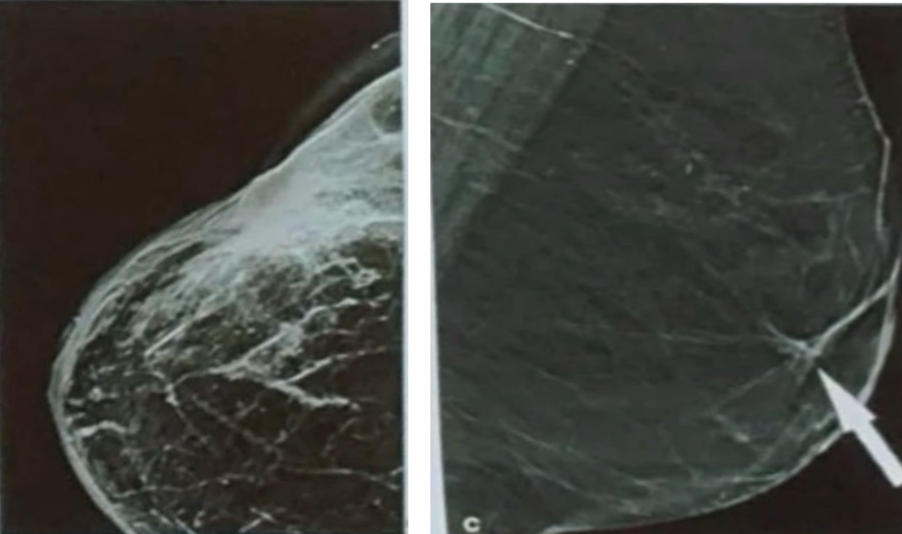
ARCHITECTURAL DISTORTIONS
201
Represents the superimposition of typical structures
ASYMMETRIES
202
BREAST BIOPSY
ULTRASOUND-GUIDED CORE NEEDLE BIOPSY, VACUUM ASSISTED DEVICE (VAD), MRI GUIDED AND STEREOTACTIC GUIDED CORE NEEDLE BIOPSY, NEEDLE WIRE LOCALIZATION
203
Always use with contrast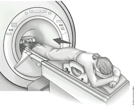
BREAST MRI
204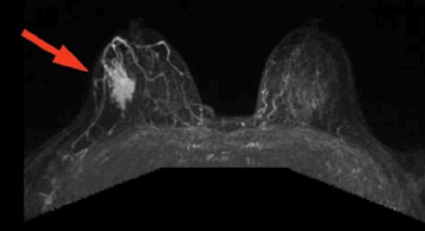
DCIS on breast MRI
205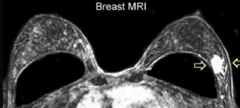
Implant in breast MRI
More Medicine & health flashcards
Hepatits CMedicine & health · EnglishKatlynKEohiMedicine & health · EnglishoozuHuman Anatomy Respiratory SystemMedicine & health · EnglishKatlynKEHypoglycemia, Nociception, and AntiemeticsMedicine & health · EnglishKaliLiver and Kidney: Lab Values and DamageMedicine & health · EnglishMatthewRespiratory Anatomy, Physiology, ARDS, PE, and COPDMedicine & health · EnglishCatLife Cycle Nutrition: Iron-Deficiency Anemia & Dental CariesMedicine & health · EnglishkiwicardiovascMedicine & health · EnglishillegitHead, Face, Neck, Lymphatics HA Exam #2Medicine & health · EnglishMadiruth2Female Reproductive Medical TerminologyMedicine & health · EnglishebradstreetVision and Lymphatic SystemMedicine & health · Englishjocyt9Chapter 20 PharmMedicine & health · EnglishMadiruth2
Study these decks in the app
Free on iPhone, Android, Mac and the web. Add any deck to your account in one tap.