Radiography Procedures - Chest
Study these flashcardsFree · spaced repetition · iPhone, Android, Mac & web
All cards
1
What is the primary disadvantage of performing an AP projection of the chest rather than a PA?
Increased magnification of the heart
2
Emphysema may require manual exposure factors.
Emphysema may require decreased manual exposure factors.
3
The xiphoid process is a reliable positioning landmark for determining the lower margin of the lungs for chest positioning.
False
4
The trachea lies to the esophagus.
The trachea lies anterior to the esophagus.
5
What are the three layers of the heart, from inner to outer?
1. Endocardium; 2. Myocardium; 3. Epicardium
6
Why is erect positioning ideal for chest radiographs?
Allows diaphragm to move down; Demonstrates air-fluid levels; Prevents engorgement of pulmonary vessels
7
What approximate breast dose was listed for an AP chest radiograph?
4 mrad
8
What adult chest exposure technique was selected as ideal (kV, mA, time, SID)?
120 kV, 800 mA, 1/40 sec, 72-inch (183 cm) SID
9
T/F Scoliosis and kyphosis may produce asymmetry of the sternoclavicular joints and rib cage as demonstrated on a PA chest radiograph.
True
10
A condition characterized as an irreversible dilation or widening of bronchi or bronchioles that may result from repeated pulmonary infection or obstruction is termed:
bronchiectasis.
11
Which structure lies posterior to the trachea and larynx in the neck?
Esophagus
12
What bony landmark is located on the superior portion of the sternum?
Jugular notch
13
Which IR orientation is generally used for asthenic, hyposthenic, and sthenic PA chest patients?
Lengthwise (portrait)
14
A PA chest radiograph reveals that the left sternoclavicular joint is closer to the spine than the right joint. What specific positioning error has been committed?
Rotation into the left anterior oblique (LAO) position
15
A patient comes to radiology for a routine chest study. On the PA projection, the radiologist sees a possible calcification near a rib, but she cannot tell whether the calcification is in the lung or on the rib. What additional projections would assist with the diagnosis?
Inspiration/expiration PA
16
Which airway structure is most inferior to the hyoid bone, vocal cords, and epiglottis?
Carina
17
The upper margin of the lungs is at the level of the:
vertebra prominens.
18
The space between the visceral and parietal pleura contains .
The space between the visceral and parietal pleura contains lubricating fluid.
19
Where is the larynx located relative to the trachea?
Superior to the trachea
20
Is tuberculosis or silicosis a nonoccupational infectious lung disease?
Tuberculosis
21
What does label E identify on this larynx diagram?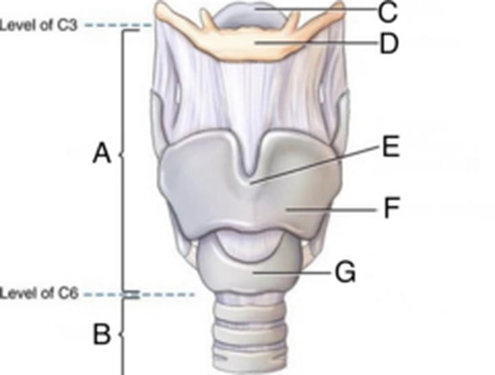
Laryngeal prominence
22
Which immune cells undergo maturation in the thymus gland?
T lymphocytes
23
The central ray (CR) for an anteroposterior (AP) supine, adult chest projection, should be centered:
3 to 4 inches (8 to 10 cm) below the jugular notch.
24
Which upper-airway structure lies outside the mediastinum: the epiglottis or trachea?
Epiglottis
25
Which patient positioning action removes most of the scapulae from the lung fields on a PA chest radiograph?
Roll shoulders forward.
26
Which commonly worn object may remain in place during a chest radiograph because it does not obscure the chest?
Glasses
27
What does label G identify on this larynx diagram?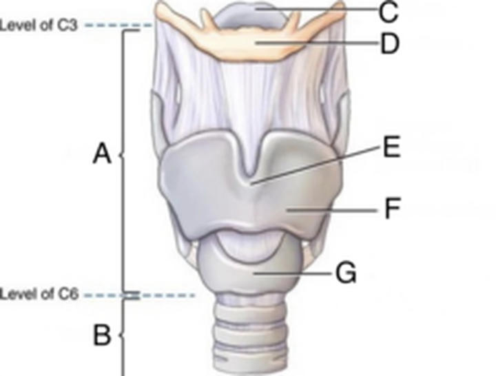
Cricoid cartilage
28
For an AP portable chest on an older or hypersthenic male patient, where should the CR be centered?
The CR should be centered 3 inches (8 cm) below the jugular notch.
29
What term describes the hilar region where bronchi and vessels enter the lung?
Hilum
30
The internal prominence or ridge in which the trachea bifurcates into the right and left bronchi is termed the:
carina.
31
What does label D identify on this larynx diagram?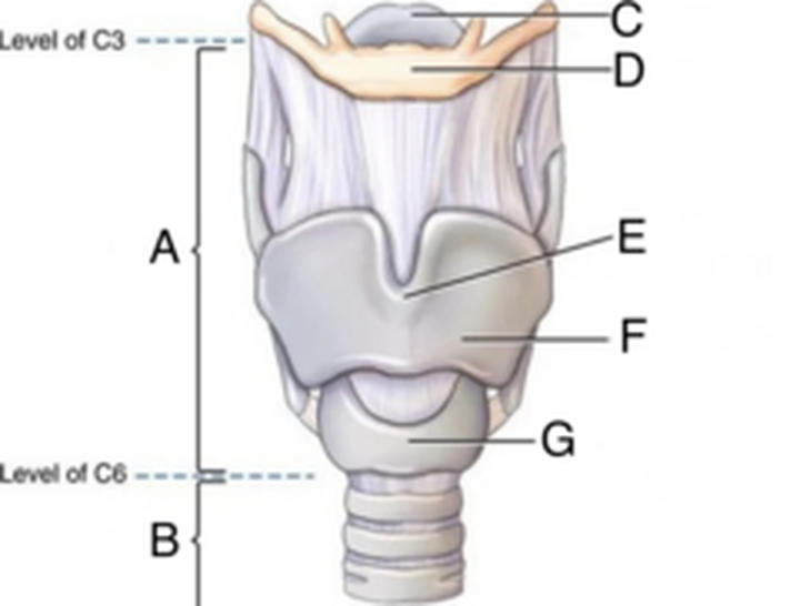
Hyoid bone
32
What is the name for the structure that serves as a lid over the larynx to prevent aspiration of food or fluid?
Epiglottis
33
The hyoid bone is located at approximately vertebral level .
The hyoid bone is located at approximately vertebral level C3.
34
The vertebra prominens corresponds to the level of:
C7.
35
List four special chest projections.
1. AP; 2. Lateral decubitus; 3. AP lordotic; 4. Obliques
36
The CR is centered to midsternum for the AP apical lordotic projection with a 14 x 17-inch (35 x 43) image receptor (IR).
True
37
For suspected left pleural effusion in an immobile patient, use positioning.
For suspected left pleural effusion in an immobile patient, use left lateral decubitus positioning.
38
Geriatric patients generally require higher central ray (CR) centering than younger patients.
True
39
What kVp range and source-to-image distance are recommended for an adult chest radiograph?
110-125 kVp, 72-inch SID
40
How do AP semierect and PA erect chest images differ?
AP semierect may magnify the heart and show less distinct air-fluid levels, whereas PA erect limits magnification and better displays fluid levels.
41
What does label B identify on this larynx diagram?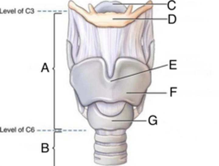
Trachea
42
Part vi refers to the: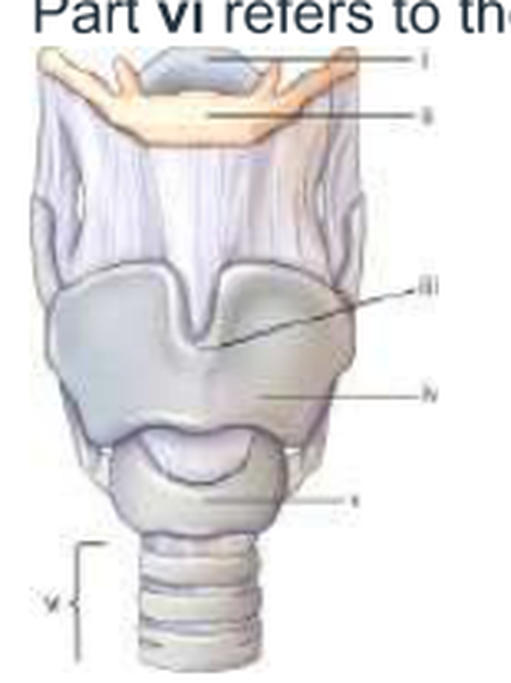
trachea.
43
The inferior angle of the scapula is located at approximately .
The inferior angle of the scapula is located at approximately T7.
44
A correctly positioned lateral chest radiograph demonstrates some separation of the posterior ribs owing to the divergent x-ray beam. But a separation of greater than ____ (insert answer) cm indicates objectionable rotation from a true lateral.
1
45
Which clinical indication often requires inspiration/expiration chest radiographs to detect a small amount of pleural air?
Small pneumothorax
46
A well-inspired average adult chest PA projection will have a minimum of ____ (insert answer) posterior ribs seen above the diaphragm.
10
47
The heart is located in the anterior chest at the level of:
T5-8.
48
For a PA chest with only seven posterior ribs above the diaphragm, which inspiration should the radiographer use to improve lung expansion?
Take exposure on the second inspiration rather than on first.
49
Pleural effusion generally requires an in manual exposure factors.
Pleural effusion generally requires an increase in manual exposure factors.
50
Which type of body habitus typically requires that the image receptor be placed crosswise rather than lengthwise for a posteroanterior (PA) chest?
Hypersthenic
51
The tracheal bifurcation, or carina, is located at approximately .
The tracheal bifurcation, or carina, is located at approximately T5–T6.
52
True or False: Occupational lung disease necessarily requires increased manual exposure factors.
False. Occupational lung disease does not necessarily require changing manual exposure factors.
53
Which positioning line must be placed perpendicular to the plane of the IR for an AP projection of the upper airway?
Acanthiomeatal
54
What does label A identify on this larynx diagram?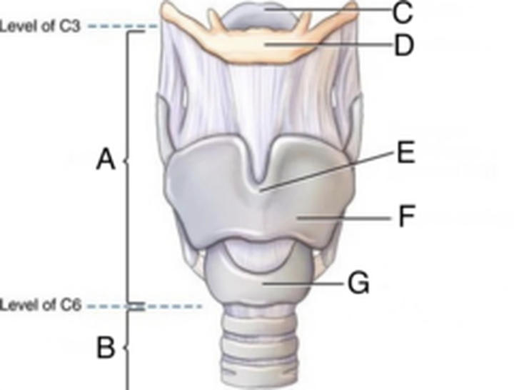
Larynx
55
Contrary to common belief, the vertical dimension of an average PA chest is greater than the horizontal dimension.
False
56
List the five major mediastinal contents.
1. Heart; 2. Great vessels; 3. Trachea; 4. Esophagus; 5. Thymus
57
What does label C identify on this larynx diagram?
Epiglottis
58
The routine projections of the upper airway are .
The routine projections of the upper airway are AP and lateral.
59
What does label F identify on this larynx diagram?
Thyroid cartilage
60
The approximate thyroid dose for both PA and AP chest is .
The approximate thyroid dose for both PA and AP chest is 1 mrad.
61
How are exposure factors generally adjusted for secondary tuberculosis?
Slight increase
62
Part iv refers to the: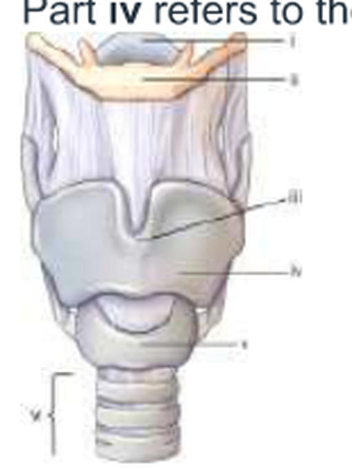
thyroid cartilage.
63
The thyroid gland is located at approximately level of: _____ (Missed on test)
C5-7. (Missed on test)
64
How many posterior ribs should ideally be visualized above the diaphragm in a PA chest radiograph?
10
65
For a suspected left pneumothorax when the patient cannot sit or stand, which lateral decubitus position places the affected lung uppermost? _____ (Missed on test)
Right lateral decubitus (Missed on test)
66
The inner-most layer of the pleura cavity is:
Visceral pleura
67
Top of image receptor placed approximately 3 inches (7.6 cm) above the shoulders is a recommended centering technique for adult chest radiography.
False
68
Should exposure factors be changed for uncomplicated pneumothorax?
No change
69
List four occupational lung diseases nameds.
1. Anthracosis; 2. Asbestosis; 3. Silicosis; 4. Byssinosis
70
The thymus gland is at its maximum size at:
puberty.
71
What is a common radiographic sign seen on a chest radiograph for a patient with respiratory distress syndrome (RDS)?
Air bronchogram
72
On a PA chest radiograph, the IR is placed about above the shoulders.
On a PA chest radiograph, the IR is placed about 1½–2 inches above the shoulders.
73
The heart is surrounded by a fibrous sac called the .
The heart is surrounded by a fibrous sac called the pericardium.
74
True or False: The left bronchus is more horizontal than the right bronchus.
False
75
How should chest collimation be balanced above the apices and below the costophrenic angles?
Approximately equal borders
76
What type of CR angle is required for the AP semiaxial projection for the lung apices?
15° to 20° cephalad
77
The lower concave area of the lung is termed the:
base.
78
For an average size female patient, where is the CR placed for a PA projection of the chest?
7 inches (18 cm) below the vertebra prominens
79
The asthenic body type makes up approximately (insert answer) ____% of the population. (Missed on test)
10 (Missed on test)
80
The membrane lining the thoracic cavity wall is the .
The membrane lining the thoracic cavity wall is the parietal pleura.
81
The laryngeal prominence is a positioning landmark located at the level of:
C5.
82
A PA chest radiograph is generally centered at vertebral level .
A PA chest radiograph is generally centered at vertebral level T7.
83
Why must the technologist slightly angle the CR caudad for most AP projections of the chest?
Prevents clavicles from obscuring apices of the lungs
84
The routine projections of the chest are .
The routine projections of the chest are PA and lateral.
85
The uppermost portion of each lung is the .
The uppermost portion of each lung is the apex.
86
What can a technologist refer to when critiquing an image, specifically when evaluation for rotation in a PA chest radiograph?
Sternoclavicular joints symmetry
87
A patient enters the emergency department (ED) to be treated for severe trauma. The physician orders an AP supine chest to evaluate the lungs. What can the technologist do to reduce the magnification of the heart?
Increase SID as much as possible.
88
Should exposure factors be increased or decreased for respiratory distress syndrome?
Increased
89
The inner layer of the pleura that encloses the lungs and heart is called the:
pulmonary pleura.
90
The two most common landmarks for chest positioning are the:
jugular notch and vertebra prominens.
91
The extreme, outermost lower corner of each lung is called the:
Costophrenic angle
92
When performing the lateral projection of the upper airway, exposure should be made during a slow, deep inspiration rather than at the end of the inspiration.
True
93
What condition involving partial or complete lung collapse may require increased manual chest exposure factors?
Atelectasis
94
Breast dose is approximately for PA chest versus 4 mrad for AP chest.
Breast dose is approximately 1 mrad for PA chest versus 4 mrad for AP chest.
95
What source-to-image receptor distance (SID) minimizes magnification of the heart on a chest radiograph?
72-inch (183 cm) SID
More Medicine & health flashcards
Breast HA Exam #4Medicine & health · EnglishMadiruth2Cerebral PalsyMedicine & health · Englishjocyt9Different types of hemagglutinationsMedicine & health · EnglishkarebearModule 1 (pgMedicine & healthEshala douleur Medicine & health · FrenchClaraBlood Pressure and AgingMedicine & health · EnglishEmmaLower Extremity Anatomy 1Medicine & health · EnglishKrisMedCardsHeart and Neck Vessels Health AssessmentMedicine & health · EnglishMadiruth2Coagulation and HemostasisMedicine & health · EnglishMontanaCardiovascular systemMedicine & health · EnglishKayleighBody PartsMedicine & health · EnglishHenry_LuisSinnesfysiologi / NervcellenMedicine & health · SwedishAlilpro
Study these decks in the app
Free on iPhone, Android, Mac and the web. Add any deck to your account in one tap.