Veterinary Coagulopathies and Hemophilia
Diese Karteikarten lernenKostenlos · Spaced Repetition · iPhone, Android, Mac & Web
Alle Karten
1
What are the two major classifications of coagulopathies?
Congenital or acquired; they may cause hemorrhage, thrombosis, or both.
2
What is hemophilia A?
The most common congenital coagulopathy in dogs and cats, caused by factor 8 deficiency.
3
How is hemophilia A inherited?
X-linked recessive; males are usually affected and females are typically carriers.
4
What determines the severity of hemophilia A?
The degree of factor 8 deficiency; signs range from spontaneous to trauma-induced hemorrhage.
5
What coagulation test pattern is expected in hemophilia A?
Prolonged aPTT, normal PT, and possibly prolonged ACT; platelet count is usually normal.
6
What is hemophilia B, and how is it inherited?
Factor 9 deficiency with X-linked recessive inheritance.
7
What coagulation test pattern is expected in hemophilia B?
Prolonged aPTT, normal PT, and sometimes prolonged ACT.
8
What is the clinical significance of factor 12 deficiency?
Prolonged aPTT without significant clinical bleeding; recognized particularly in cats.
9
What coagulation test abnormality occurs with factor 7 deficiency?
Prolonged PT, typically with normal aPTT; mild to moderate bleeding may occur.
10
How do deficiencies of factors 1, 2, and 10 affect coagulation tests?
They can prolong both PT and aPTT because they affect the common pathway.
11
Why can factor 13 deficiency cause bleeding despite normal PT and aPTT?
Factor 13 stabilizes fibrin after clot formation, which PT and aPTT do not assess.
12
A young male dog has post-castration bruising, normal platelets, prolonged aPTT, and factor 8 activity of 4%. Diagnosis?
Hemophilia A.
13
Which coagulation factors require vitamin K?
Factors 2, 7, 9, and 10, plus anticoagulant proteins C and S.
14
Why is vitamin K required for coagulation?
It enables y-flutamyl carboxylation of clotting factors so they can interact with calcium.
15
Where does vitamin K come from, and what is required for its absorption?
Vitamin K1 comes from plants, K2 from gut microbiota; absorption requires bile and pancreatic enzymes.
16
What bloodwork pattern is expected with vitamin K-dependent coagulopathy?
Regenerative anemia, hypoproteinemia, normal platelets, and prolonged PT/aPTT.
17
Why does PT become prolonged before aPTT in early vitamin K antagonism?
Factor 7 has the shortest half-life and is depleted first.
18
What happens to PIVKA testing with vitamin K antagonism?
PIVKA/Thrombotest PT becomes prolonged due to reduced vitamin K-dependent factor activity.
19
How does anticoagulant rodenticide toxicity cause hemorrhage?
It inhibits vitamin K recycling, preventing functional production of factors 2, 7, 9, and 10.
20
What is thrombosis?
Inappropriate formation of a blood clot inside a blood vessel.
21
How do arterial and venous thrombosis differ?
Arterial thrombosis causes ischemia/necrosis and platelet-rich white clots; venous thrombosis causes congestion and RBC-rich red clots.
22
What is an embolus?
A detached thrombus fragment that travels and lodges in a smaller blood vessel.
23
What are the three components of Virchow's triad?
Endothelial injury/dysfunction, abnormal blood flow or stasis, and hypercoagulability.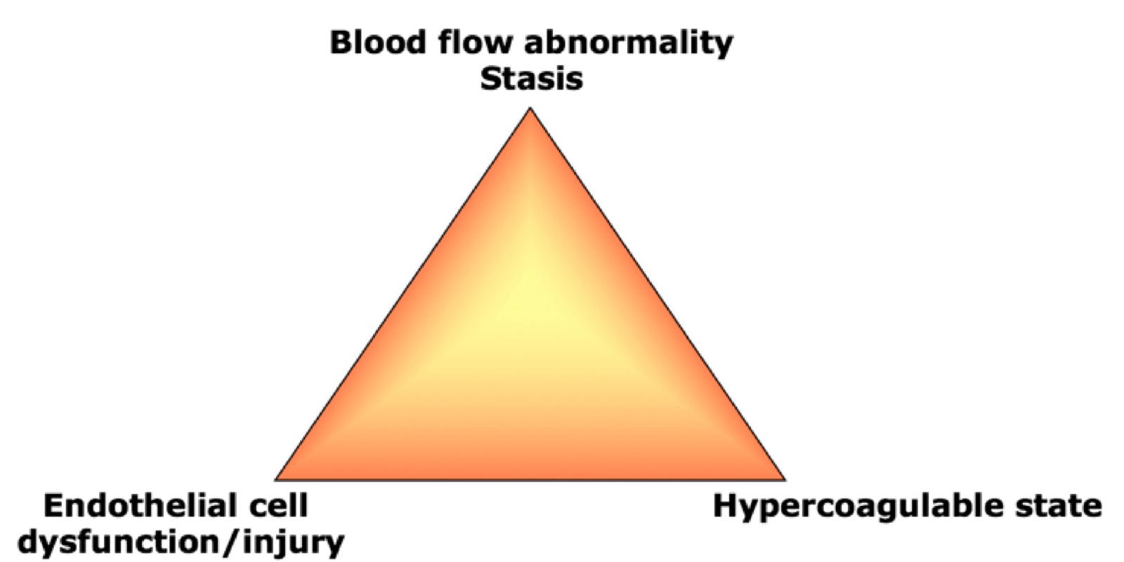
24
What is feline arterial thromboembolism (FATE)?
A thrombus, often associated with cardiomyopathy, that embolizes and commonly lodges at the aortic bifurcation.
A blood clot that often forms in the heart, breaks off, and gets stuck where the aorta splits into the hind legs, blocking blood flow.
A blood clot that often forms in the heart, breaks off, and gets stuck where the aorta splits into the hind legs, blocking blood flow.
25
What are the classic clinical signs of a feline saddle thrombus?
Acute hindlimb pain/paraparesis, weak or absent femoral pulses, and cold cyanotic paws.
26
What is important about coagulation tests and treatment in feline arterial thromboembolism?
Routine PT/aPTT, fibrinogen, D-dimer, and AT may be normal; clopidogrel is used for prevention.
27
Why does protein-losing nephropathy (PLN) predispose dogs to thrombosis?
Antithrombin is lost through the kidneys, creating a hypercoagulable state.
28
What findings characterize nephrotic syndrome associated with PLN?
Proteinuria, hypoalbuminemia, hypercholesterolemia, and edema.
29
How may hemostasis testing appear in dogs with PLN?
Decreased antithrombin, increased fibrinogen, and hypercoagulability on TEG.
30
Why does immune-mediated hemolytic anemia (IMHA) increase thrombosis risk?
Increased tissue factor, platelet activation, endothelial injury, microparticles, and NETs promote coagulation.
31
What thrombotic complications are associated with IMHA?
Pulmonary thromboembolism and infarcts involving the spleen, kidneys, or heart.
32
What other diseases predispose animals to thrombosis?
Acute necrotizing pancreatitis, sepsis, cancer, and hyperadrenocorticism.
33
A Doberman has pulmonary thrombosis, proteinuria, hypoalbuminemia, hypercholesterolemia, and low AT. Most likely cause?
Acquired antithrombin loss from protein-losing nephropathy.
34
Why can severe liver disease cause both bleeding and thrombosis?
The liver produces clotting factors and natural anticoagulants, so both systems become impaired.
35
What coagulation abnormalities may occur with liver failure?
Prolonged PT/aPTT, thrombocytopenia, decreased antithrombin/protein C, and sometimes hyperfibrinolysis.
36
A puppy has hemorrhagic diarrhea, prolonged PT/aPTT, low fibrinogen, and marked liver dysfunction. Most likely diagnosis?
Acute liver failure-associated coagulopathy.
37
What is disseminated intravascular coagulation (DIC)?
A secondary thrombohemorrhagic syndrome involving widespread coagulation activation and possible consumption of platelets and factors.
38
What are the two major phases of DIC?
Compensated/non-overt DIC and uncompensated/overt DIC.
39
What characterizes non-overt DIC?
Early hypercoagulability that is partially controlled by natural anticoagulant and fibrinolytic mechanisms.
40
What test results are expected in non-overt DIC?
PT/aPTT and fibrinogen may be normal; D-dimers may increase and TEG may show hypercoagulability.
41
What happens during progression from non-overt to overt DIC?
Uncontrolled thrombosis consumes platelets and coagulation factors, eventually causing hemorrhage.
42
What are the diagnostic hallmarks of overt DIC?
Thrombocytopenia, prolonged PT/aPTT, decreased fibrinogen, increased D-dimers, and decreased antithrombin.
43
What conditions can initiate DIC in veterinary patients?
Sepsis, inflammation, pancreatitis, IMHA, cancer, heatstroke, envenomation, and endotoxemia.
44
What species-specific causes of DIC are emphasized?
Dogs/cats: sepsis, IMHA, pancreatitis;
horses: colic/endotoxemia or foal sepsis;
ruminants: mastitis/metritis.
horses: colic/endotoxemia or foal sepsis;
ruminants: mastitis/metritis.
45
How does sepsis promote DIC?
PAMPs and cytokines increase tissue factor, activate platelets/contact pathways, injure endothelium, and suppress anticoagulants.
46
How does cancer promote DIC?
Tumor tissue factor expression, inflammatory cytokines, procoagulant secretion, and tumor lysis activate coagulation.
47
How does aspirin impair primary hemostasis?
It irreversibly inhibits platelet cyclooxygenase, reducing platelet function for several days.
48
How do clopidogrel and heparin differ in their anticoagulant effects?
Clopidogrel blocks platelet ADP receptors; heparin enhances antithrombin activity.
49
What mechanisms contribute to trauma-induced coagulopathy (TIC)?
Hypothermia, acidosis, dilution, activated protein C, platelet dysfunction, low fibrinogen, and increased fibrinolysis.
50
How does activated protein C contribute to bleeding after trauma?
It inhibits factors 5a and 8a and promotes fibrinolysis, reducing clot formation.
51
Can trauma-induced coagulopathy cause both bleeding and thrombosis?
Yes; early hypocoagulability may be followed by hypercoagulability as protein C becomes depleted.
52
What bleeding pattern suggests primary versus secondary hemostatic dysfunction?
Primary: petechiae, ecchymoses, mucosal bleeding; secondary: hematomas, hemarthrosis, and body-cavity hemorrhage.
53
Which tests evaluate secondary hemostasis, fibrinolysis, and global coagulation?
PT/aPTT, fibrinogen, factor activity, D-dimers/FDPs, antithrombin, and TEG/ROTEM.
54
What test pattern suggests von Willebrand disease or thrombocytopathy?
Normal platelet count and PT/aPTT, with possible prolonged buccal mucosal bleeding time and abnormal specialized testing.
55
How can vitamin K antagonism be distinguished from overt DIC using laboratory findings?
Vitamin K antagonism usually has normal platelets/fibrinogen; overt DIC often has low platelets/fibrinogen and high D-dimers.
Mehr Karteikarten: Medizin und Gesundheit
section1_quizlet_flashcardsMedizin und Gesundheit · EnglischJoshMicosis Endémicas Medizin und Gesundheit · SpanischMajoHeart CirculationMedizin und Gesundheit · EnglischLynInflammationMedizin und Gesundheit · EnglischBarzoGI Class/IndicationsMedizin und Gesundheit · Lateinebradstreet4- sys. lymphatiqueMedizin und Gesundheit · FranzösischOcéaneInnate Immunity: First Line DefensesMedizin und Gesundheit · EnglischalexNeck AnatomyMedizin und Gesundheit · EnglischSaraPediatric Surgery SCORE 22-24Medizin und Gesundheit · Englischyyl109anatomy lab finalMedizin und Gesundheit · EnglischGreysonLab 6: Anatomy StructuresMedizin und Gesundheit · EnglischKailecture 1 -intro to MS systemsMedizin und Gesundheit · EnglischNia_D
Alle Decks: Medizin und Gesundheit
Lerne diese Decks in der App
Kostenlos auf iPhone, Android, Mac und im Web. Füge jedes Deck mit einem Tipp zu deinem Konto hinzu.