Lower Limb Positioning: Foot Anatomy
Diese Karteikarten lernenKostenlos · Spaced Repetition · iPhone, Android, Mac & Web
Alle Karten
1
The foot has bones in total: 14 phalanges, 5 metatarsals, and 7 tarsals.
The foot has 26 bones in total: 14 phalanges, 5 metatarsals, and 7 tarsals.
2
The 14 phalanges are divided so that toe 1 has phalanges and toes 2–5 have 3 each.
The 14 phalanges are divided so that toe 1 has 2 phalanges and toes 2–5 have 3 each.
3
Digits and metatarsals are numbered 1–5, starting on the side of the foot.
Digits and metatarsals are numbered 1–5, starting on the medial side of the foot.
4
What are the three parts of a metatarsal?
1. Head; 2. Body (shaft); 3. Base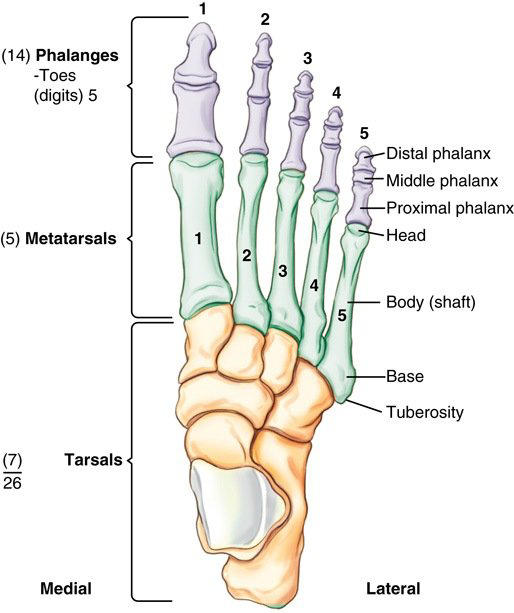
5
What are the seven tarsal bones? (Mnemonic: “Come to Colorado next 3 Christmases”)
1. Calcaneus; 2. Talus; 3. Cuboid; 4. Navicular; 5. Medial (1st) cuneiform; 6. Intermediate (2nd) cuneiform; 7. Lateral (3rd) cuneiform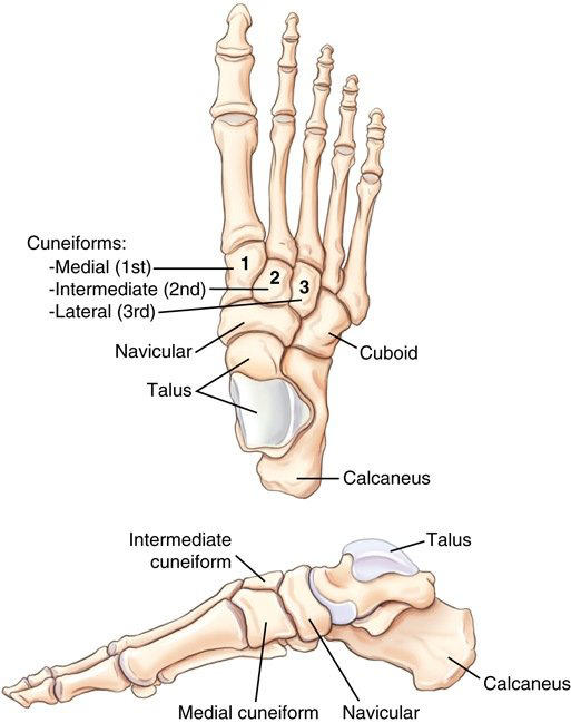
6
The is the largest and strongest tarsal.
The calcaneus is the largest and strongest tarsal.
7
The is the second largest tarsal and the only tarsal in the ankle joint.
The talus is the second largest tarsal and the only tarsal in the ankle joint.
8
Which tarsal lies on the medial side of the foot, between the talus and the three cuneiforms?
Navicular
9
Which cuneiform is the largest, lying between the navicular and the base of the 1st metatarsal?
Medial (1st) cuneiform
10
Which cuneiform is the smallest?
Intermediate (2nd) cuneiform
11
Which tarsal lies on the lateral side, between the calcaneus and the 4th and 5th metatarsals?
Cuboid
12
The calcaneal is at the posterior-inferior heel and is a common site of bone spurs.
The calcaneal tuberosity is at the posterior-inferior heel and is a common site of bone spurs.
13
The lateral ridge of the calcaneus is the trochlear process, also called the .
The lateral ridge of the calcaneus is the trochlear process, also called the peroneal trochlea.
14
What calcaneal structure provides medial support for the talus?
Sustentaculum tali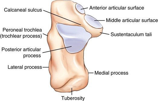
15
The subtalar joint, formed by the calcaneus and talus, is also called the joint.
The subtalar joint, formed by the calcaneus and talus, is also called the talocalcaneal joint.
16
What is the opening between the calcaneus and talus, in the middle of the subtalar joint, called?
Sinus tarsi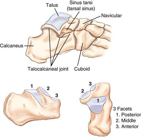
17
True or False: The distal part of the 5th metatarsal is the common fracture site.
False. The proximal portion (base or tuberosity) of the 5th metatarsal is the common fracture site.
18
What is the difference between the metatarsophalangeal (MTP) and tarsometatarsal (TMT) joints?
MTP joints are at the heads of the metatarsals, whereas TMT joints are at the bases of the metatarsals.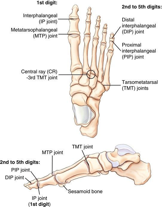
19
The sesamoid bones of the foot lie on the plantar surface at the head of the metatarsal (1st MTP joint).
The sesamoid bones of the foot lie on the plantar surface at the head of the 1st metatarsal (1st MTP joint).
20
The is the largest sesamoid bone in the body.
The patella is the largest sesamoid bone in the body.
21
The ankle joint is formed by the tibia, fibula, and .
The ankle joint is formed by the tibia, fibula, and talus.
22
What is the distal tibial surface that forms the roof of the ankle mortise called?
Tibial plafond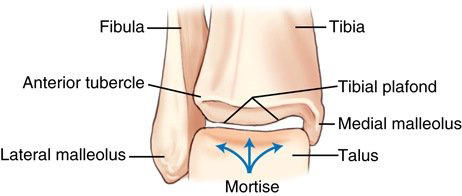
23
What socket, formed by the distal tibia and fibula, does the talus fit into?
Mortise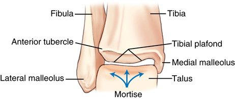
24
The lateral malleolus (fibula) sits about more posterior and more distal than the medial malleolus (tibia).
The lateral malleolus (fibula) sits about 1 cm more posterior and more distal than the medial malleolus (tibia).
25
The intermalleolar plane is from the coronal plane.
The intermalleolar plane is 15°–20° from the coronal plane.
26
The is on the distal anterior-lateral tibia.
The anterior tubercle is on the distal anterior-lateral tibia.
27
The is the weight-bearing bone of the lower leg.
The tibia is the weight-bearing bone of the lower leg.
28
The intercondylar eminence of the tibia is also called the , and it has medial and lateral intercondylar tubercles.
The intercondylar eminence of the tibia is also called the tibial spine, and it has medial and lateral intercondylar tubercles.
29
The tibial plateau (articular facets) slopes posteriorly.
The tibial plateau (articular facets) slopes 10°–20° posteriorly.
30
True or False: The fibula forms part of the knee joint.
False. The fibula is not part of the knee joint.
31
The intercondylar fossa (notch) is a deep depression on the distal femur.
The intercondylar fossa (notch) is a deep depression on the posterior distal femur.
32
The patellar surface of the distal femur, on its anterior side, is also called the intercondylar sulcus or .
The patellar surface of the distal femur, on its anterior side, is also called the intercondylar sulcus or trochlear groove.
33
The adductor tubercle is on the condyle of the femur.
The adductor tubercle is on the medial condyle of the femur.
34
The base of the patella is and its apex is inferior.
The base of the patella is superior and its apex is inferior.
35
The cruciate ligaments (anterior cruciate ligament, ACL, and posterior cruciate ligament, PCL) stabilize the knee .
The cruciate ligaments (anterior cruciate ligament, ACL, and posterior cruciate ligament, PCL) stabilize the knee front-to-back.
36
The collateral ligaments (medial, MCL, and lateral, LCL) prevent movement of the knee.
The collateral ligaments (medial, MCL, and lateral, LCL) prevent side-to-side movement of the knee.
37
The are shock absorbers between the femur and tibia.
The menisci are shock absorbers between the femur and tibia.
38
The protects the front of the knee.
The infrapatellar fat pad protects the front of the knee.
39
The two joints of the knee are the femorotibial joint and the joint.
The two joints of the knee are the femorotibial joint and the patellofemoral joint.
40
What type of joint is the interphalangeal (IP) joint of the toes?
Hinge
41
What type of joint is the metatarsophalangeal (MTP) joint?
Modified condyloid (ellipsoidal)
42
What type of joint is the tarsometatarsal (TMT) joint?
Gliding (plane)
43
What type of joint is the ankle joint?
Saddle (sellar)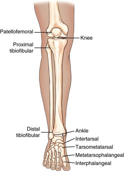
44
What type of joint is the knee (femorotibial) joint?
Bicondylar
45
What type of joint is the patellofemoral joint?
Saddle
46
What type of joint is the distal tibiofibular joint?
Syndesmosis (fibrous)
47
The minimum source-to-image-receptor distance (SID) for lower limb radiography is .
The minimum source-to-image-receptor distance (SID) for lower limb radiography is 40″ (100 cm).
48
The kVp range for the lower limb is , and for the knee it is 65–80.
The kVp range for the lower limb is 50–85, and for the knee it is 65–80.
49
A grid is used for lower limb parts measuring over .
A grid is used for lower limb parts measuring over 4″.
50
True or False: A grid is required for foot and ankle radiography.
False. A grid is not required for foot and ankle radiography.
51
Does close collimation replace gonadal shielding in lower limb radiography?
No. Gonadal shielding is still used.
52
For digital lower limb imaging, center the anatomy to the image receptor (IR) and use collimation.
For digital lower limb imaging, center the anatomy to the image receptor (IR) and use four-sided collimation.
53
For a pediatric patient, instead of holding the child, and lower the exposure.
For a pediatric patient, immobilize instead of holding the child, and lower the exposure.
54
Why should a bariatric patient not wear pants for lower limb imaging?
Clothing creates artifacts on digital images.
55
For AP toes, the central ray (CR) is angled toward the calcaneus.
For AP toes, the central ray (CR) is angled 10°–15° toward the calcaneus.
56
Where is the CR centered for AP toes?
The MTP joint of the toe in question
57
On an AP toe image, what shows that there is no rotation?
The shafts of the phalanges are equally concave.
58
For oblique toes, rotate with the CR perpendicular to the MTP joint.
For oblique toes, rotate 30°–45° with the CR perpendicular to the MTP joint.
59
For oblique toes, toes 1–3 are rotated and toes 4–5 are rotated .
For oblique toes, toes 1–3 are rotated medially and toes 4–5 are rotated laterally.
60
What is the difference between the lateral toe projections for toes 1–3 and toes 4–5?
Toes 1–3 are imaged lateromedial, whereas toes 4–5 are imaged mediolateral.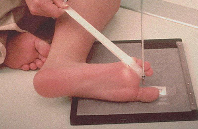
61
For a lateral toe, the CR is centered to the of toe 1 or the of toes 2–5.
For a lateral toe, the CR is centered to the IP joint of toe 1 or the PIP joint of toes 2–5.
62
For tangential sesamoids, the patient is positioned with the foot dorsiflexed.
For tangential sesamoids, the patient is positioned prone with the foot dorsiflexed.
63
For tangential sesamoids, the foot is dorsiflexed so the plantar surface is from vertical.
For tangential sesamoids, the foot is dorsiflexed so the plantar surface is 15°–20° from vertical.
64
How is the CR directed for the tangential sesamoid projection?
Perpendicular to the IR, tangential to the posterior 1st MTP joint
65
True or False: The supine alternative for tangential sesamoids gives less object-to-image-receptor distance (OID) and a sharper image.
False. The supine alternative for tangential sesamoids gives more OID and a less sharp image.
66
The AP foot is also called , and the PA foot is plantodorsal (PD).
The AP foot is also called dorsoplantar (DP), and the PA foot is plantodorsal (PD).
67
For an AP foot, the CR is angled posterior (toward the heel), perpendicular to the metatarsals.
For an AP foot, the CR is angled 10° posterior (toward the heel), perpendicular to the metatarsals.
68
Where is the CR centered for the AP and oblique foot?
The base of the 3rd metatarsal
69
How does the AP foot CR angle change for a high arch and for a low arch?
High arch: 15°; low arch: 5°
70
What 2 findings on an AP foot show there is no rotation?
Near-equal distance between the 2nd–5th metatarsals; 1st–2nd cuneiform joint open
71
For the AP oblique foot, rotate the foot medially with the CR perpendicular to the IR.
For the AP oblique foot, rotate the foot medially 30°–40° with the CR perpendicular to the IR.
72
The AP oblique foot with medial rotation is the best projection for the and most tarsal bones.
The AP oblique foot with medial rotation is the best projection for the cuboid and most tarsal bones.
73
True or False: The lateral oblique foot (30°) best shows the cuboid.
False. The lateral oblique foot (30°) best shows the navicular and the 1st–2nd cuneiforms.
74
What foot projection is shown?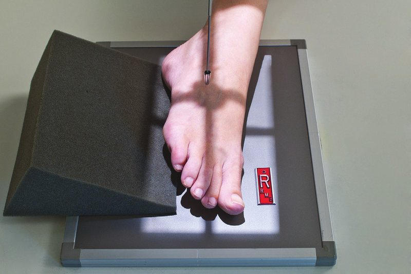
AP oblique foot with medial rotation (30°–40°), CR perpendicular at the base of the 3rd metatarsal
75
For a lateral foot, the knee is flexed about and the foot is dorsiflexed.
For a lateral foot, the knee is flexed about 45° and the foot is dorsiflexed.
76
Which lateral foot projection is the routine one: mediolateral or lateromedial?
Mediolateral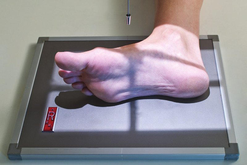
77
What 4 findings show a correct lateral foot image?
Tibiotalar joint open; distal fibula superimposed by the posterior tibia; metatarsal heads superimposed; 5th metatarsal tuberosity in profile
78
Weight-bearing foot studies evaluate the under full body weight.
Weight-bearing foot studies evaluate the longitudinal arches under full body weight.
79
The weight-bearing AP and lateral foot is the best study for a possible injury.
The weight-bearing AP and lateral foot is the best study for a possible Lisfranc injury.
80
For the weight-bearing AP foot, the CR is angled posterior to the midpoint between the feet at the base of the metatarsals.
For the weight-bearing AP foot, the CR is angled 15° posterior to the midpoint between the feet at the base of the metatarsals.
81
How is the CR directed for the weight-bearing lateral foot?
Horizontal, to the base of the 3rd metatarsal
82
For the plantodorsal (axial) calcaneus, the CR is angled cephalad to the base of the 3rd metatarsal.
For the plantodorsal (axial) calcaneus, the CR is angled 40° cephalad to the base of the 3rd metatarsal.
83
If the foot is not perpendicular to the IR on the axial calcaneus, the CR angle (30° → 40° fixes foreshortening).
If the foot is not perpendicular to the IR on the axial calcaneus, increase the CR angle (30° → 40° fixes foreshortening).
84
On the axial calcaneus, the sustentaculum tali shows .
On the axial calcaneus, the sustentaculum tali shows medially.
85
What calcaneus projection is shown?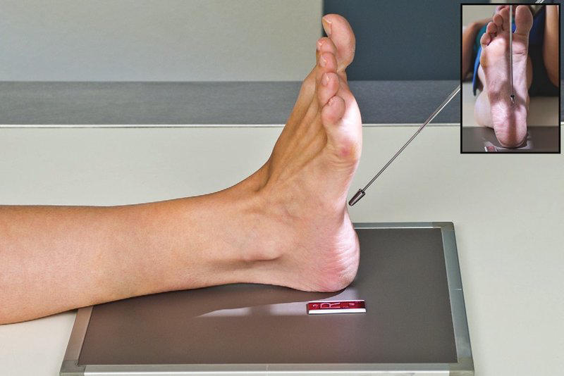
Plantodorsal (axial) calcaneus: CR 40° cephalad to the base of the 3rd metatarsal
86
For the lateral calcaneus, the CR is perpendicular, inferior to the medial malleolus.
For the lateral calcaneus, the CR is perpendicular, 1″ (2.5 cm) inferior to the medial malleolus.
87
On an AP ankle, which malleolus is about 15° more posterior?
The lateral malleolus
88
On a true AP ankle, the lateral mortise should look .
On a true AP ankle, the lateral mortise should look closed.
89
If the lateral mortise is open on a true AP ankle, suspect .
If the lateral mortise is open on a true AP ankle, suspect ligament rupture.
90
For an AP ankle, the CR is perpendicular and centered .
For an AP ankle, the CR is perpendicular and centered midway between the malleoli.
91
For the AP mortise, rotate the leg and foot medially until the intermalleolar line is parallel to the IR.
For the AP mortise, rotate the leg and foot 15°–20° medially until the intermalleolar line is parallel to the IR.
92
Which ankle projection is best for the ankle mortise?
AP mortise (15°–20° medial rotation)
93
For the AP mortise, the foot stays , which shows the base of the 5th metatarsal.
For the AP mortise, the foot stays plantar flexed, which shows the base of the 5th metatarsal.
94
On a correct AP mortise, the space around the talus is an even .
On a correct AP mortise, the space around the talus is an even 3–4 mm.
95
What 3 findings on an AP mortise indicate insufficient medial rotation?
Lateral joint space not open; lateral malleolus over the talus; talus distorted (looks like an AP)
96
What ankle projection is shown?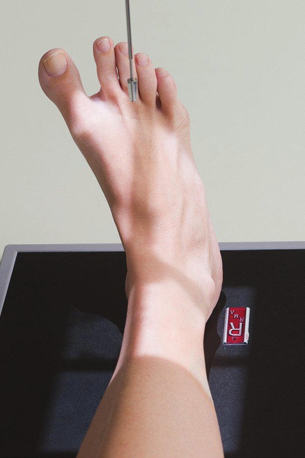
AP mortise: leg and foot rotated 15°–20° medially, foot plantar flexed
97
For the 45° oblique ankle, rotate the leg and foot medially.
For the 45° oblique ankle, rotate the leg and foot 45° medially.
98
Why is the foot dorsiflexed (plantar surface 10°–15° from vertical) for the 45° oblique ankle?
So the calcaneus does not overlap the lateral malleolus
99
The 45° oblique ankle is best for fractures of which two sites?
Lateral malleolus; base of the 5th metatarsal
100
What is the difference between the AP mortise and the 45° oblique ankle?
The AP mortise rotates 15°–20° medially with the foot plantar flexed to open the mortise, whereas the 45° oblique rotates 45° medially with the foot dorsiflexed to open the distal tibiofibular joint.
101
What ankle projection is shown?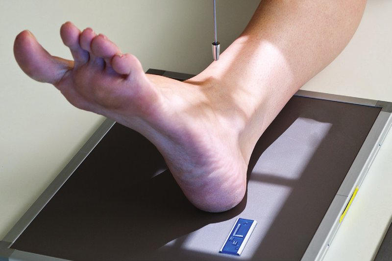
45° oblique ankle: leg and foot rotated 45° medially, foot dorsiflexed
102
On a lateral ankle, the distal fibula should lie over the half of the tibia.
On a lateral ankle, the distal fibula should lie over the posterior half of the tibia.
103
True or False: Superimposed malleoli on a lateral ankle indicate a true lateral position.
False. Superimposed malleoli on a lateral ankle make a partial oblique, not a true lateral.
104
The routine lateral ankle projection is , and lateromedial is the alternative.
The routine lateral ankle projection is mediolateral, and lateromedial is the alternative.
105
Which structure should be included on lateral and 45° oblique ankle views?
The base of the 5th metatarsal
106
True or False: AP stress views of the ankle show stress fractures.
False. AP stress views of the ankle show ligament tear or rupture (joint separation).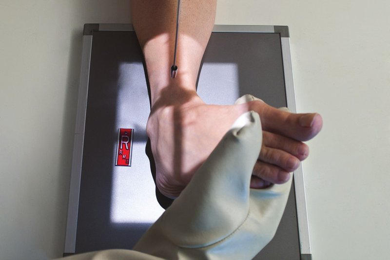
107
What 2 forced motions are used for AP ankle stress views?
Inversion; eversion
108
Who holds the foot for AP ankle stress views, and what protection is used if it is held by hand?
A physician or other health professional (or weights or straps); lead gloves and apron
109
For the AP knee, rotate the leg internally so the interepicondylar line is parallel to the IR.
For the AP knee, rotate the leg internally 3°–5° so the interepicondylar line is parallel to the IR.
110
Where is the CR centered for the AP knee?
½″ (1.25 cm) distal to the apex of the patella
111
For the AP knee, what CR angle is used when the anterior superior iliac spine (ASIS)-to-table distance is under 19 cm; 19–24 cm; over 24 cm?
Under 19 cm: 5° caudad; 19–24 cm: 0°; over 24 cm: 5° cephalad
112
On a correct AP knee, about of the fibular head is superimposed by the tibia.
On a correct AP knee, about half of the fibular head is superimposed by the tibia.
113
For the AP oblique knee, the whole leg is rotated , with the CR 0° on an average patient.
For the AP oblique knee, the whole leg is rotated 45°, with the CR 0° on an average patient.
114
True or False: If only one knee oblique is routine, it is usually the lateral oblique.
False. If only one knee oblique is routine, it is usually the medial oblique.
115
“Medial” and “lateral” oblique name the direction the rotates.
“Medial” and “lateral” oblique name the direction the front of the knee rotates.
116
What is the difference between the medial and lateral oblique knee views?
In the medial (internal) oblique, the proximal tibiofibular joint is open and the lateral condyles are in profile, whereas in the lateral (external) oblique, the proximal fibula is superimposed by the tibia and the medial condyles are in profile.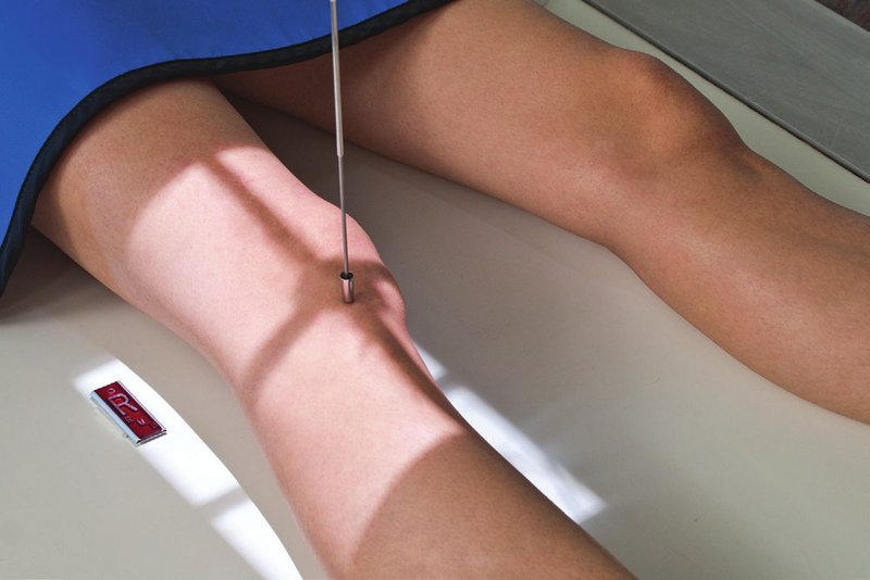
117
For the lateral knee, the patient is in lateral recumbent with the knee flexed .
For the lateral knee, the patient is in lateral recumbent with the knee flexed 20°–30°.
118
For the lateral knee, the CR is angled cephalad to superimpose the distal femoral condyles.
For the lateral knee, the CR is angled 5°–7° cephalad to superimpose the distal femoral condyles.
119
A lateral knee needs a cephalad CR because the femoral condyle sits lower.
A lateral knee needs a cephalad CR because the medial femoral condyle sits lower.
120
For the lateral knee, the CR is centered distal to the medial epicondyle.
For the lateral knee, the CR is centered 1″ (2.5 cm) distal to the medial epicondyle.
121
How does fibular head overlap differ with over-rotation versus under-rotation on a lateral knee?
Over-rotation gives less fibular head overlap, whereas under-rotation gives more.
122
What 3 findings show a correct lateral knee image?
Posterior condyles superimposed; patella in profile; patellofemoral joint open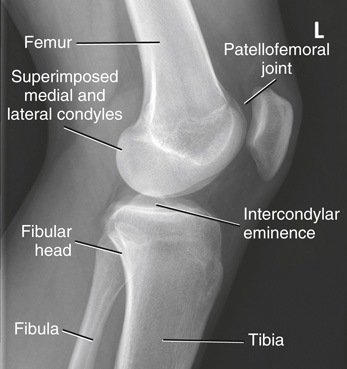
123
If the knee cannot flex for a lateral knee, use a beam.
If the knee cannot flex for a lateral knee, use a horizontal beam.
124
For the AP weight-bearing bilateral knees, the CR is centered ½″ (1.25 cm) the apex of the patellae.
For the AP weight-bearing bilateral knees, the CR is centered ½″ (1.25 cm) below the apex of the patellae.
125
For the AP weight-bearing knees, the CR is perpendicular for an average patient, or on a thin patient.
For the AP weight-bearing knees, the CR is perpendicular for an average patient, or 5°–10° caudad on a thin patient.
126
What does the AP weight-bearing bilateral knee study show?
The femorotibial joint spaces (cartilage degeneration), with both knees for comparison
127
The Rosenberg method is a weight-bearing projection of the knees.
The Rosenberg method is a PA axial weight-bearing projection of the knees.
128
In the Rosenberg method, the knees are flexed with the patellae against the upright bucky.
In the Rosenberg method, the knees are flexed 45° with the patellae against the upright bucky.
129
True or False: In the Rosenberg method, the CR is angled 10° cephalad.
False. In the Rosenberg method, the CR is angled 10° caudad, parallel to the tibial facets.
130
What does the Rosenberg method show?
The joint spaces and the intercondylar fossa
131
What knee method is shown?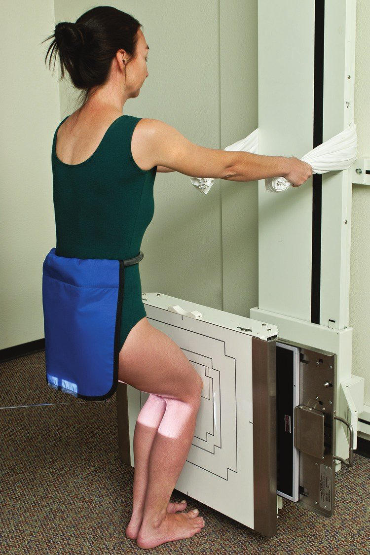
Rosenberg method (PA axial weight-bearing): knees flexed 45°, CR 10° caudad
132
For the Camp-Coventry method, the patient is prone with the knee flexed .
For the Camp-Coventry method, the patient is prone with the knee flexed 40°–50°.
133
In the Camp-Coventry method, the CR is perpendicular to the lower leg, which is , matching the flexion.
In the Camp-Coventry method, the CR is perpendicular to the lower leg, which is 40°–50° caudad, matching the flexion.
134
What 4 structures does the Camp-Coventry method show?
Intercondylar fossa; condyles; tibial plateaus; intercondylar eminence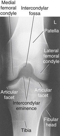
135
What knee method is shown?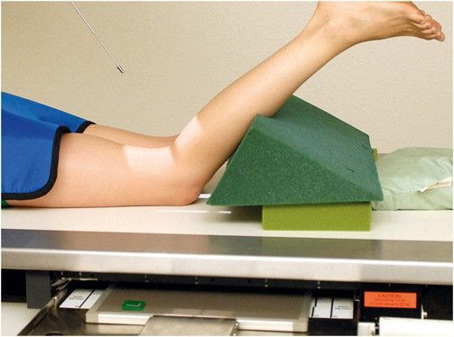
Camp-Coventry method (PA axial, prone): knee flexed 40°–50°, CR perpendicular to the lower leg
136
In the Holmblad method, leaning forward gives 60°–70° of knee flexion.
In the Holmblad method, leaning forward 20°–30° gives 60°–70° of knee flexion.
137
In the Holmblad method, the CR is perpendicular to the IR and lower leg, centered to the .
In the Holmblad method, the CR is perpendicular to the IR and lower leg, centered to the mid-popliteal crease.
138
What are 2 Holmblad variations for a cooperative, ambulatory patient?
Straddle a lowered table; rest the affected knee on a stool or chair
139
What knee method is shown?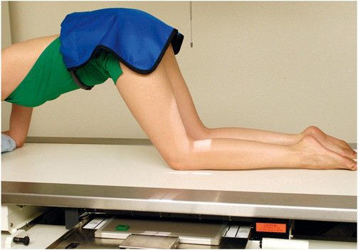
Holmblad method: kneeling on all fours, IR under the knee, CR perpendicular to the lower leg
140
The Béclère method is an projection for patients who cannot lie prone.
The Béclère method is an AP axial projection for patients who cannot lie prone.
141
In the Béclère method, the knee is flexed and the CR is perpendicular to the lower leg.
In the Béclère method, the knee is flexed 40°–45° and the CR is perpendicular to the lower leg.
142
What 3 problems make the Béclère method not preferred?
CR angle distorts; OID increased; gonadal exposure rises
143
What knee method is shown?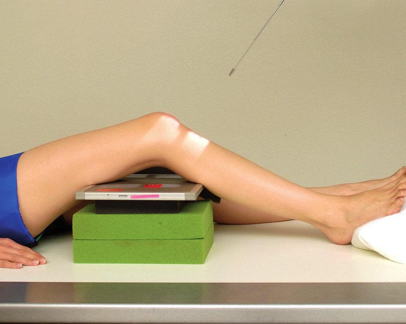
Béclère method (AP axial): supine, knee flexed 40°–45°, IR against the posterior thigh and leg
144
Why is the PA patella projection preferred over the AP?
Less OID; less magnification; less distortion
145
The PA patella is used to check for fracture flexing the knee.
The PA patella is used to check for fracture before flexing the knee.
146
Where is the CR centered for the PA patella?
The mid-patella
147
Why is the knee flexed only 5°–10° on a lateral patella?
More flexion can separate fracture fragments.
148
The lateral patella is used with the projection to evaluate patellar fractures.
The lateral patella is used with the PA projection to evaluate patellar fractures.
149
For the bilateral Merchant method, the CR is angled caudad from horizontal, midway between the patellae.
For the bilateral Merchant method, the CR is angled 30° caudad from horizontal, midway between the patellae.
150
For the bilateral Merchant method, the knees are flexed over the table end on a leg support.
For the bilateral Merchant method, the knees are flexed 40° over the table end on a leg support.
151
The bilateral Merchant method uses an SID of to reduce magnification.
The bilateral Merchant method uses an SID of 48–72″ to reduce magnification.
152
What 2 findings show correct positioning on a bilateral Merchant image?
Patellofemoral joint spaces open; patellae and intercondylar sulcus symmetric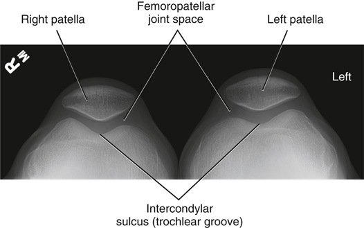
153
What patellar method is shown?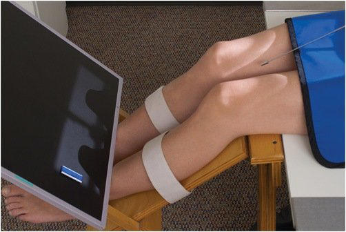
Bilateral Merchant method: supine, knees flexed 40°, CR 30° caudad
154
For the inferosuperior axial (tangential) patella, the CR is from the lower legs, tangential to the patellofemoral joint.
For the inferosuperior axial (tangential) patella, the CR is 10°–15° from the lower legs, tangential to the patellofemoral joint.
155
What are 3 advantages of the inferosuperior axial patella?
No special equipment; comfortable; quadriceps relaxed
156
What is the disadvantage of the inferosuperior axial patella?
The IR is hard to hold or support if the patient cannot cooperate fully.
157
What patellar method is shown?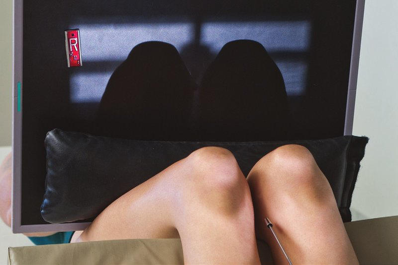
Inferosuperior axial (tangential) patella: supine, knees flexed 40°–45°, IR on the thighs
158
For the Hughston method, the patient is prone and the knee is flexed .
For the Hughston method, the patient is prone and the knee is flexed 50°–60°.
159
For the Hughston method, the CR is cephalad, tangential to the patellofemoral joint.
For the Hughston method, the CR is 45° cephalad, tangential to the patellofemoral joint.
160
What patellar method is shown?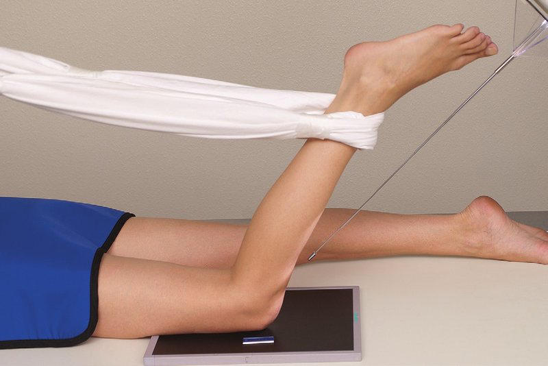
Hughston method: prone, knee flexed 50°–60°, CR 45° cephalad
161
For the Settegast method, the CR is from the lower leg, tangential to the patellofemoral joint.
For the Settegast method, the CR is 15°–20° from the lower leg, tangential to the patellofemoral joint.
162
True or False: The Settegast method uses knee flexion of 50°–60°.
False. The Settegast method uses knee flexion of 90° or more.
163
What is the major disadvantage of the Settegast method?
Acute flexion tightens the quadriceps and draws the patella into the intercondylar sulcus.
164
Before acute flexion for the Settegast or Hobbs method, must be ruled out.
Before acute flexion for the Settegast or Hobbs method, patellar fracture must be ruled out.
165
What patellar method is shown?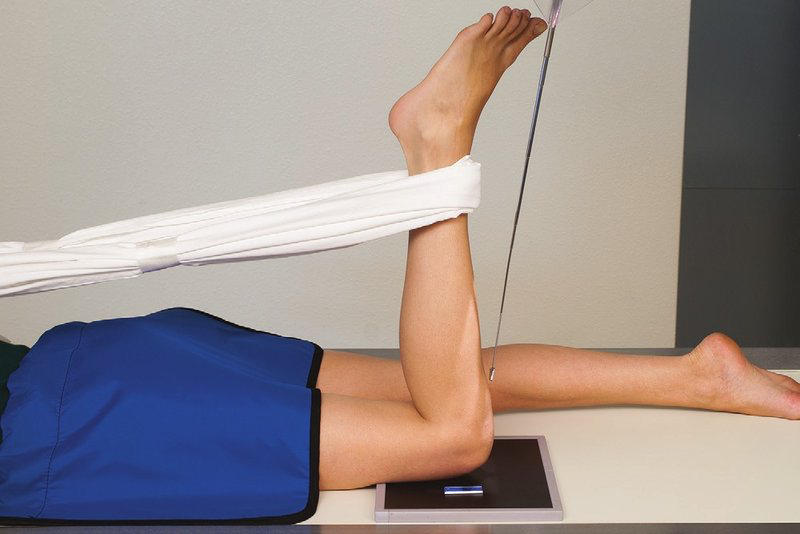
Settegast method: prone, knee flexed 90° or more, CR 15°–20° from the lower leg
166
What is the difference between the Hughston and Settegast methods?
Hughston is prone with 50°–60° flexion and a CR of 45° cephalad, whereas Settegast is prone with 90° or more flexion and a CR 15°–20° from the lower leg.
167
Which tangential patellar method is performed seated, with the CR perpendicular to the IR?
Hobbs modification (superoinferior)
168
The Hobbs modification uses an SID of to reduce magnification from the increased OID.
The Hobbs modification uses an SID of 48–50″ to reduce magnification from the increased OID.
169
What patellar method is shown?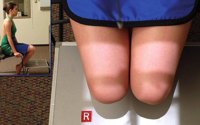
Hobbs modification: seated superoinferior, IR under the knees on a step stool, CR perpendicular to the IR
170
Which injury involves the large band ligament from the medial cuneiform to the bases of the 1st and 2nd metatarsals?
Lisfranc injury (sprain to fracture-dislocation)
171
What condition occurs when the tibial tuberosity is pulled away from the tibial shaft, usually in boys 10–15?
Osgood–Schlatter disease
172
What is the softening and erosion of the cartilage under the patella called?
Chondromalacia patellae (“runner’s knee”)
173
is a highly malignant primary bone tumor seen from childhood to young adult.
Osteogenic sarcoma is a highly malignant primary bone tumor seen from childhood to young adult.
174
Osteomalacia is bone softening from lack of mineralization, and it is called in children.
Osteomalacia is bone softening from lack of mineralization, and it is called rickets in children.
175
True or False: Osteomalacia is called rickets in adults.
False. Osteomalacia is called rickets in children.
176
Gout is caused by deposits in joints, mostly in men.
Gout is caused by uric acid deposits in joints, mostly in men.
177
The common first site of gout is the joint.
The common first site of gout is the 1st MTP joint.
178
On an AP knee image, the joint spaces are unequal and the proximal fibula is superimposed over the tibia. What is the error?
Lateral rotation of the lower limb
179
A Camp-Coventry image shows the fossa not open and foreshortened (prone, 45° flexion, CR 30° caudad). How is it fixed?
Increase the CR to 45° caudad (match the flexion).
180
Which patellar method is used for a bilateral patellofemoral study on a paraplegic patient who cannot stand?
Hobbs modification (seated superoinferior)
Mehr Karteikarten: Medizin und Gesundheit
High Yield ConditionsMedizin und Gesundheit · EnglischKrisMedCardsChapter 20 PharmMedizin und Gesundheit · EnglischMadiruth2Hepatits CMedizin und Gesundheit · EnglischKatlynKENervous System - Brain FunctionsMedizin und Gesundheit · EnglischAlexHemolytic AnemiasMedizin und Gesundheit · Englischeinem OutLearn-StudierendenPathophysiology Final ReviewMedizin und Gesundheit · EnglischMadiruth2Oral Pathology: Lesions 16-30Medizin und Gesundheit · EnglischoozuEarly Effects of Ionizing RadiationMedizin und Gesundheit · EnglischillegitPericarditis, Endocarditis and VHDMedizin und Gesundheit · EnglischcathsimsBloodMedizin und Gesundheit · EnglischBobertIde-celMedizin und Gesundheit · EnglischkrishanCH- Leading Causes of Death in the USMedizin und Gesundheit · EnglischRobo
Alle Decks: Medizin und Gesundheit
Lerne diese Decks in der App
Kostenlos auf iPhone, Android, Mac und im Web. Füge jedes Deck mit einem Tipp zu deinem Konto hinzu.

