Radiographic Procedures: Femur and Pelvic Girdle
Réviser ces flashcardsGratuit · répétition espacée · iPhone, Android, Mac et web
Toutes les cartes
1
The notch directly superior to the ischial spine is the .
The notch directly superior to the ischial spine is the greater sciatic notch.
2
What four joint groups are described for the pelvis and proximal femora?
1. Sacroiliac joints; 2. Symphysis pubis; 3. Unions of the acetabula; 4. Hip joints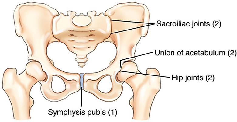
3
How does lesser trochanter visibility differ with internal versus external rotation of the hip?
With internal rotation the lesser trochanter is obscured or barely visible, whereas with external rotation it is seen in profile.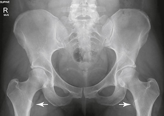
4
The thin, flared upper portion of the ilium is its .
The thin, flared upper portion of the ilium is its ala (wing).
5
The femoral head and neck project approximately relative to the femoral shaft.
The femoral head and neck project approximately 15–20° anteriorly relative to the femoral shaft.
6
The is formed where the superior pubic rami meet in the midline.
The symphysis pubis is formed where the superior pubic rami meet in the midline.
7
On this AP pelvis radiograph, what structure is labeled E?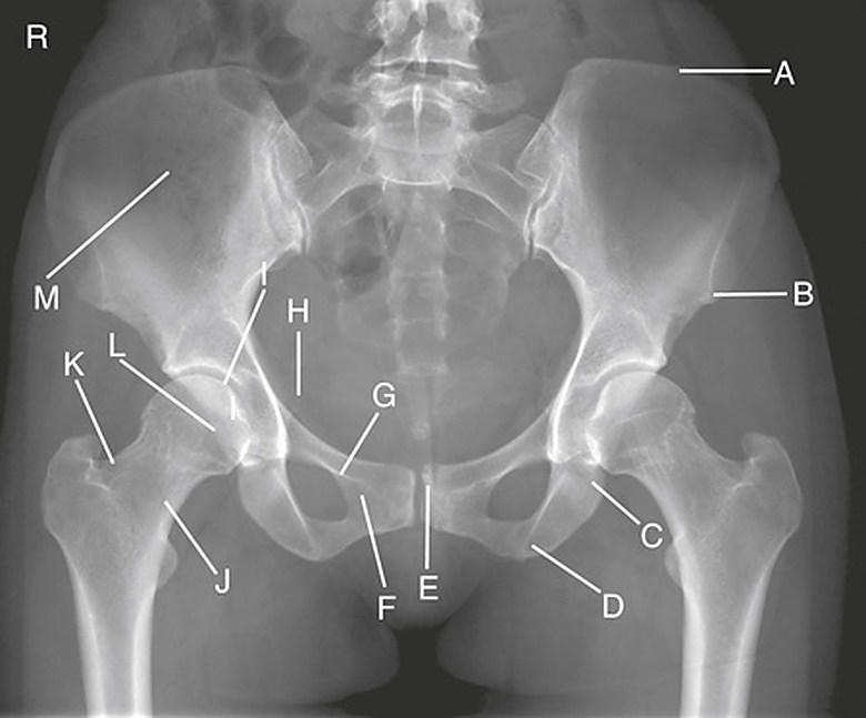
Symphysis pubis
8
Which pelvic cavity forms the actual birth canal?
Lesser (true) pelvis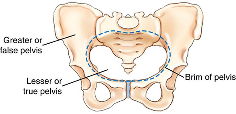
9
For bariatric patients, a grid may be used when the anatomic structure exceeds in thickness.
For bariatric patients, a grid may be used when the anatomic structure exceeds 4 in (10 cm) in thickness.
10
On this AP pelvis radiograph, what structure is labeled I?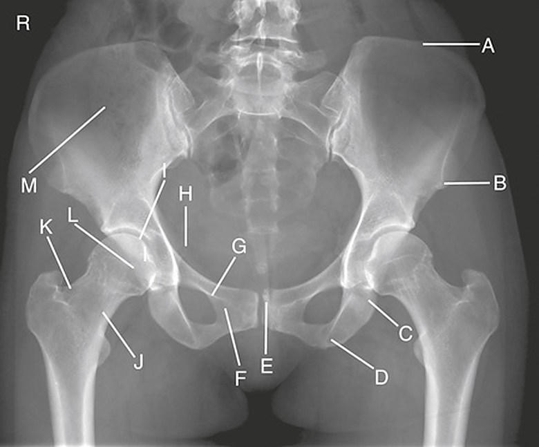
Right acetabulum
11
The average adult femoral neck-to-shaft angle is approximately .
The average adult femoral neck-to-shaft angle is approximately 125° (±15°).
12
The depression near the center of the femoral head that anchors a major ligament is the .
The depression near the center of the femoral head that anchors a major ligament is the fovea capitis.
13
On this axiolateral hip radiograph, what structure is labeled F?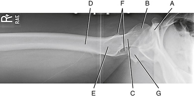
Area of the greater trochanter
14
Between which two spines does the iliac crest extend?
Anterior superior iliac spine (ASIS); posterior superior iliac spine (PSIS)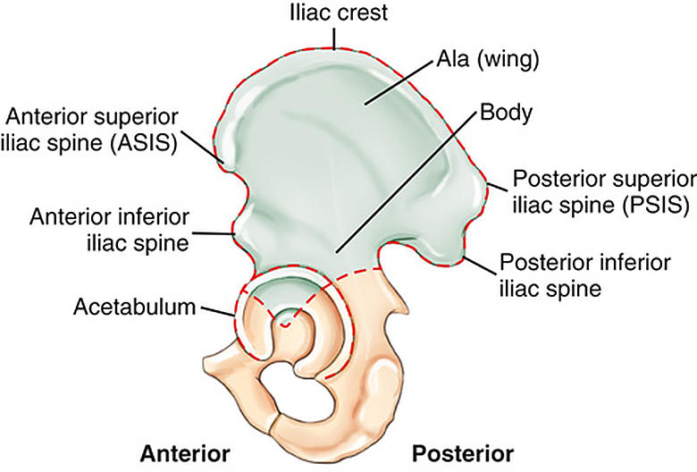
15
On this AP pelvis radiograph, what structure is labeled B?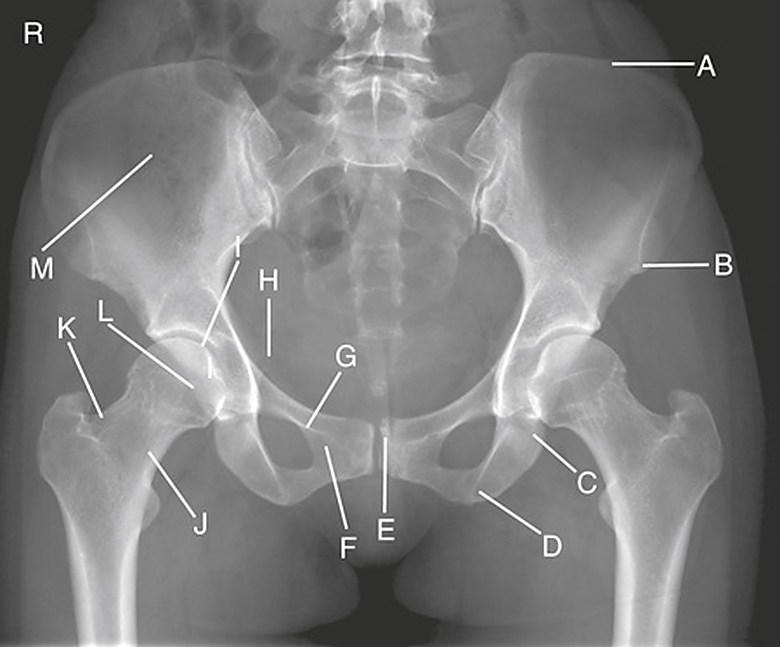
Anterior superior iliac spine (ASIS)
16
What posterior ridge joins the greater and lesser trochanters?
Intertrochanteric crest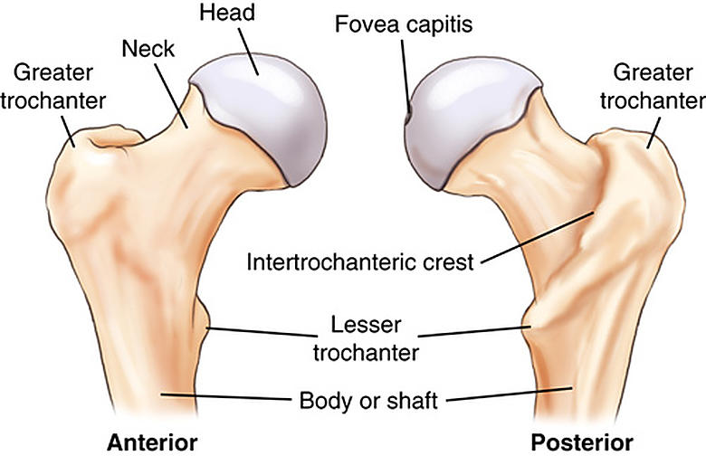
17
True or False: A platypelloid pelvic inlet is wider anterior to posterior than right to left.
False. A platypelloid pelvic inlet is wider right to left than anterior to posterior.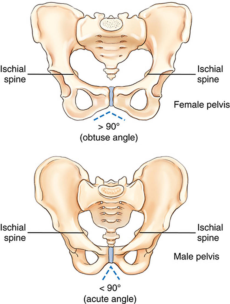
18
What movement type describes the freely movable, synovial hip joint?
Ball-and-socket (spheroidal)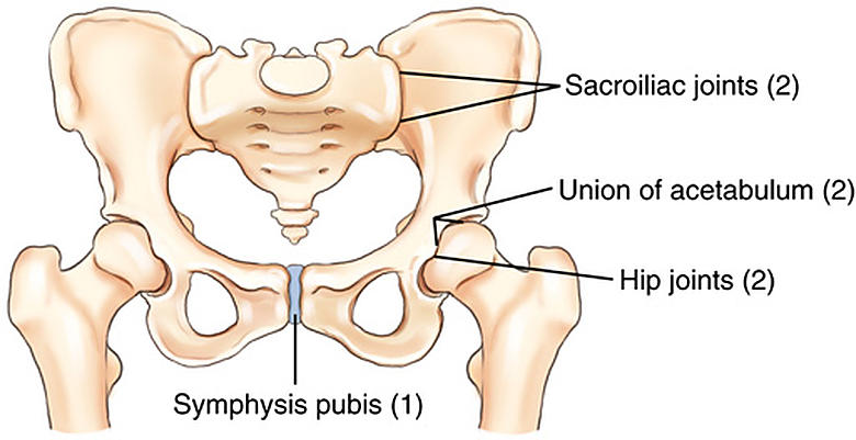
19
The ischial tuberosity lies about below the symphysis pubis.
The ischial tuberosity lies about 1½–2 in (4–5 cm) below the symphysis pubis.
20
The temporary growth joint where the acetabular divisions unite is a cartilaginous .
The temporary growth joint where the acetabular divisions unite is a cartilaginous synchondrosis.
21
The is the largest foramen in the human skeletal system.
The obturator foramen is the largest foramen in the human skeletal system.
22
After hip replacement surgery, patients should not be placed in the position.
After hip replacement surgery, patients should not be placed in the modified Cleaves (frog-leg) position.
23
True or False: A geriatric patient with suspected hip fracture should have an AP image of both hips first without moving the affected limb.
True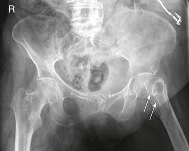
24
On this AP pelvis radiograph, what structure is labeled J?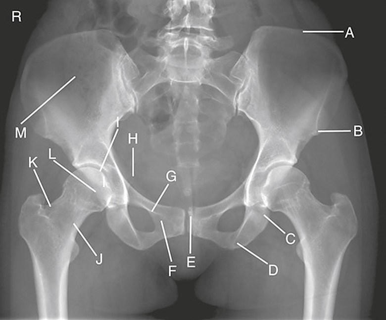
Right femoral neck
25
On this AP pelvis radiograph, what structure is labeled H?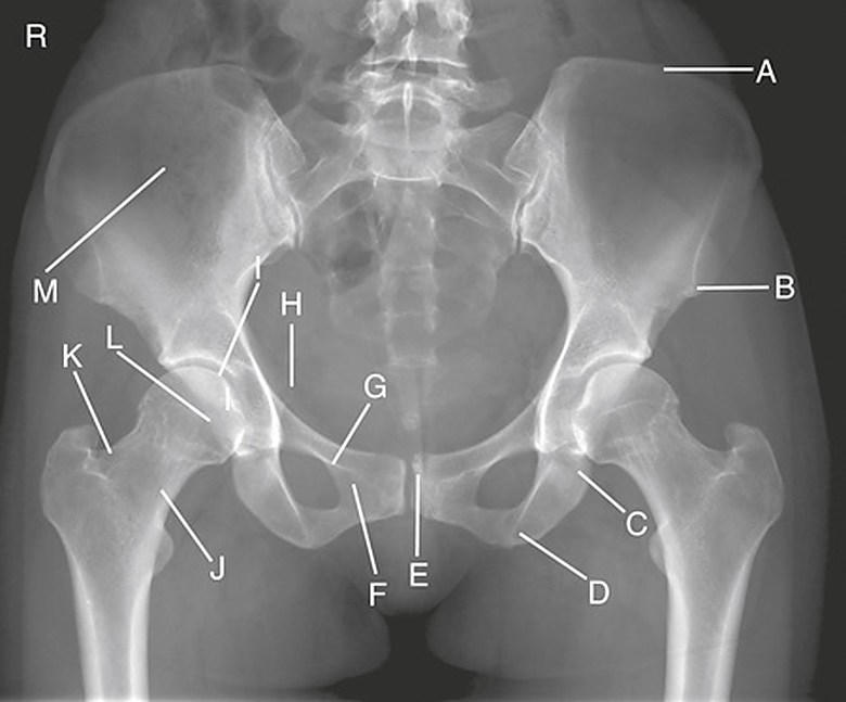
Right ischial spine
26
What four bones make up the complete pelvis?
1. Right hip bone; 2. Left hip bone; 3. Sacrum; 4. Coccyx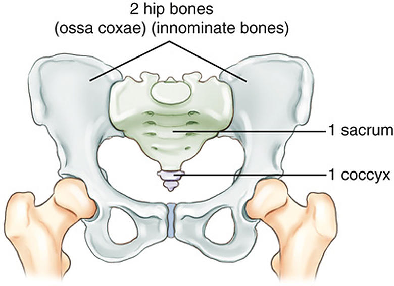
27
The lies inferior and posterior to the acetabulum.
The ischium lies inferior and posterior to the acetabulum.
28
With correct AP hip internal rotation, the lesser trochanter is .
With correct AP hip internal rotation, the lesser trochanter is not visible or only slightly visible.
29
Which two easily palpated bony landmarks are especially important in pelvic positioning?
Iliac crest; anterior superior iliac spine (ASIS)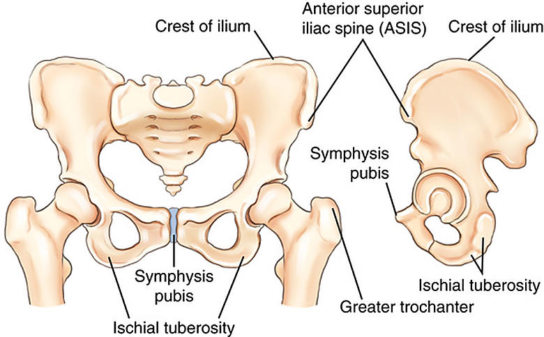
30
For hip and pelvis imaging, a higher kVp range of with lower mAs may reduce patient dose.
For hip and pelvis imaging, a higher kVp range of 80–90 kVp with lower mAs may reduce patient dose.
31
The projects medially and posteriorly from the junction of the femoral neck and shaft.
The lesser trochanter projects medially and posteriorly from the junction of the femoral neck and shaft.
32
How are the ASIS landmarks used to check for pelvic rotation?
Compare the distance from each ASIS to the tabletop; the distances should be equal.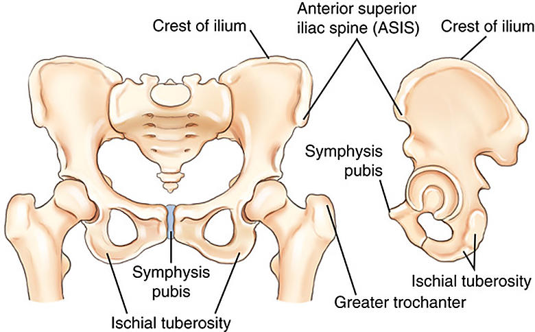
33
How do typical female and male pelves differ in overall shape?
The female pelvis is wider, shallower, and more flared, whereas the male pelvis is narrower, deeper, and less flared.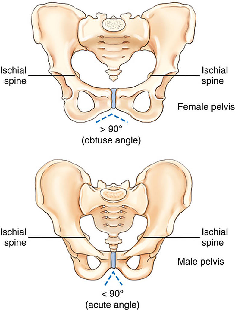
34
For digital hip and pelvis imaging, ALARA calls for the that still produce a diagnostic image.
For digital hip and pelvis imaging, ALARA calls for the lowest exposure factors that still produce a diagnostic image.
35
True or False: Ovarian shielding is possible on every AP pelvis projection.
False. It may not be possible when the entire pelvis, including the sacrum and coccyx, must be shown.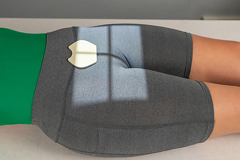
36
Ischial spines generally project farther medially into the pelvic cavity in than in .
Ischial spines generally project farther medially into the pelvic cavity in males than in females.
37
A true AP view of the proximal femur requires of the femur and lower limb.
A true AP view of the proximal femur requires 15–20° of internal rotation of the femur and lower limb.
38
A notable reason for pelvic or hip radiography in newborns is .
A notable reason for pelvic or hip radiography in newborns is developmental dysplasia of the hip (DDH).
39
What two factors are particularly important for digital imaging of the hip and pelvis?
Close collimation to the body part; accurate centering
40
In an average adult, the long axis of the femur is about from vertical in anatomic position.
In an average adult, the long axis of the femur is about 10° from vertical in anatomic position.
41
The separates the greater (false) from lesser (true) pelvis.
The pelvic brim separates the greater (false) from lesser (true) pelvis.
42
True or False: The greater trochanter projects superiorly and laterally from the femoral shaft.
True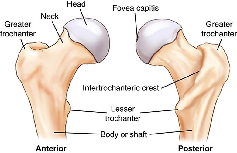
43
Where should the top of a male gonadal shield be placed for an AP pelvis image?
At the inferior margin of the symphysis pubis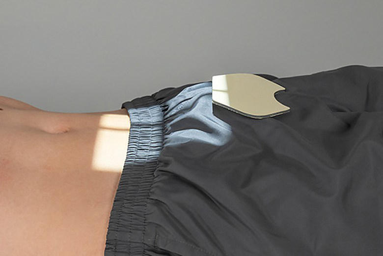
44
The bear most of the body’s weight when seated.
The ischial tuberosities bear most of the body’s weight when seated.
45
The symphysis pubis is a joint permitting limited (amphiarthrodial) movement.
The symphysis pubis is a cartilaginous symphysis joint permitting limited (amphiarthrodial) movement.
46
What are the four essential parts of the proximal femur?
1. Head; 2. Neck; 3. Greater trochanter; 4. Lesser trochanter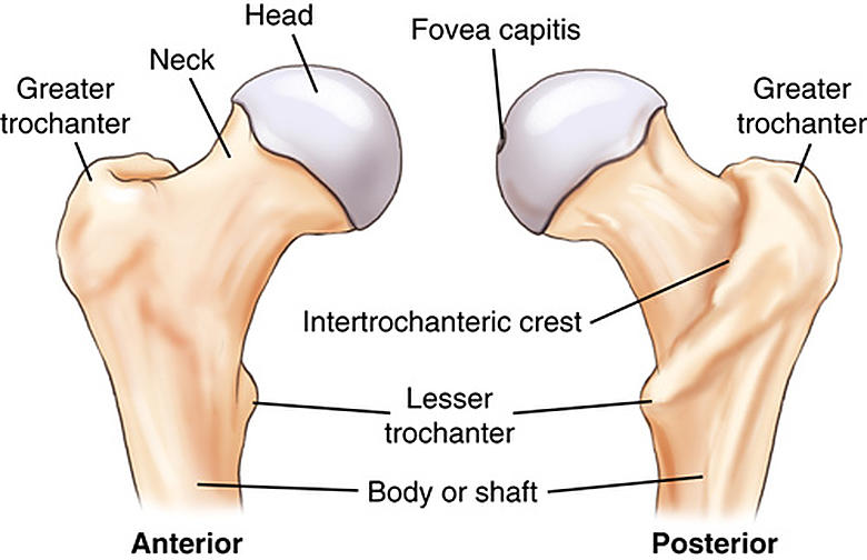
47
True or False: In anatomic position, femoral necks appear partially foreshortened on an AP pelvis image.
True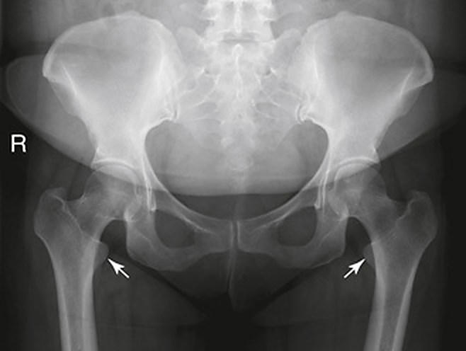
48
What deep, cup-shaped cavity accepts the femoral head to form the hip joint?
Acetabulum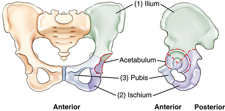
49
How do the typical female and male pubic arch angles differ?
The female pubic arch is 80–85°, whereas the male pubic arch is 50–60°.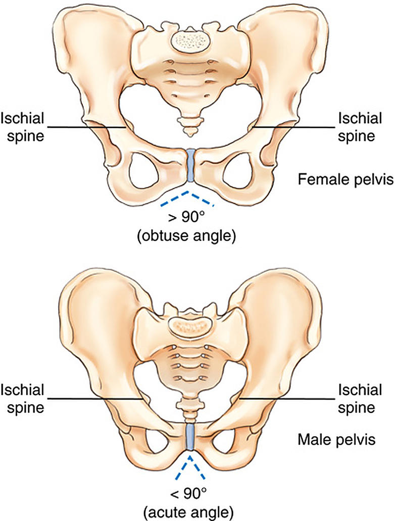
50
The sacroiliac joint is classified as with limited, irregular gliding movement.
The sacroiliac joint is classified as synovial with limited, irregular gliding movement.
51
During the middle teens, the three divisions of the hip bone fuse in the area of the .
During the middle teens, the three divisions of the hip bone fuse in the area of the acetabulum.
52
The greater trochanter is at about the same level as the superior border of the .
The greater trochanter is at about the same level as the superior border of the symphysis pubis.
53
The is the largest hip-bone division and lies superior to the acetabulum.
The ilium is the largest hip-bone division and lies superior to the acetabulum.
54
What are the four common pelvic inlet shapes and their shapes?
1. Gynecoid—round; 2. Platypelloid—wider right to left than anterior to posterior; 3. Android—heart shaped; 4. Anthropoid—wider anterior to posterior than right to left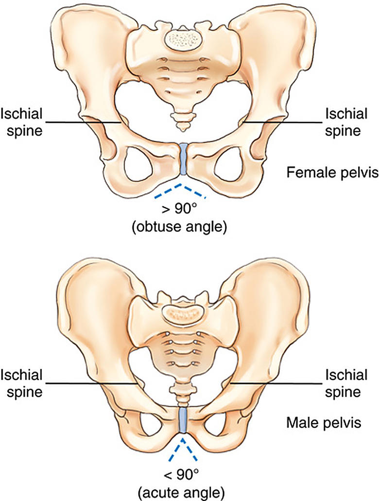
55
Which bony landmarks define the outlet of the true pelvis?
The two ischial tuberosities; tip of the coccyx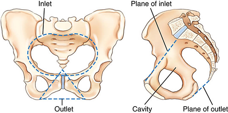
56
What three divisions make up each hip bone?
1. Ilium; 2. Ischium; 3. Pubis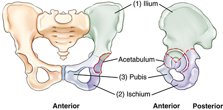
57
On this axiolateral hip radiograph, what structure is labeled C?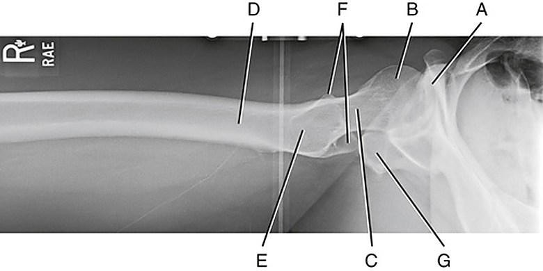
Femoral neck
58
Why may very high kVp be undesirable when imaging an older patient with osteoporosis?
It decreases subject contrast and visibility of bony detail.
59
From the midpoint of the ASIS-to-symphysis-pubis line, how far distal are the femoral head and neck?
Femoral head: 1½ in (4 cm); femoral neck: 2½ in (6–7 cm)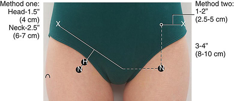
60
Using only the ASIS, where is the femoral neck located relative to it?
1–2 in (3–5 cm) medial; 3–4 in (8–10 cm) distal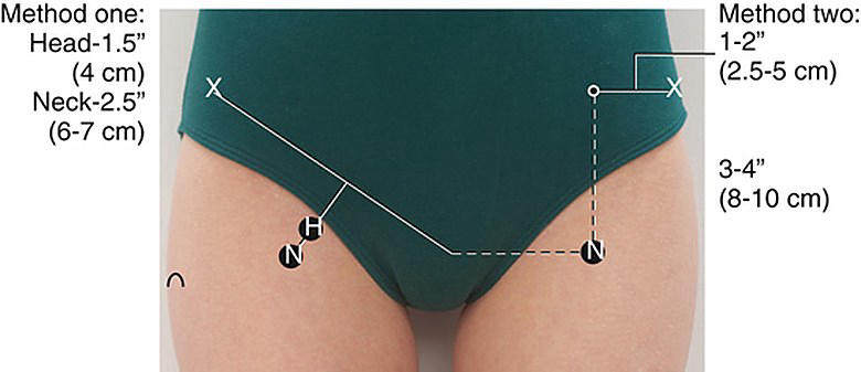
61
What three regions must the baby pass through during birth, in order?
1. Pelvic inlet; 2. Pelvic cavity (midcavity); 3. Pelvic outlet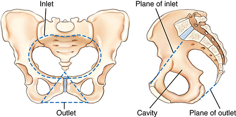
62
The lies inferior and anterior to the acetabulum.
The pubis lies inferior and anterior to the acetabulum.
63
What should you do with the affected leg when external foot rotation suggests a hip fracture?
Do not internally rotate it; obtain the AP pelvis radiograph as is.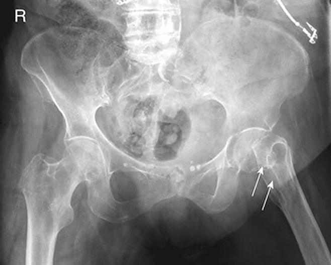
64
On this AP pelvis radiograph, what structure is labeled M?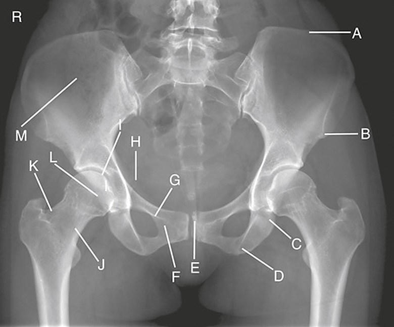
Ala (wing) of right ilium
Plus de flashcards : Médecine et santé
Terminologie médicale 2 : l'œil et les paupièresMédecine et santé · FrançaisbrabantImmunopharmacologyMédecine et santé · AnglaisCoryAbdominal RadiologyMédecine et santé · AnglaiscathsimsHepatic System and CirrhosisMédecine et santé · AnglaisEshaAmniotic FluidMédecine et santé · AnglaisJESSQTRANMed Term Prefix & Suffix ReviewMédecine et santé · AnglaisKatlynKEMyocardial InfarctionMédecine et santé · AnglaisAveryToxidromesMédecine et santé · LituanienCoryBlood supply to the brain and spinal cordMédecine et santé · AnglaisSammie16 - classification of bonesMédecine et santé · AnglaiselijahCH- HeartMédecine et santé · AnglaisRoboURINALYSIS OVERVIEWMédecine et santé · Anglaiskarebear
Tous les decks : Médecine et santé
Révise ces decks dans l’app
Gratuit sur iPhone, Android, Mac et le web. Ajoute n’importe quel deck à ton compte en un tap.