Histology Exam 2
Study these flashcardsFree · spaced repetition · iPhone, Android, Mac & web
All cards
1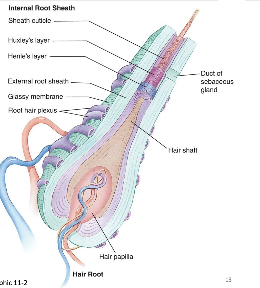
Image occlusion: name the hidden labels
2
Identify the structure + function at the pointer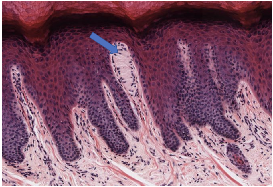
Meissner's corpuscle, trnasmit sensations of fine, discriminative touch and vibration
3
Identify the nucleated cell + function at the upper left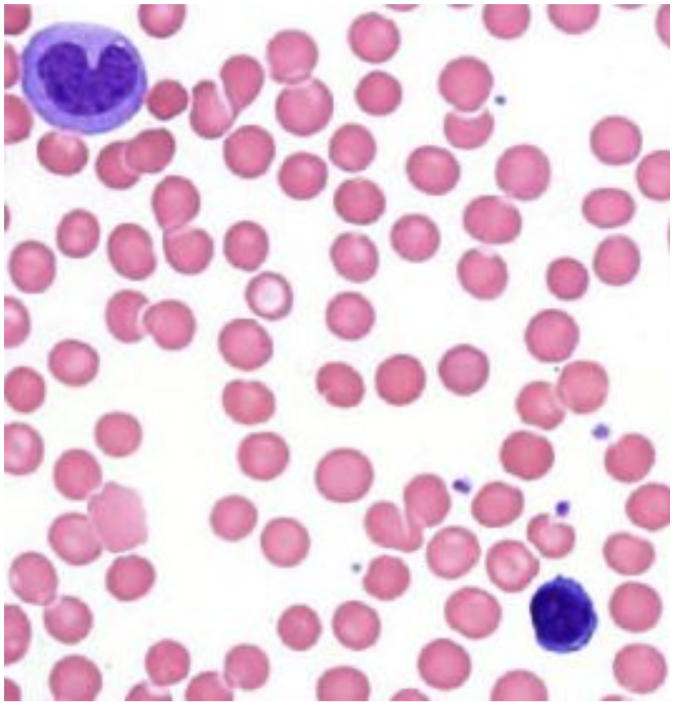
Monocyte, immune response via inflammatory and anti-inflammatory processes
4
Identify the deeply basophilic layer at the pointer.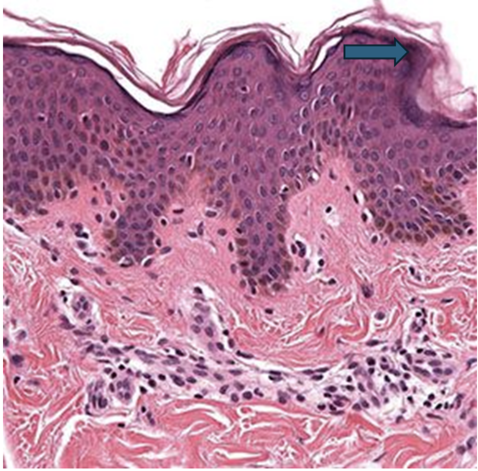
Stratum granulosum
5
Identify the tissue layer at the pointer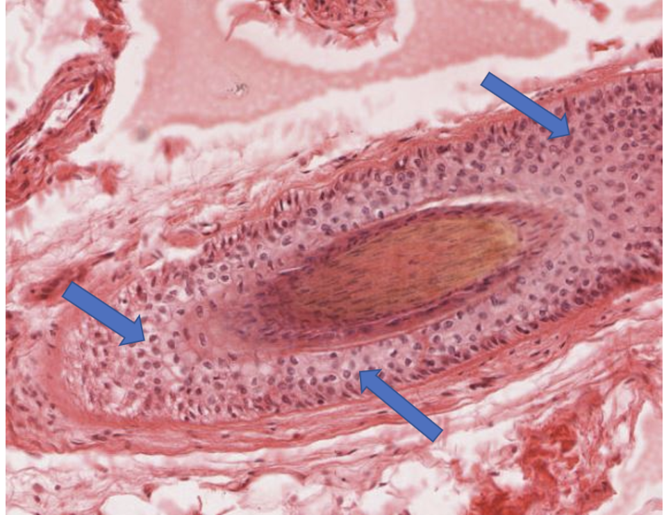
Outer root sheath
6
Identify the structures indicated by the pointers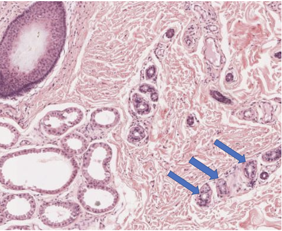
Eccrine sweat glands
7
Identify the layer indicated by the pointer. Be specific. 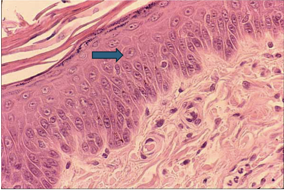
Stratum spinosum
8
Identify the layer indicated by the pointer. Be specific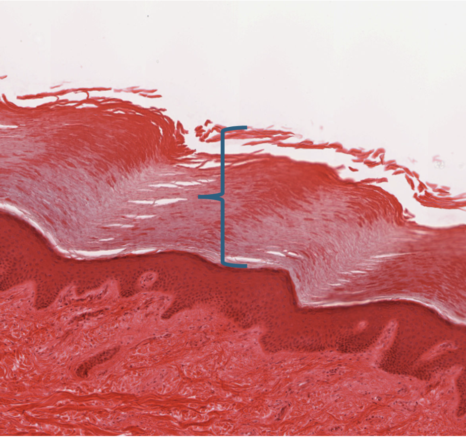
Stratum corneum
9
Identify the structure indicated by the pointer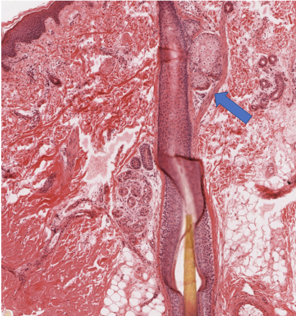
Arrector pili muscle
10
Identify the nucleated cell + its function at the lower right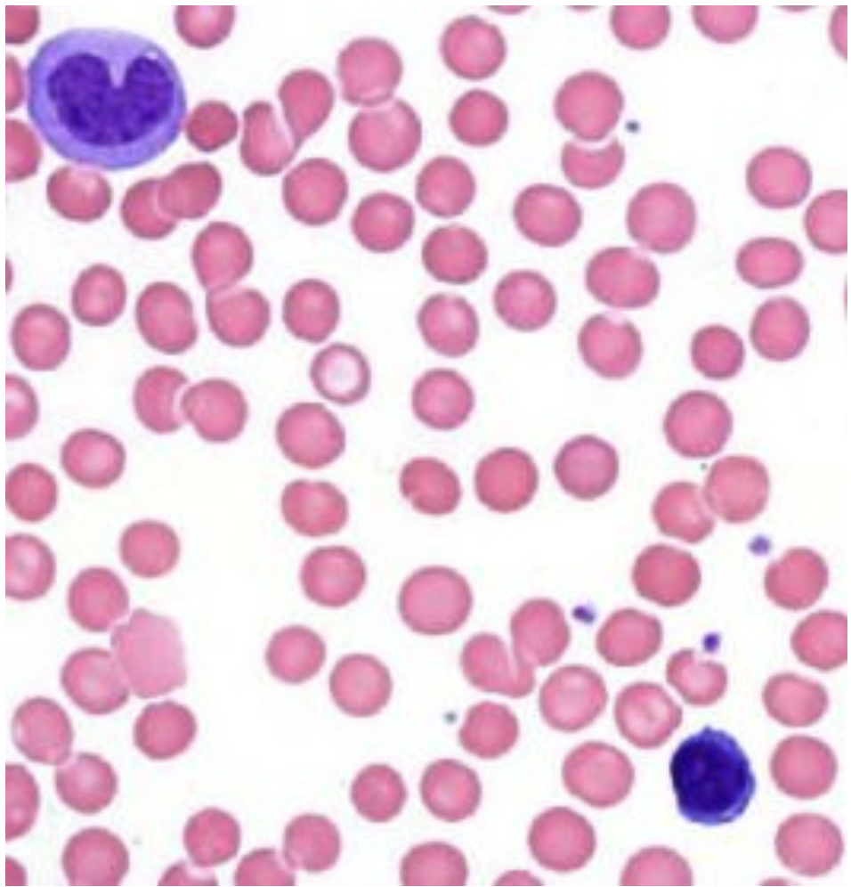
Lymphocyte, antibody production and regulation of immune response
11
Identify the tissue layer indicated by the bracket. Be specific.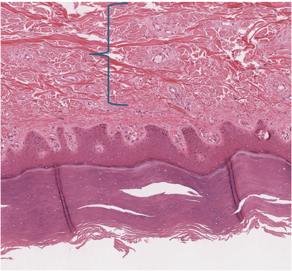
Reticular layer of the dermis
12
Identify the structure (clump of cells indicated by the pointer. 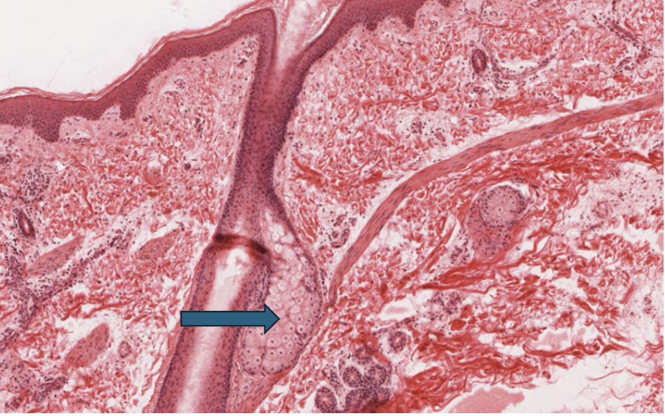
Sebaceous gland
13
Name the cell and its function at the pointer.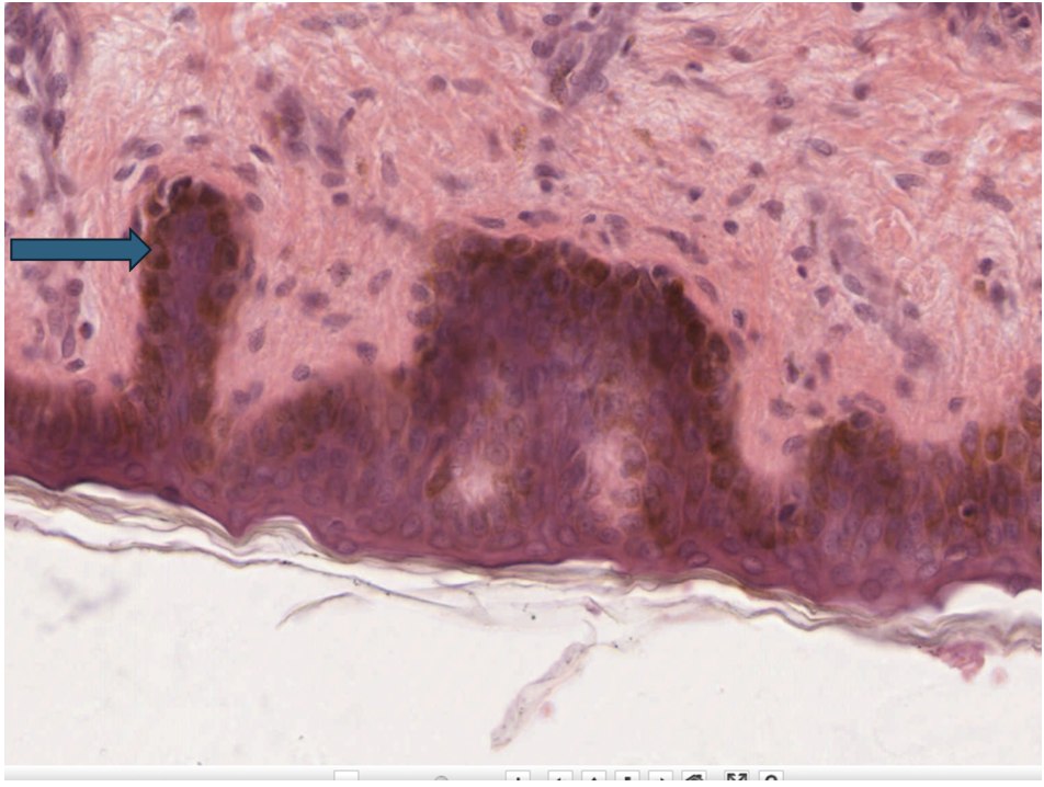
Melanocyte, melanin production/UV protection
14
Name the cell and its function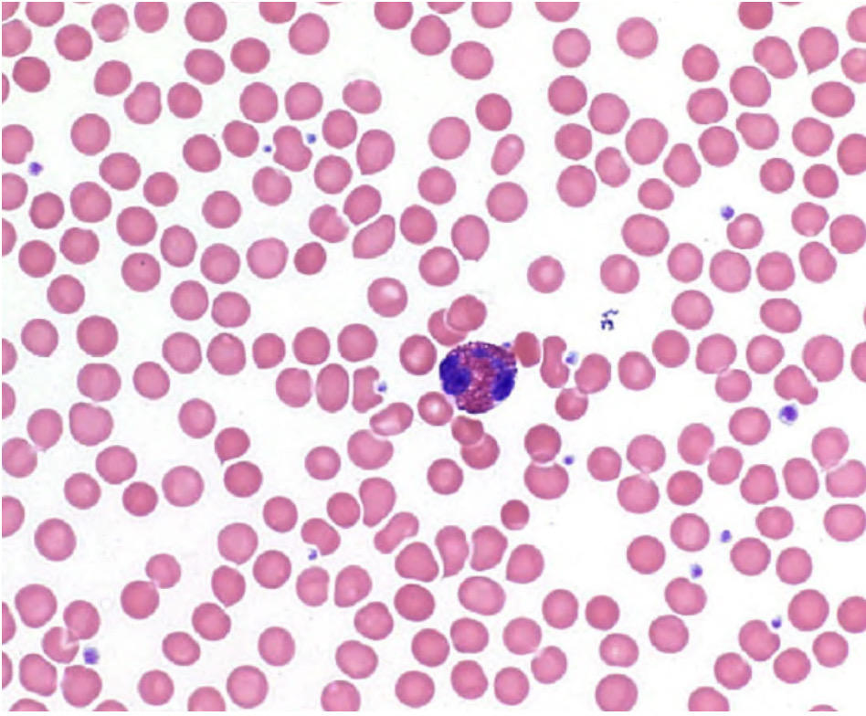
Eosinophil, immune response - eukaryotic pathogens
15
Identify the layer indicated by the pointer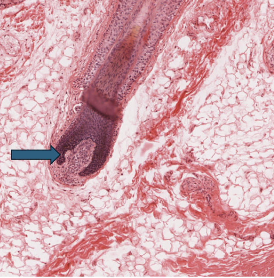
Hair bulb
16
Name the cell and its function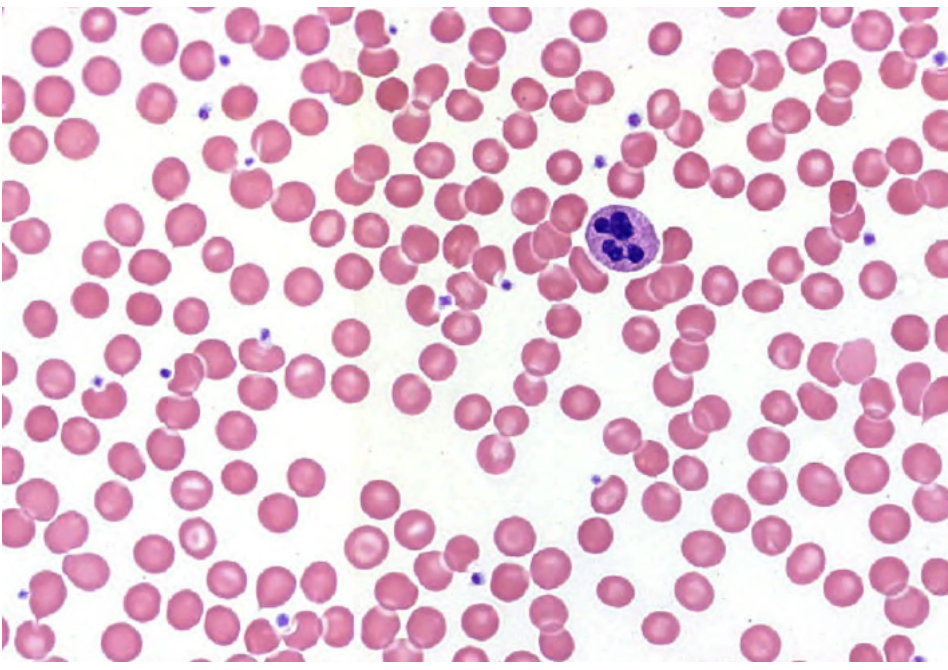
Neutrophil, immune response - prokaryotic pathogen/ bacteria
17
Identify the cell and its function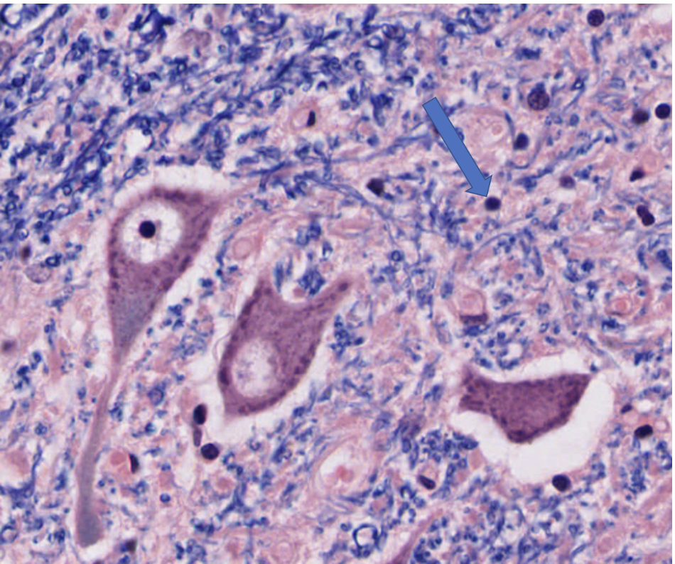
Glial cell (or neuroglia), support, connect, and portect neurons.
18
Identify the function of the substance represneted by the clear space at the pointer. 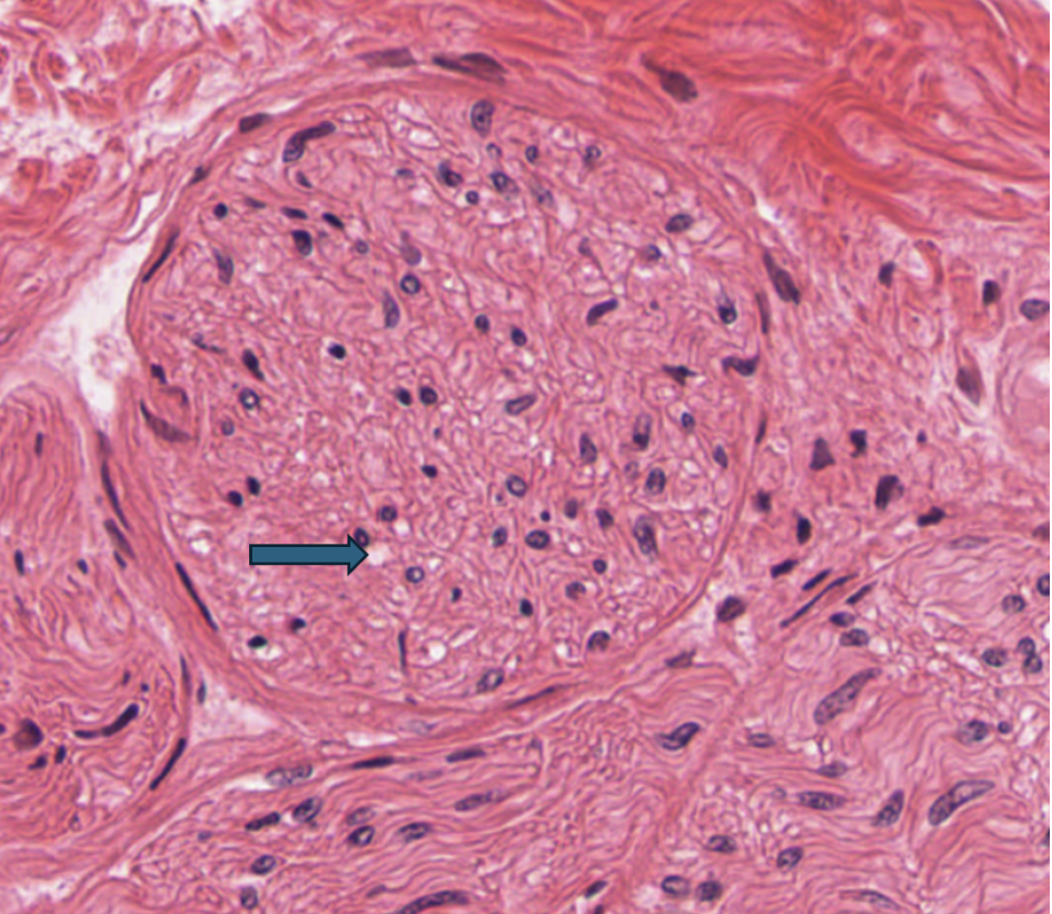
Aids in the transmission of electrical synapses down the axon.
19
Identify the large, circular structures indicated by the blue pointers.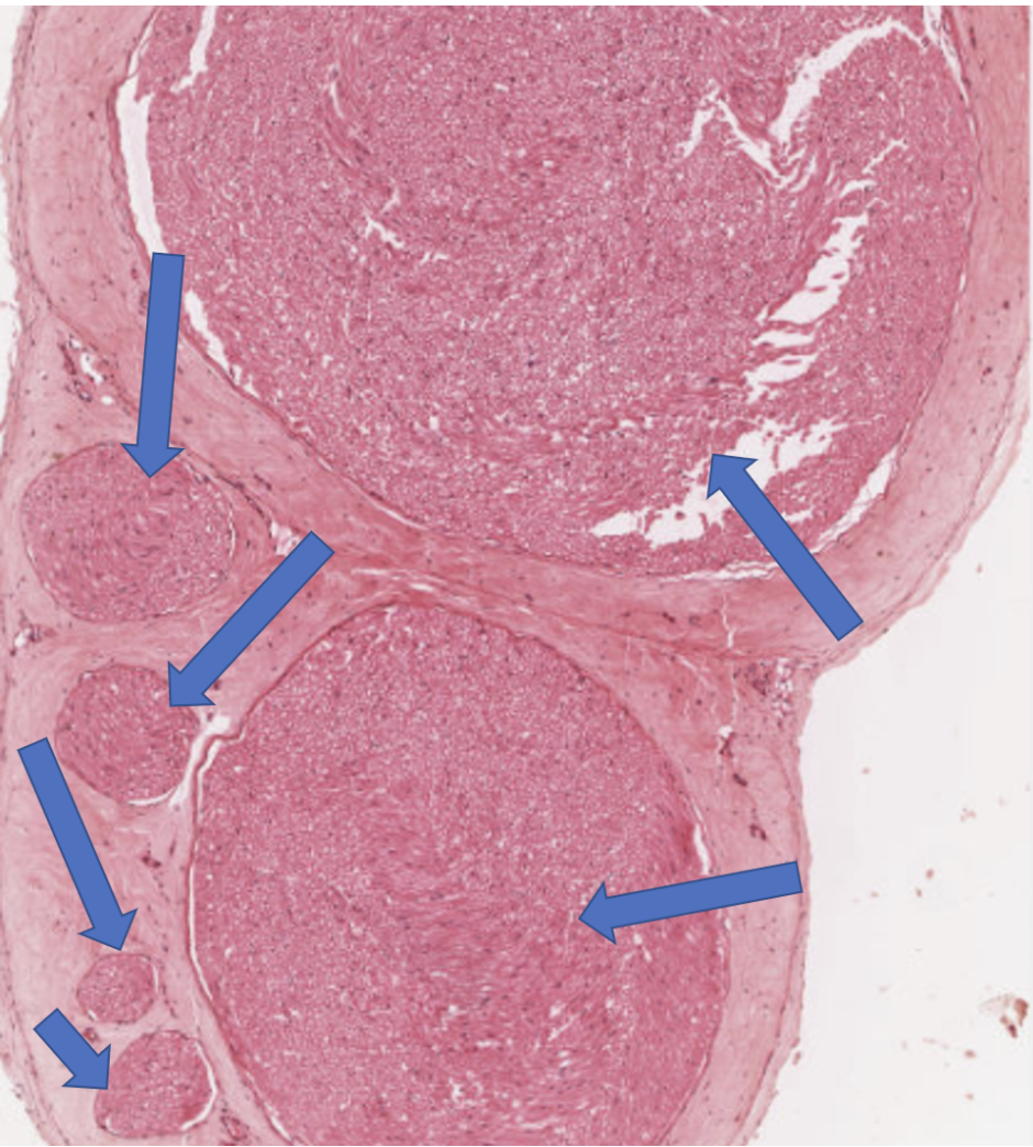
nerve fascicles
20
Identify the large, circular structure shown in this image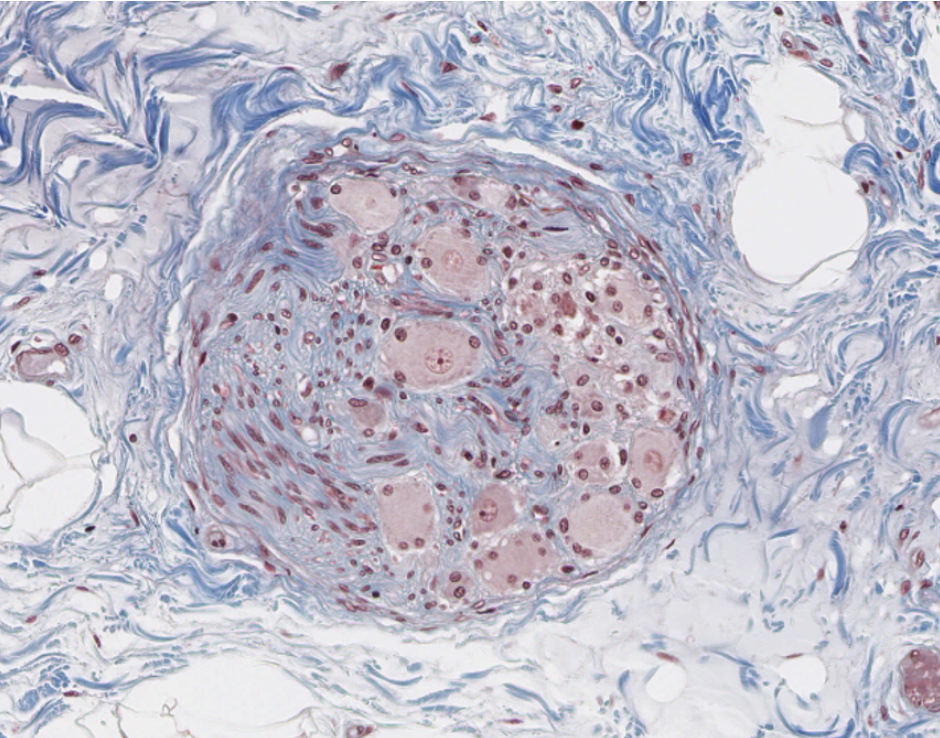
Peripheral ganglion
21
Identify the layer surrounding the pointer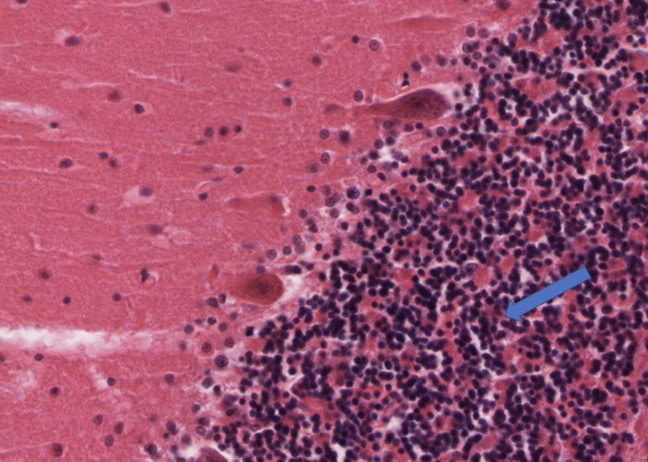
granular layer of the cerebellum
22
Identify the location-specific tissue type indicated by the pointer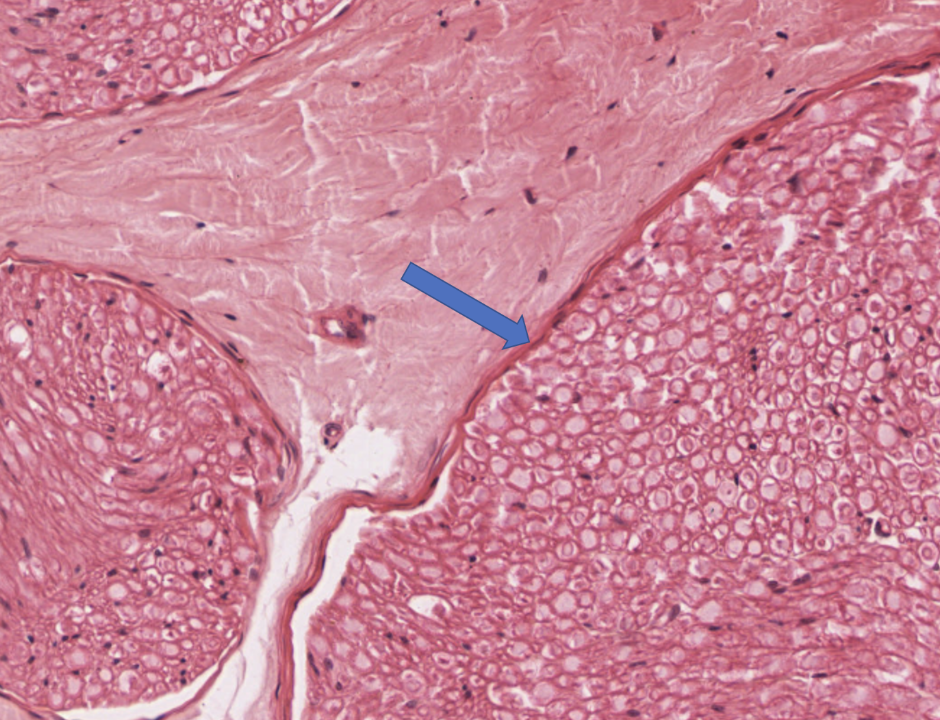
Perineurium
23
Identify the cell indicated by the blue pointers. Be specific.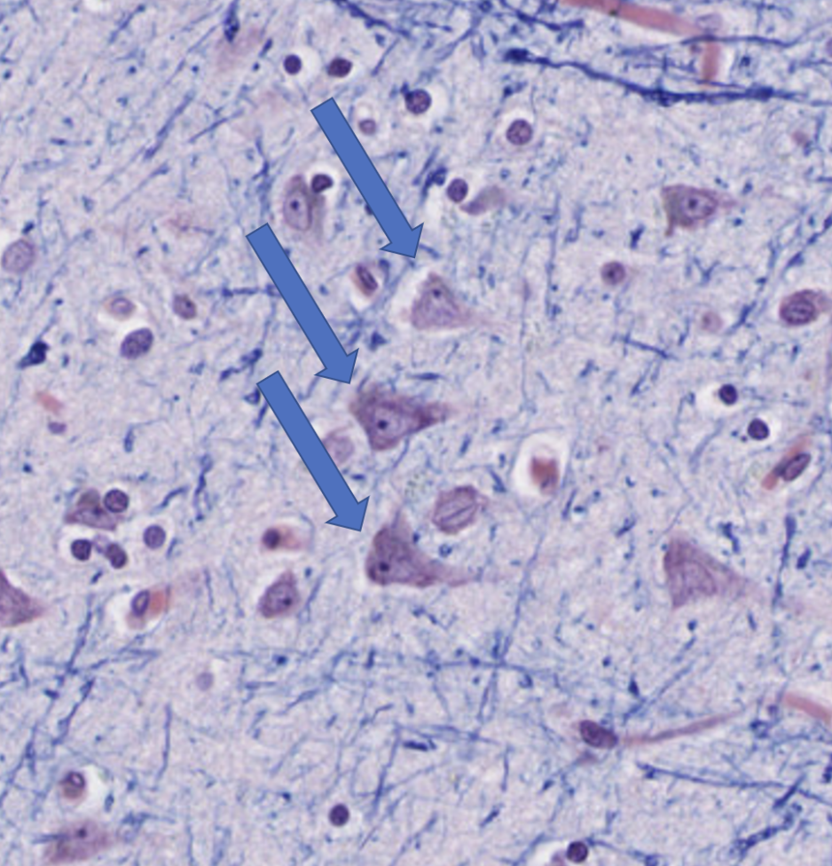
Pyramidal cell of the cerebrum
24
Identify the cell indicated by the blue pointer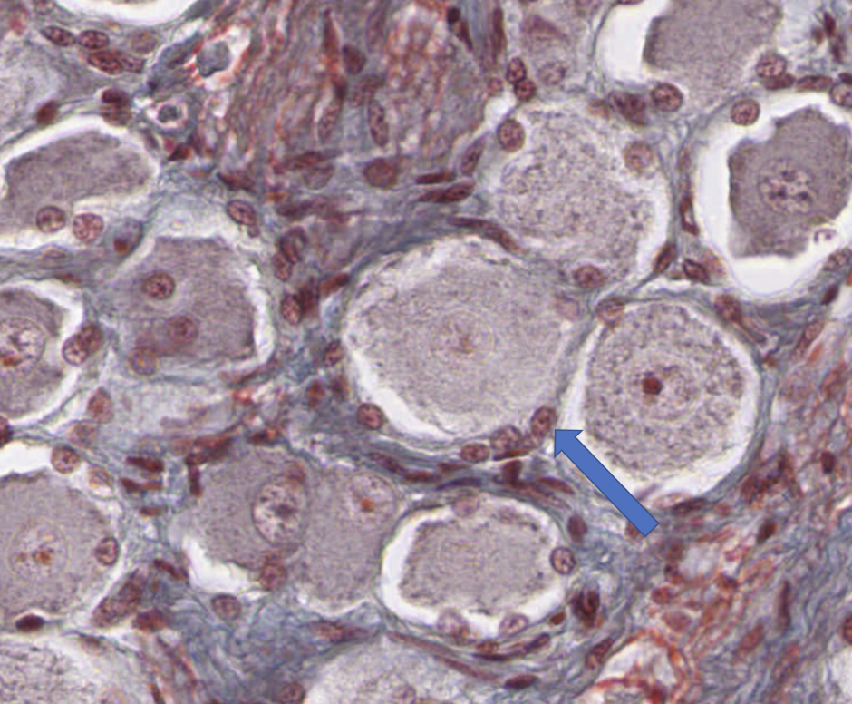
Satellite cell
25
Identify the function of the cell at the pointer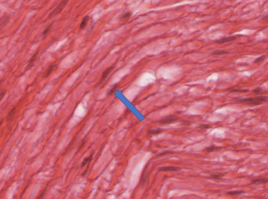
Myelination of axons
26
Identify the function of the cell indicated by the blue pointer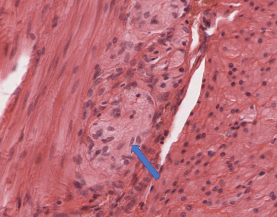
Myelination of axons
27
Identify the region indicated by the pointer. Be as specific as possible. 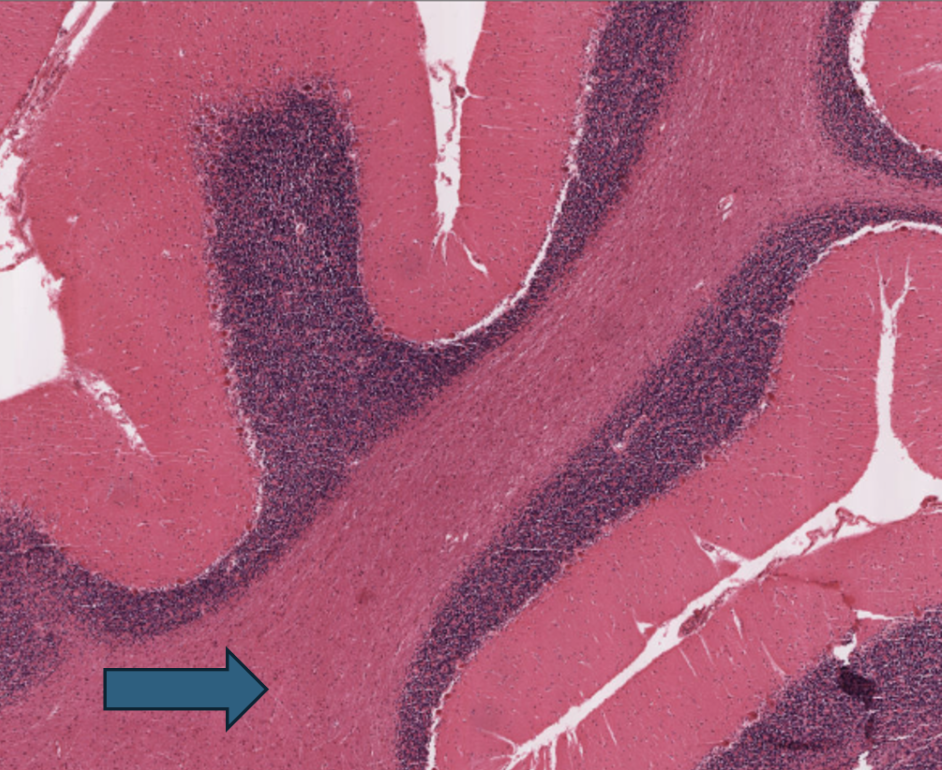
Cerebellar medulla/white matter
28
Describe the fiber type and neuron type populating the region at the pointer.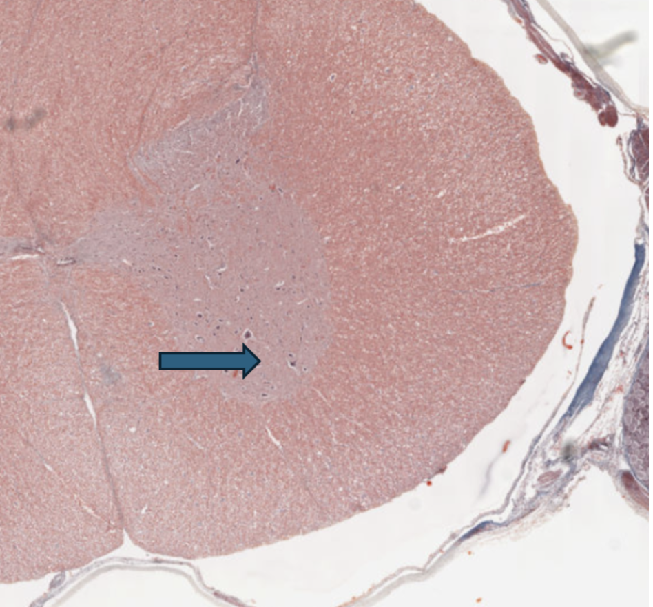
Motor neurons & axons (descending)
29
Identify the region indicated by the pointer. Be specific as possible. 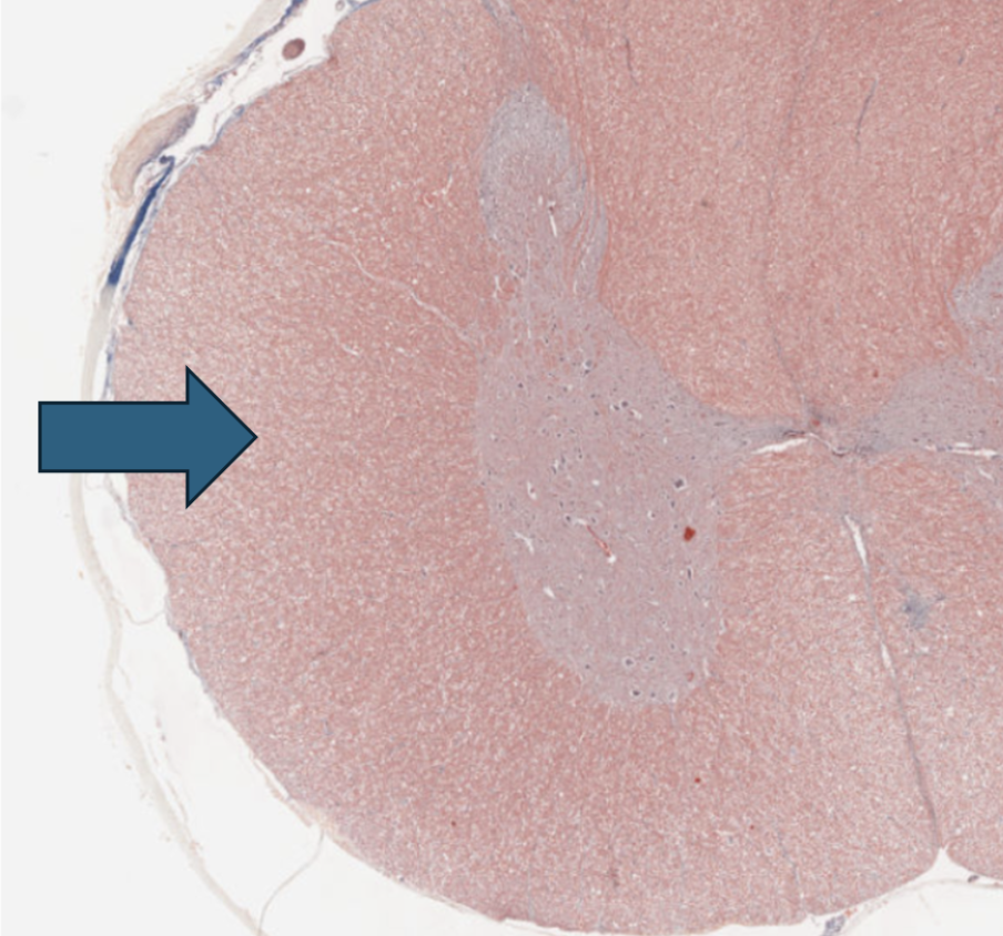
White matter of the spinal cord
30
Identify the cell indicated by the blue pointer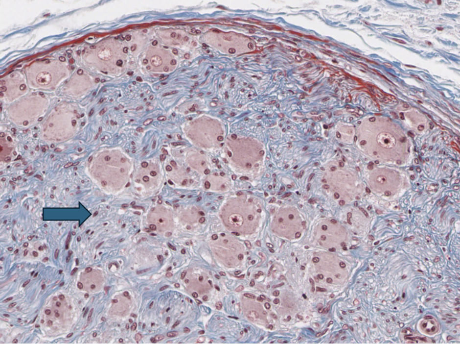
Schwann cell
31
Identify the function of the thin eosinophilic structure at the pointer.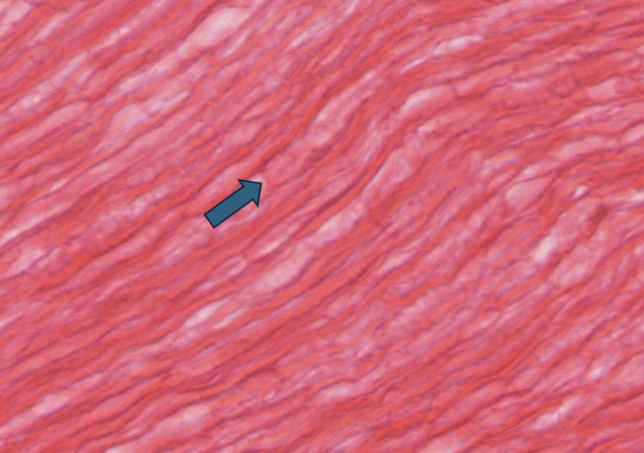
Speed up propagation of action potentials along the axon via saltatory conduction.
32
Identify the region of the nervous system present here and indicate any/all structures, cells, and sub-regions. (Not a representative exam question, but good practice).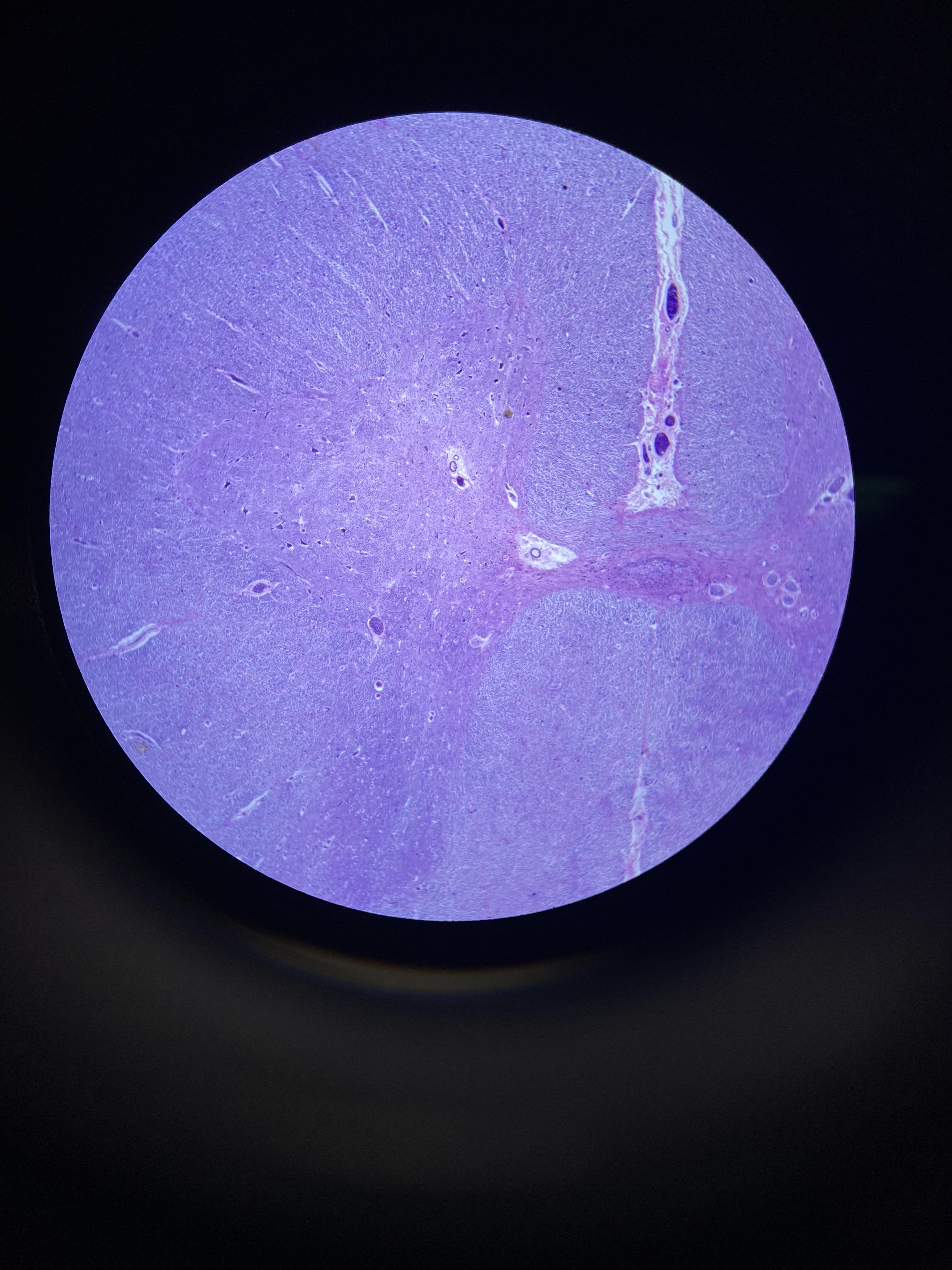
Spinal cord. Can see gray matter (pinkish) and white matter (purple/ blue). In gray matter, neruon cell bodies and neuropil. Central canal to the right. Dorsal horn up, posterior down. In white matter, neuroglia and myelinated/non-myelinated axons.
33
Identify the four layers shown here. Indicate the sub-region of the nervous system shown. (Not a representative exam question, but good practice)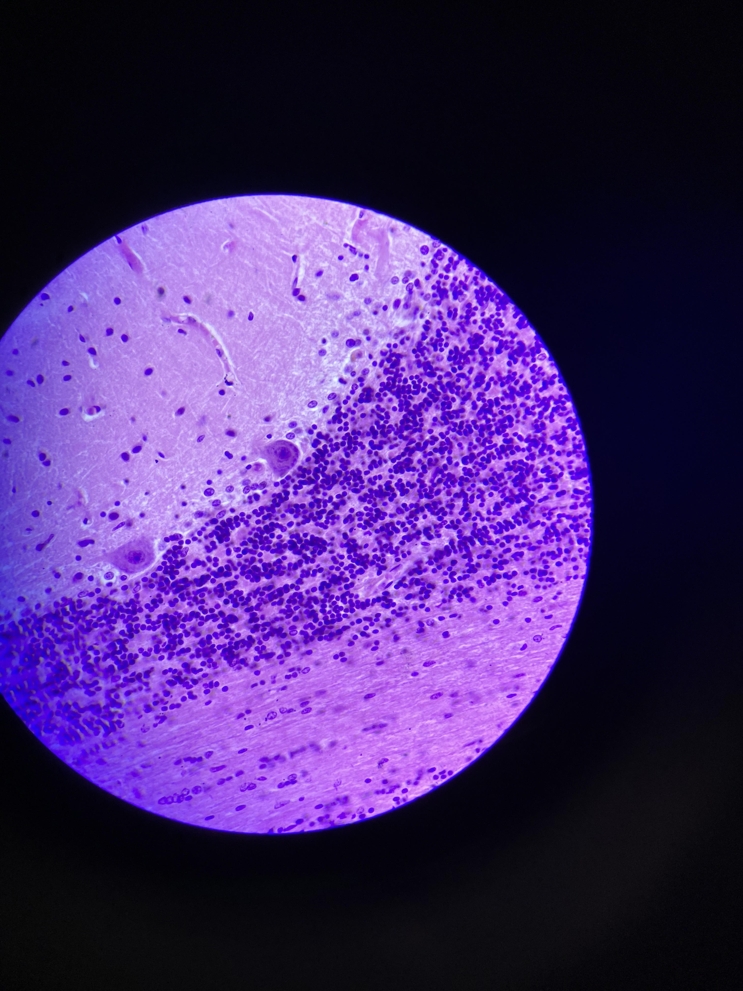
From up to down: cortex: molecular, Purkinje, granular. Bottom: medulla (white matter)
34
Identify the cell indicated by the pointer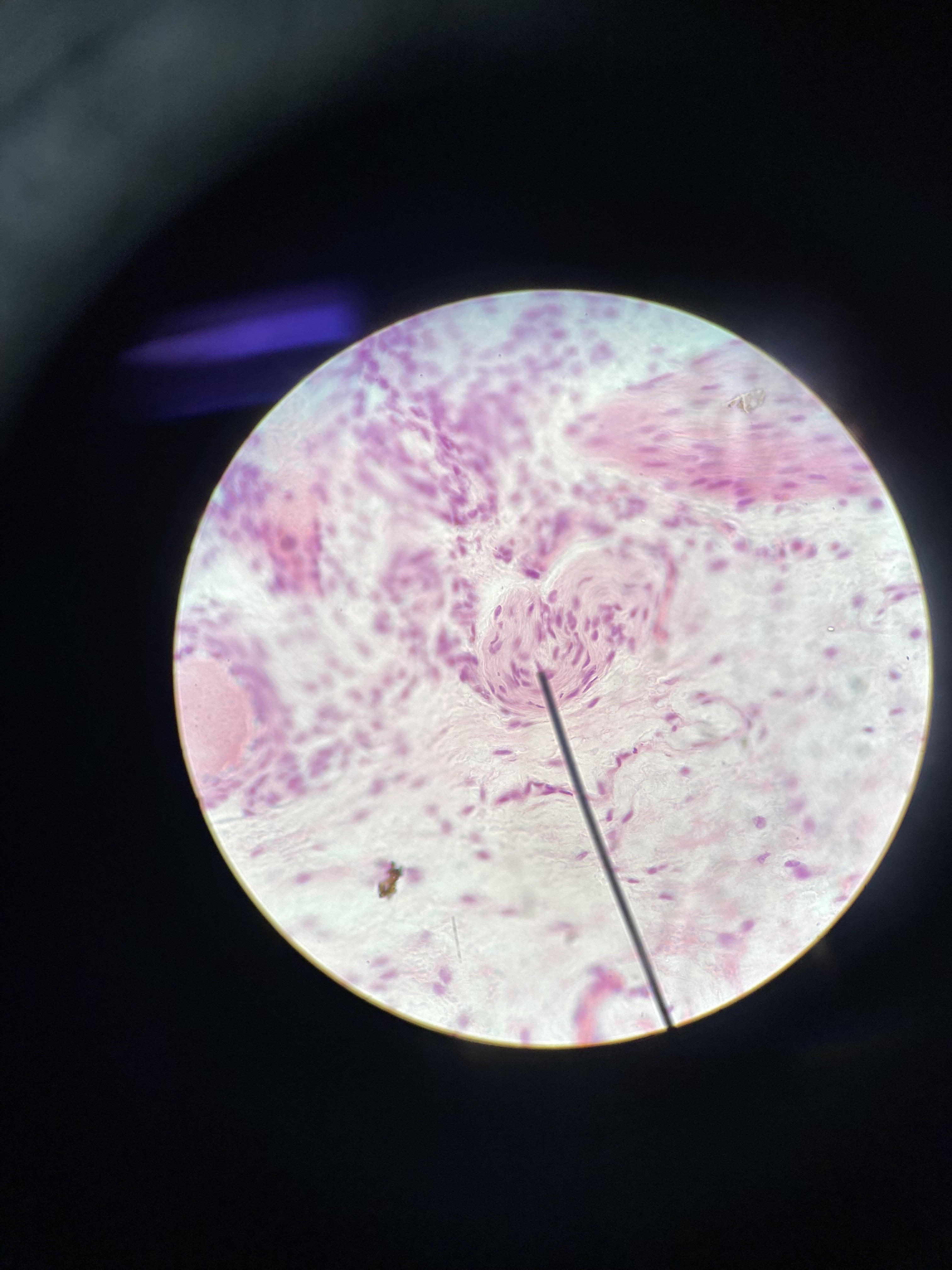
Schwann cell
35
Identify any/all structures visible in the field of view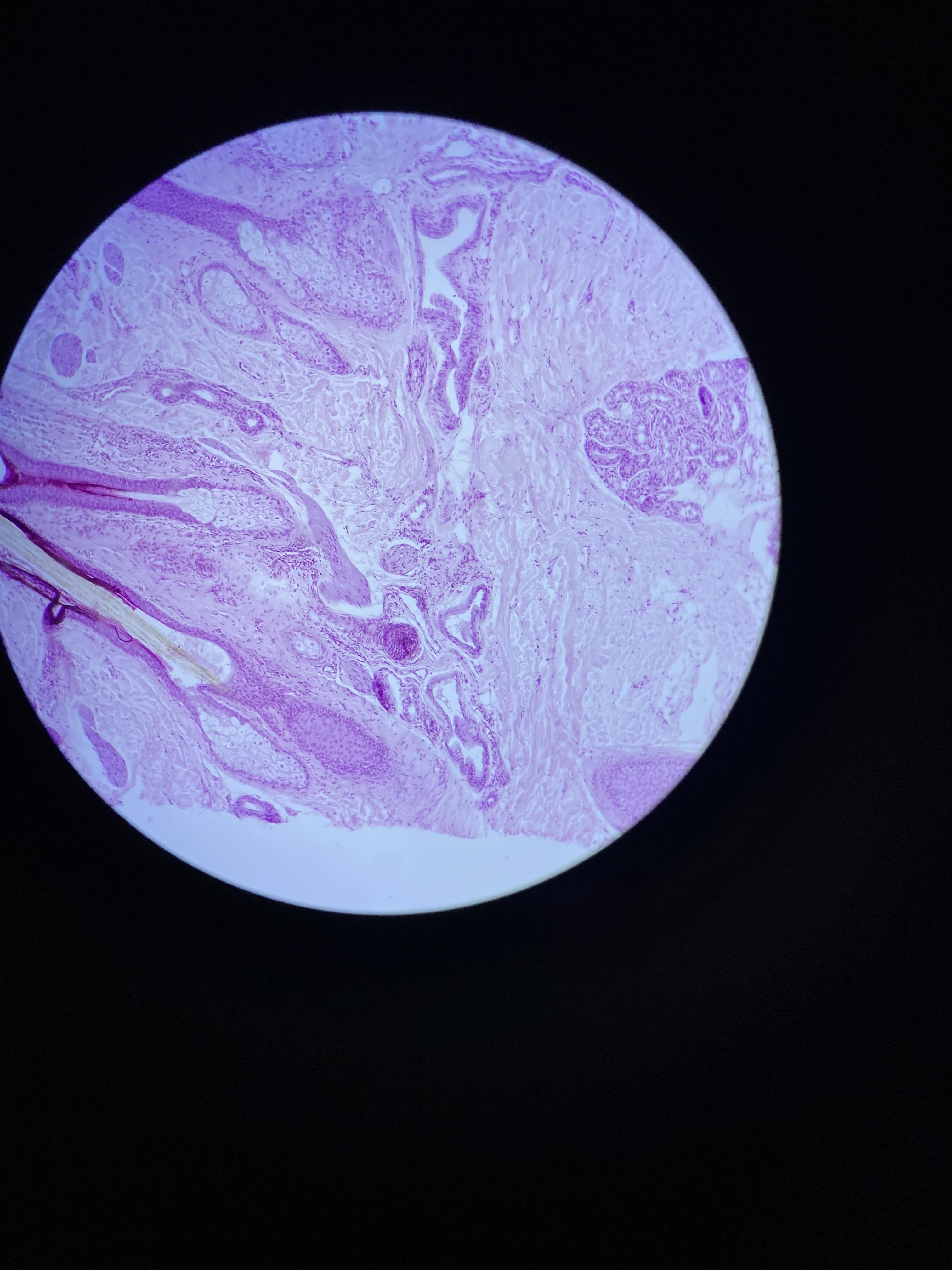
Hair follicles, sebaceous gland, arrector pili, sweat glands + ducts, nerves
36
Identify the function of the cell indicated by the pointer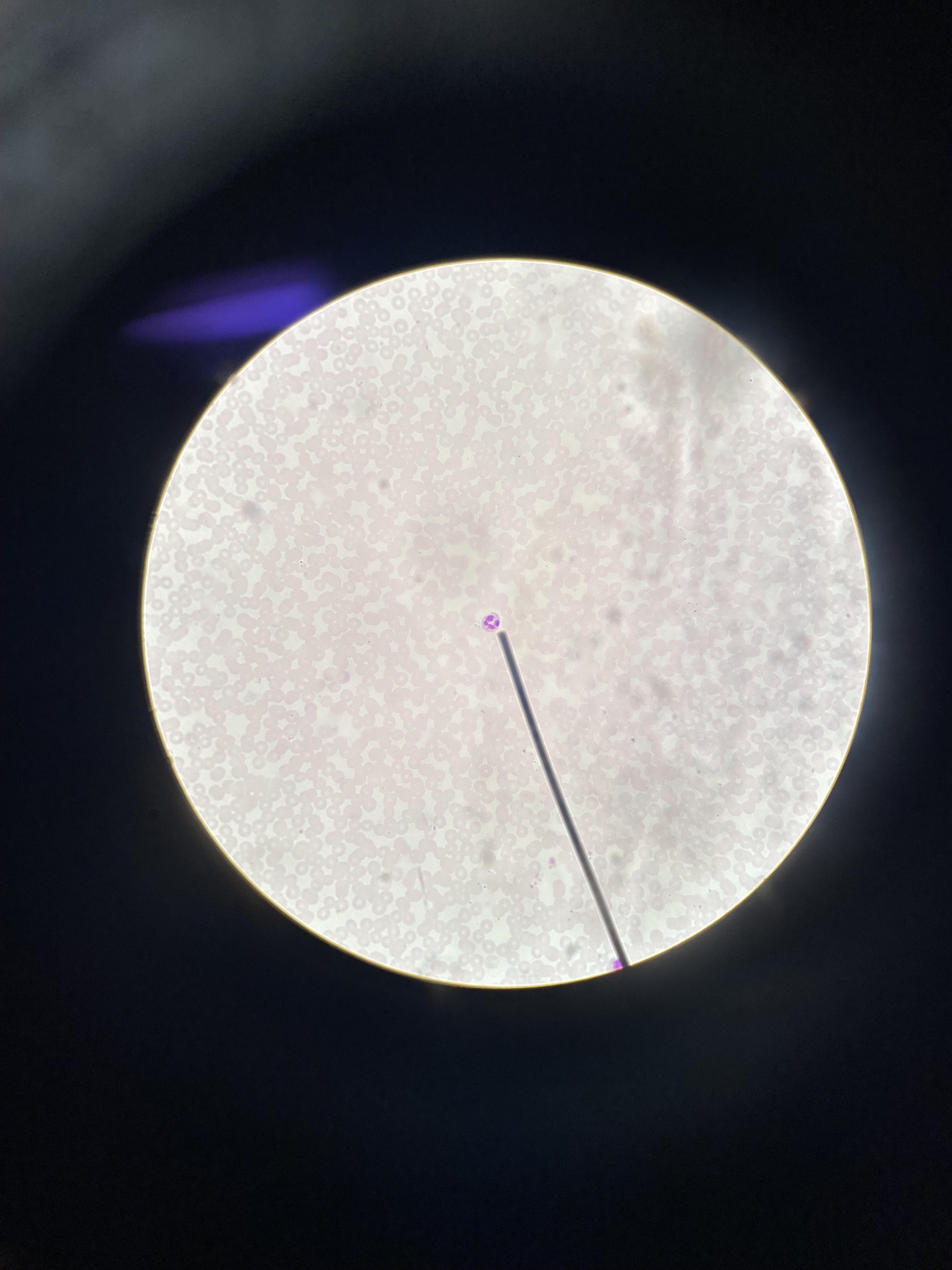
Neutrophils travel to the site of inflammation and destroy foreign material, especially bacteria. They have several other functions as well.
37
Identify 7 layers of the integument we can see here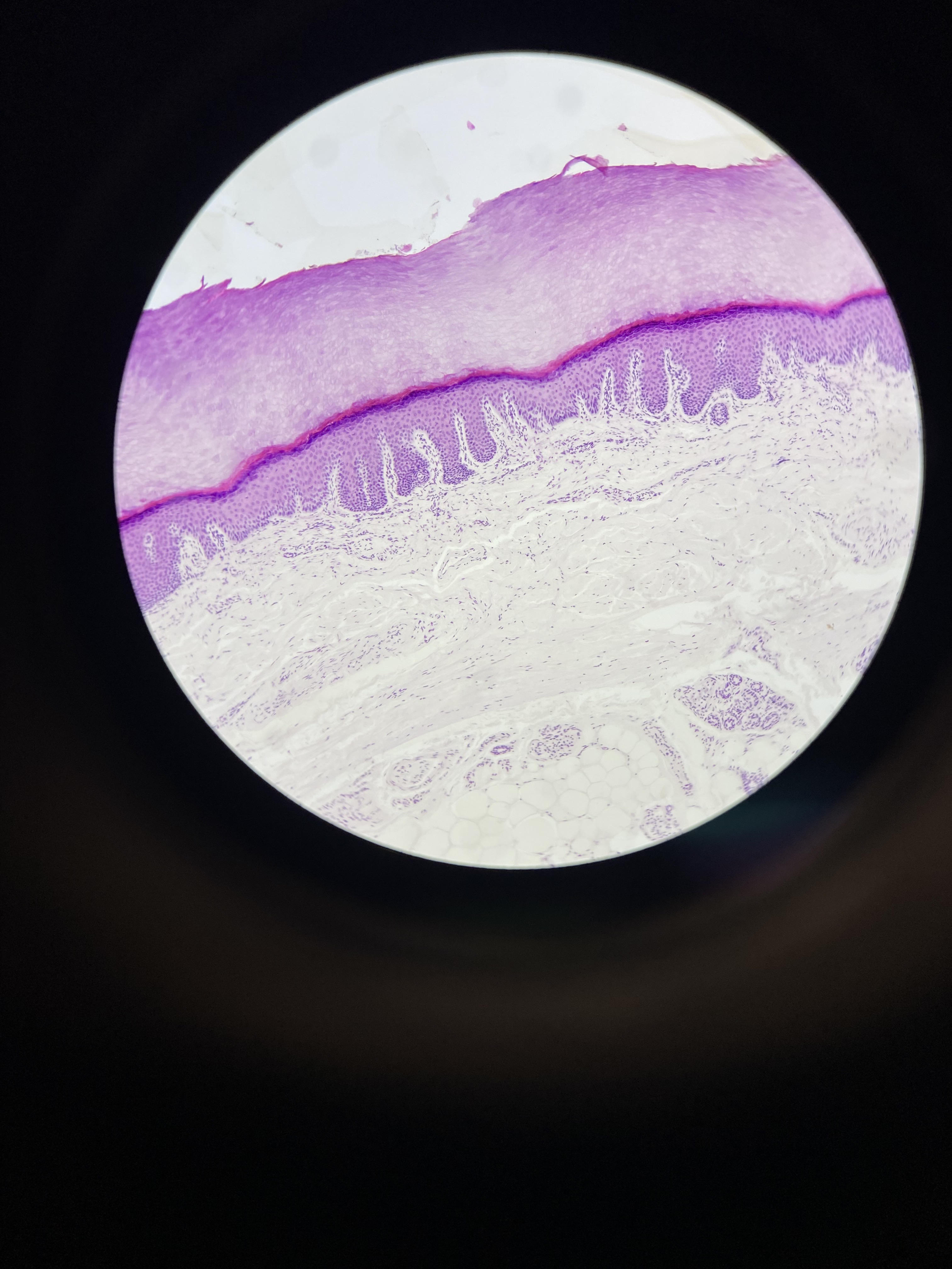
Epidermis containing stratum corneum, stratum lucidum (not apparent), stratum granulosum, stratum spinosum and stratum basale, Dermis containing papillary layer (LCT) and reticular layer (DICT)
38
Identify the function of the cell layer at the center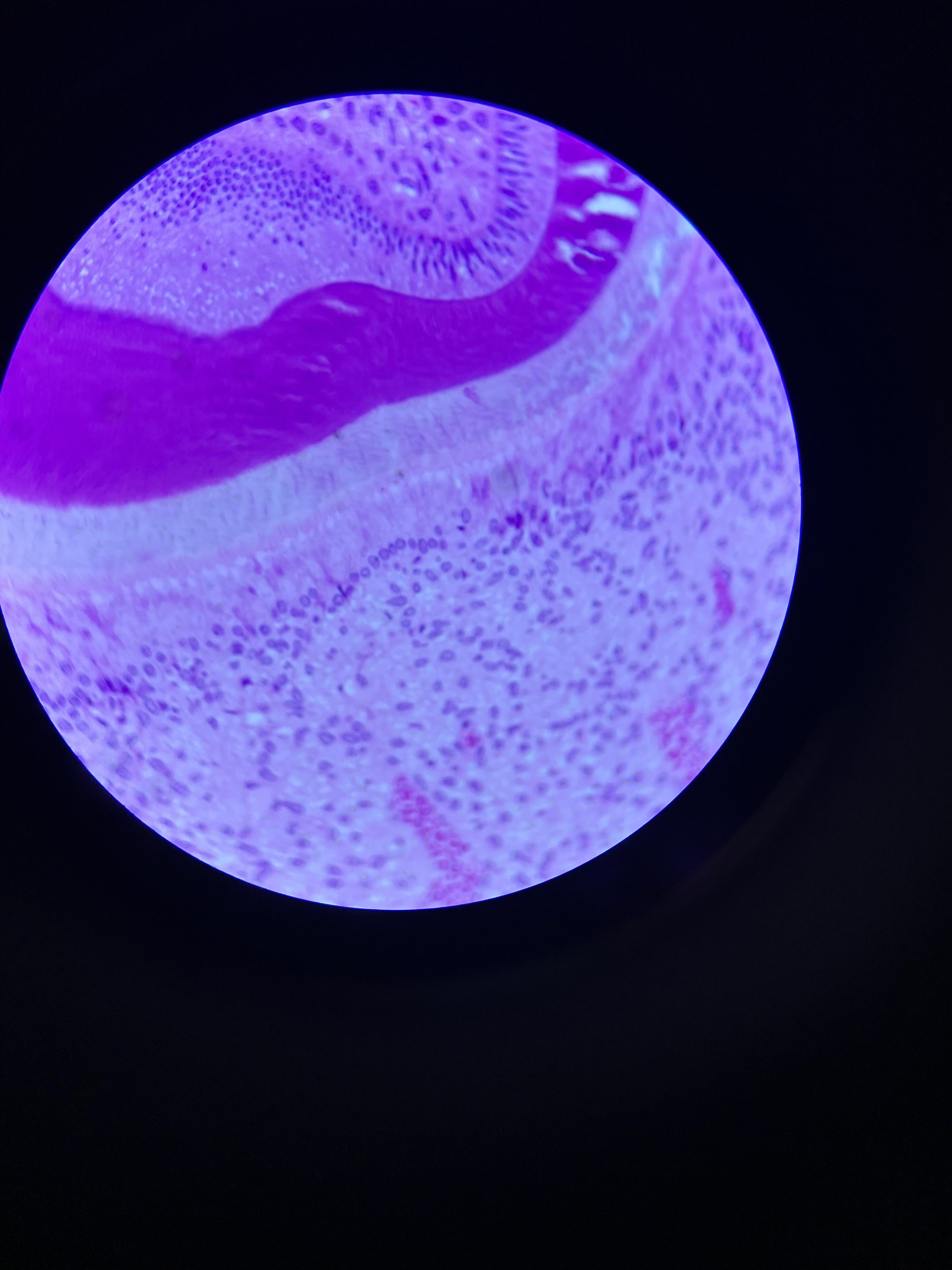
Synthesis of dentin
39
Identify the region of the GI tract shown here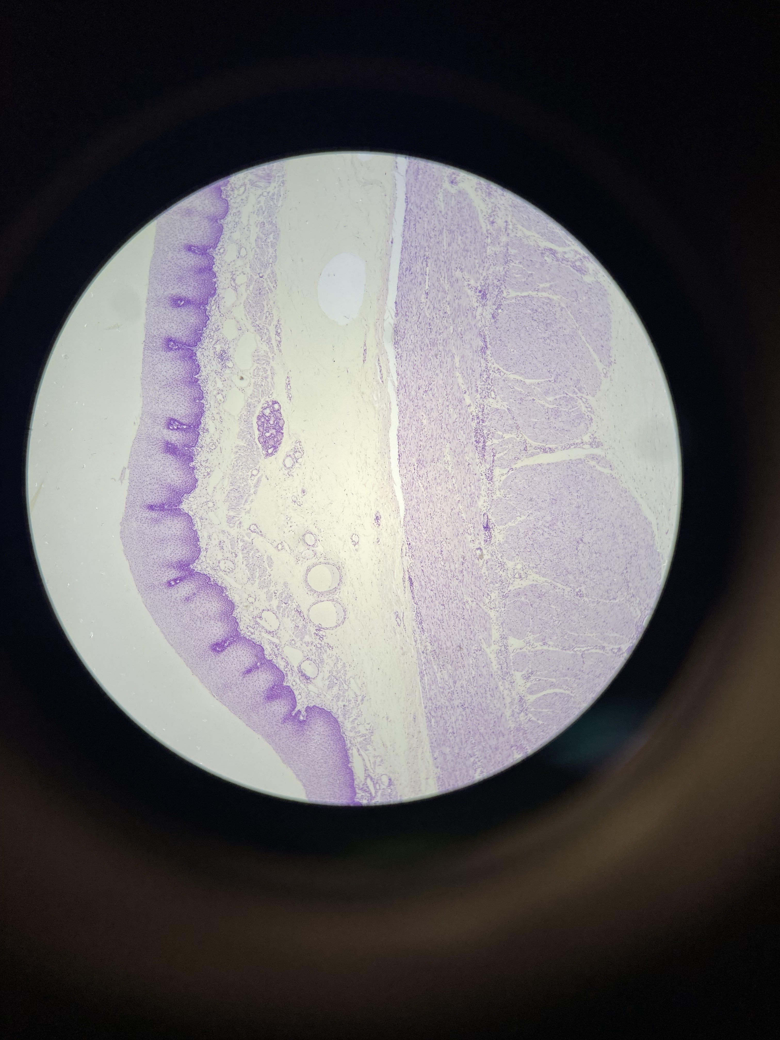
Esophagus
40
Identify the clump of cells indicated at the pointer. Be specific (type and gland) 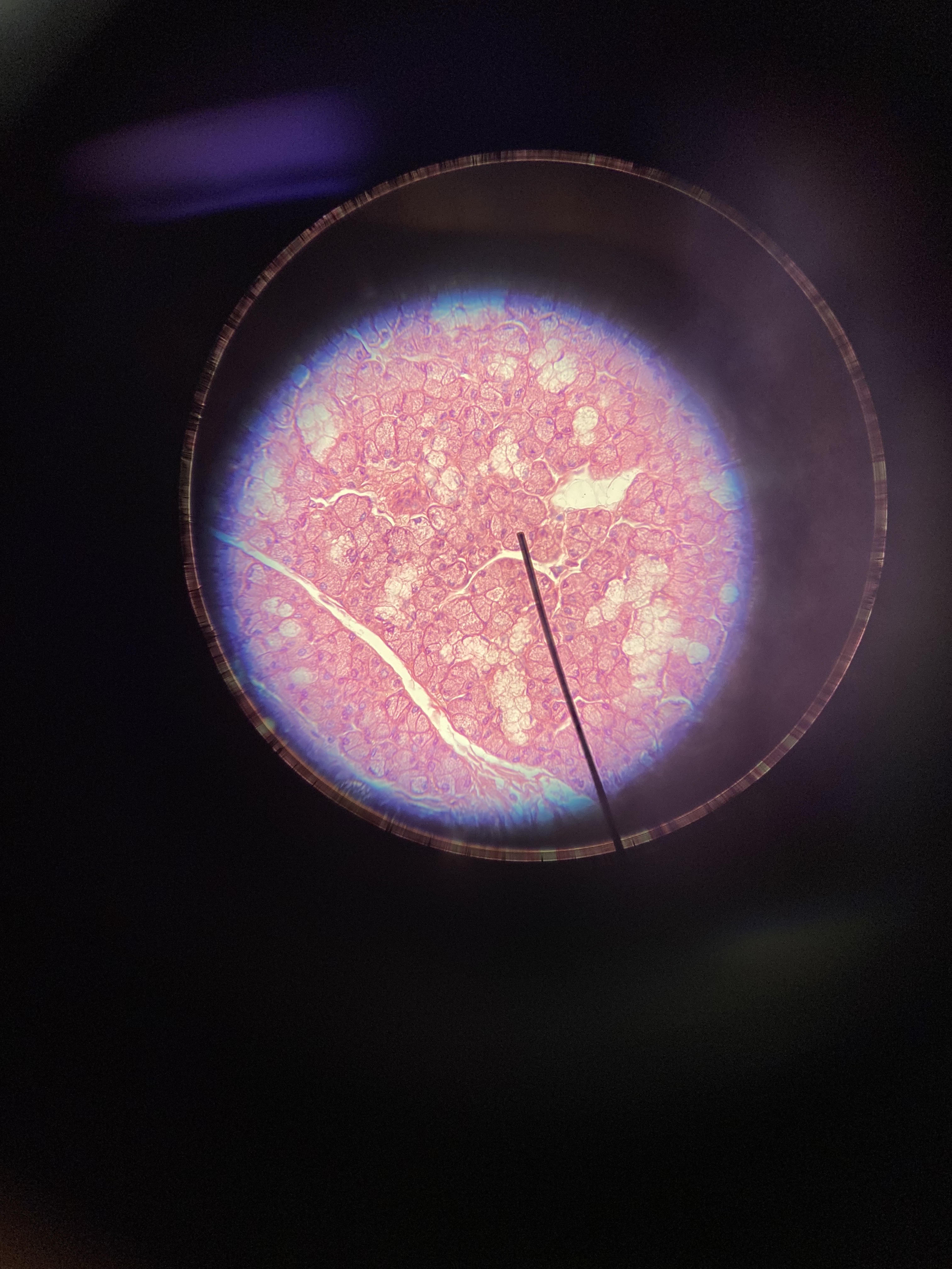
Serous acini of the submandibular gland
41
Identify the vessel shown (center, not left)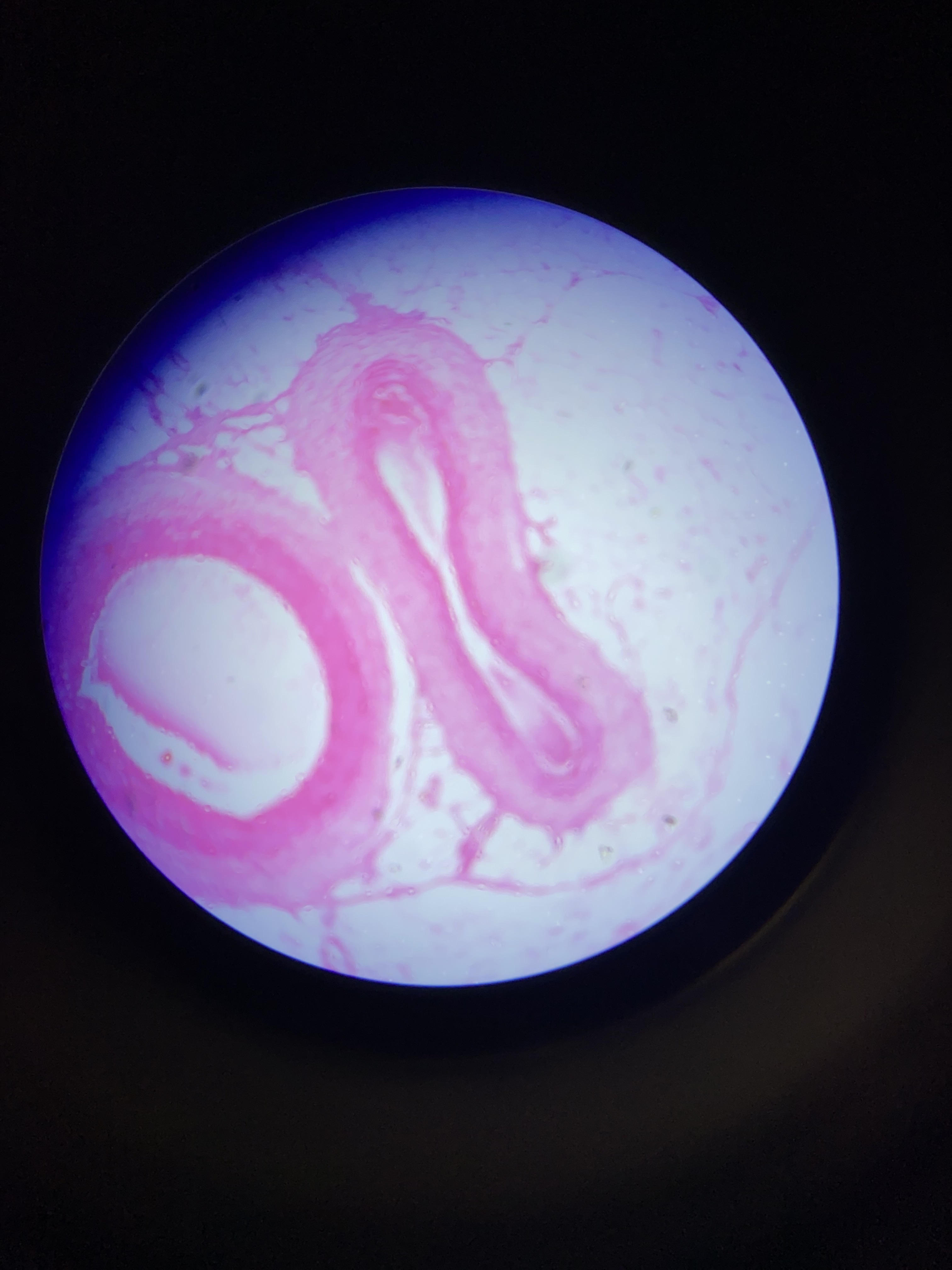
The size, disorganization, and collagen fibers in the large adventitia lead us to determine this is a vein
42
Identify the vessel shown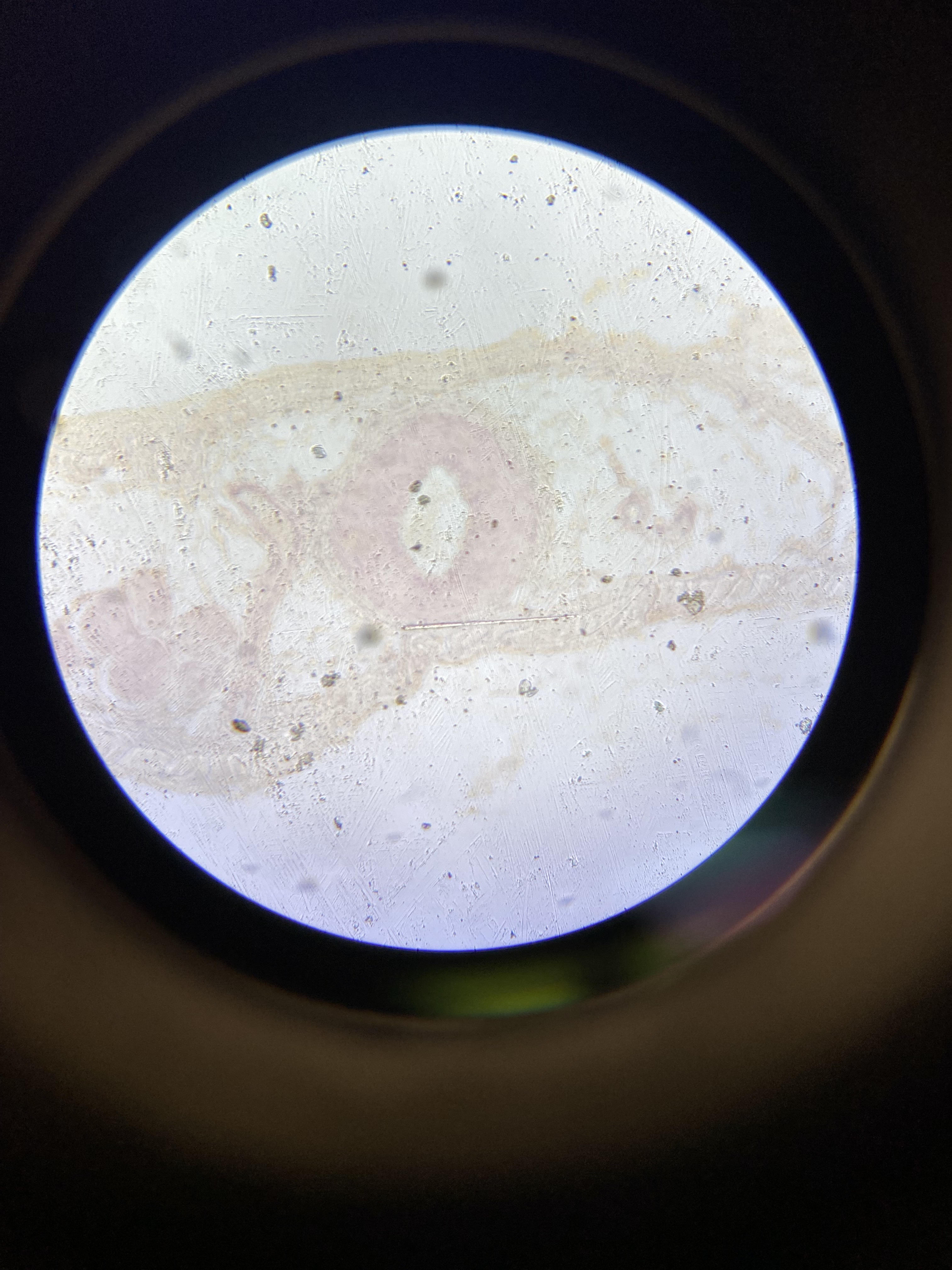
The presence of a significant tunic media, the distinct IEL, the retention of shape, and absence of blood lead us to determine this is an artery
43
Identify the structure indicated by the pointer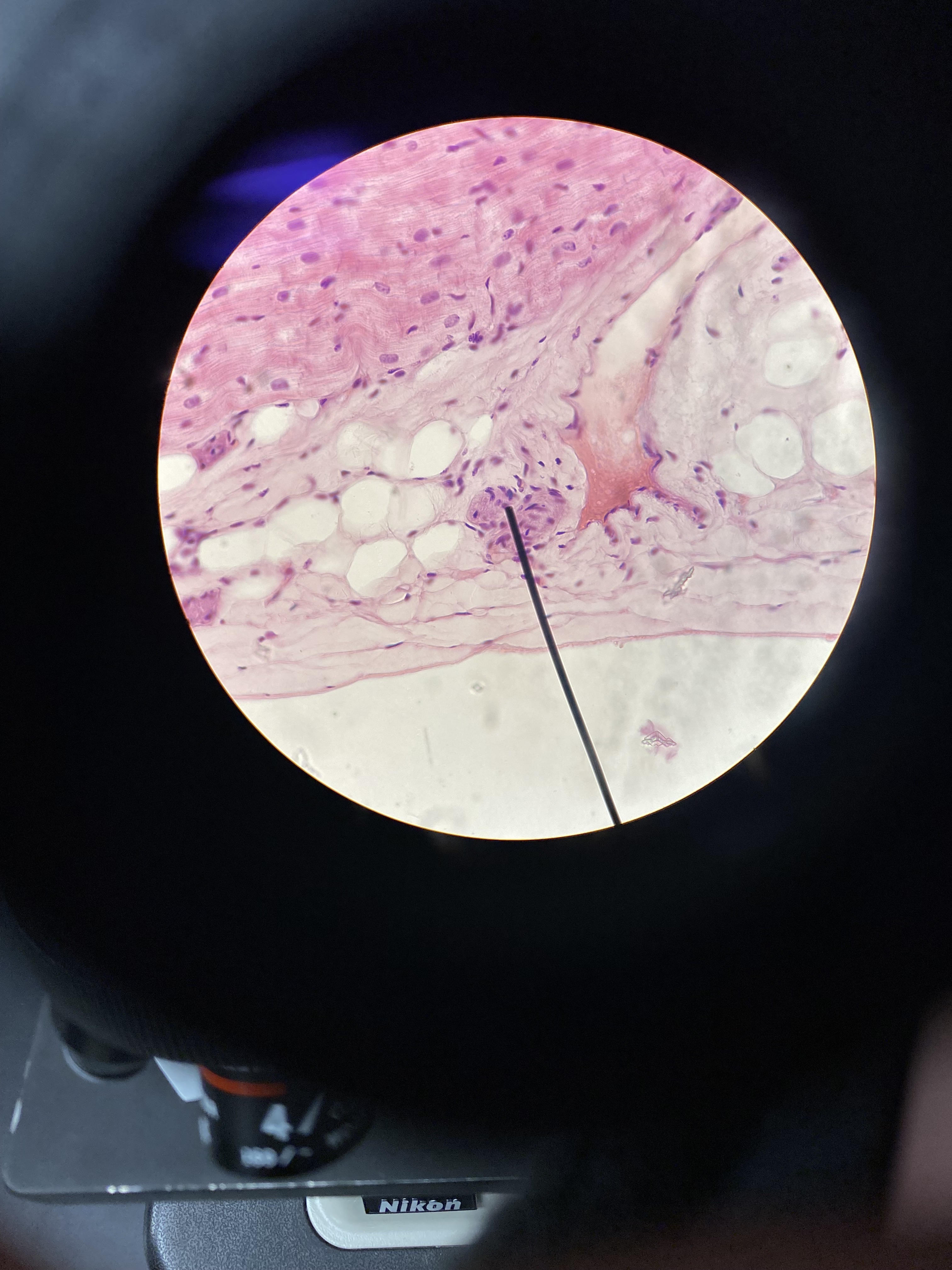
The presence of significant fat, a clear mesothelium, and the obvious myocardium lead us to determine this is a cardiac nerve.
44
Identify the layer within the blue bracket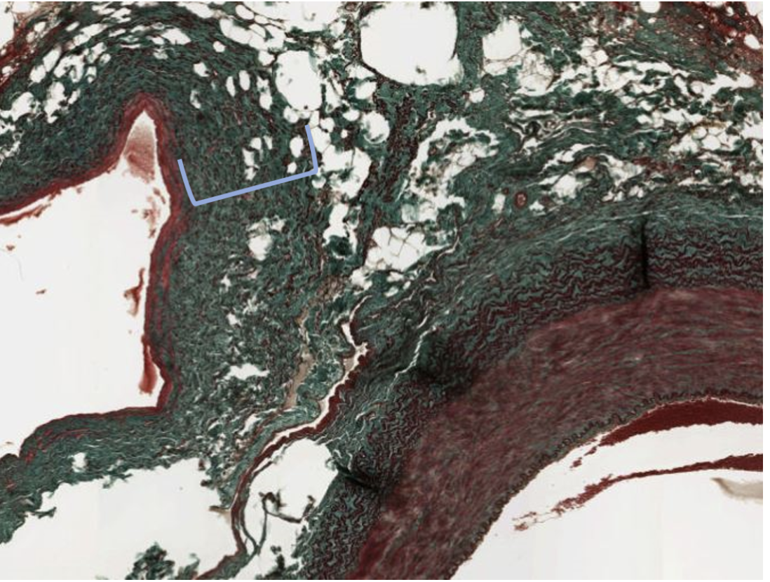
Tunica adventitia of vein
45
Identify the layer indicated by the pointer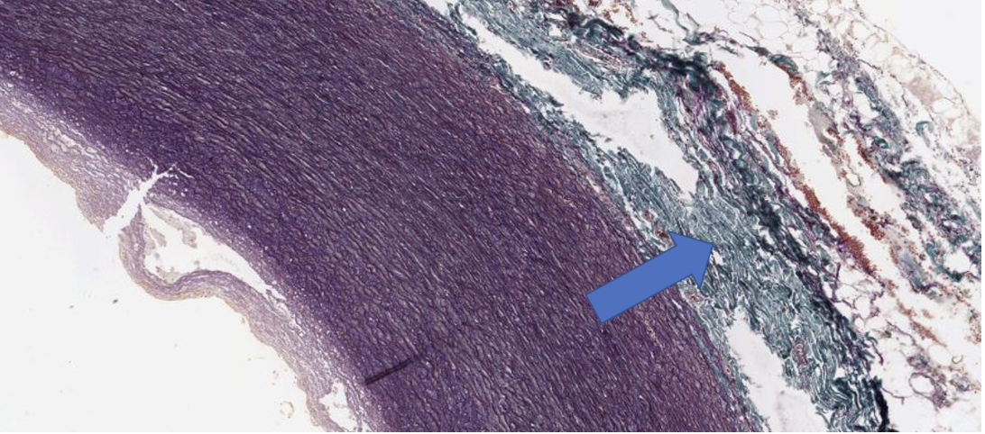
Tunica adventitia of large artery
46
Identify the organ and its function shown in the image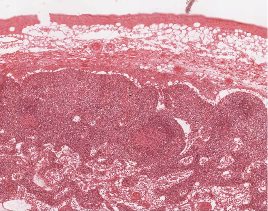
Lymph node, filtering of lymph for survelliance of foreign material (full answer needed for full credit)
47
Identify the organ and its function shown in the image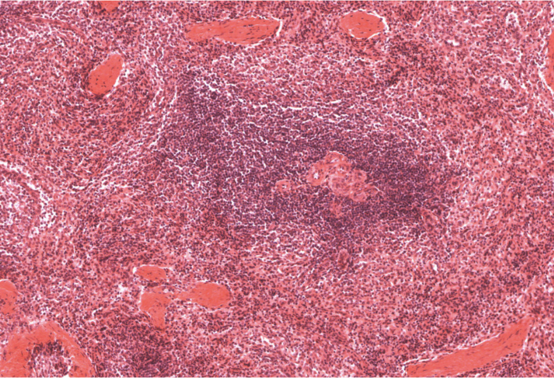
Spleen, filtering of blood for surveillance of foreign material and also RBC turnover and iron recycling (full answer needed for full credit)
48
Identify the fine layer indiated by the pointer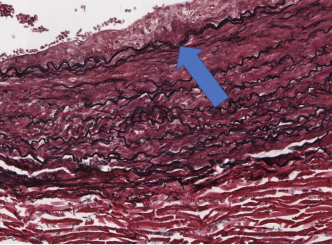
Internal elastic lamina
49
Identify the name of the region indicated by the pointer. Be specific and include organ name.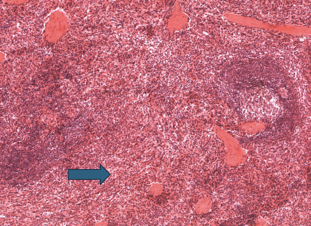
Red pulp of spleen
50
Identify the layer indicated by the bracket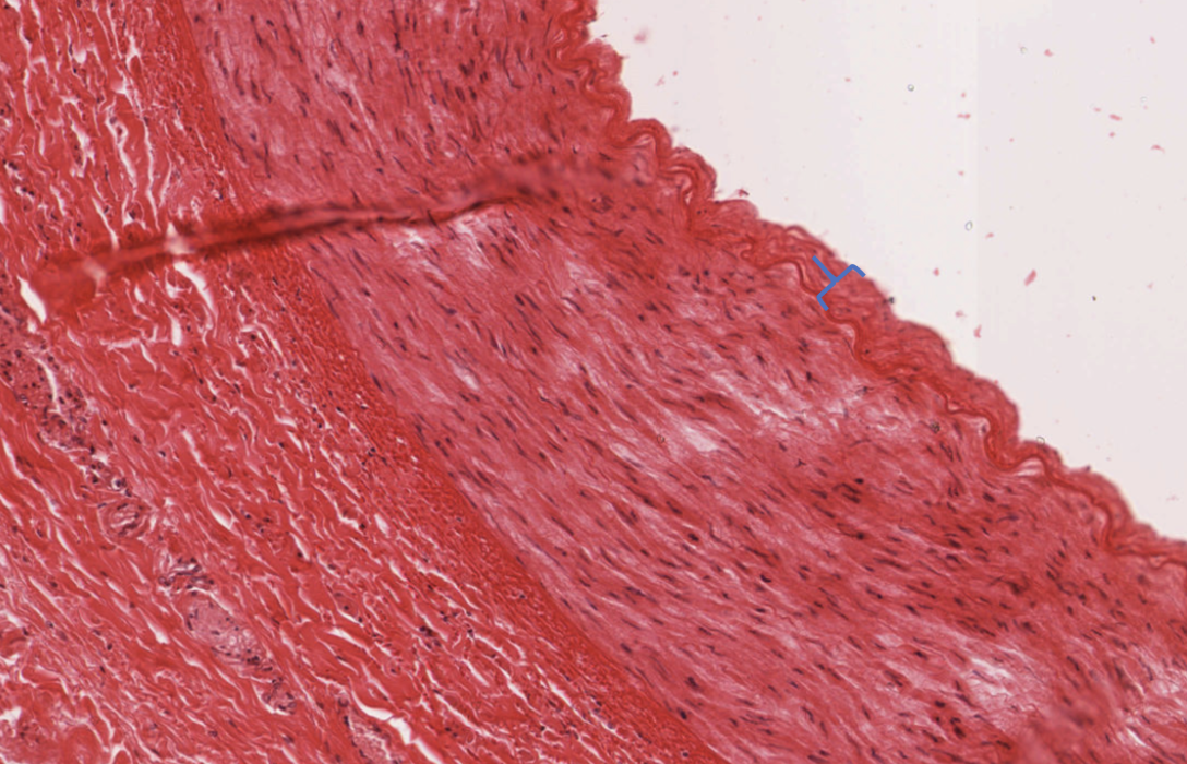
Tunica intima
51
Identify the thick chain of cells indicated by the pointer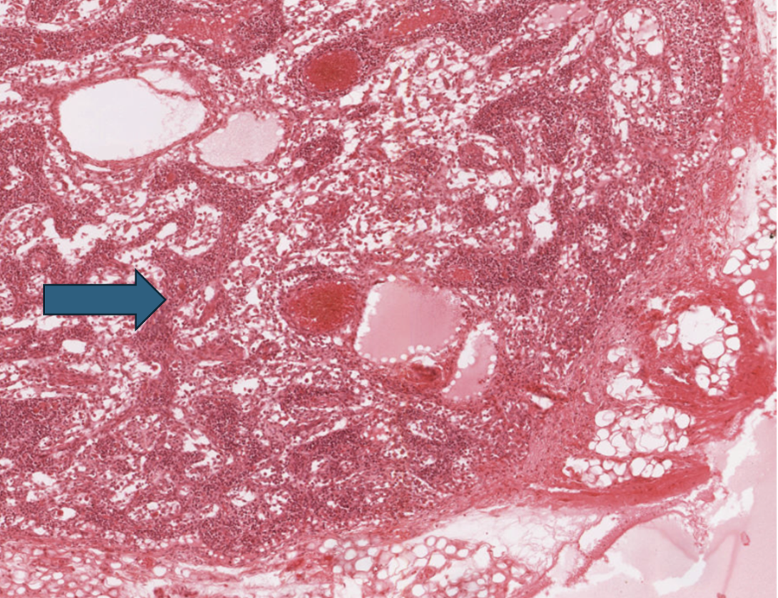
Medullary cords (containing plasma cells, B cells, and macrophages)
52
Identify the tissue layer indicated by the bracket 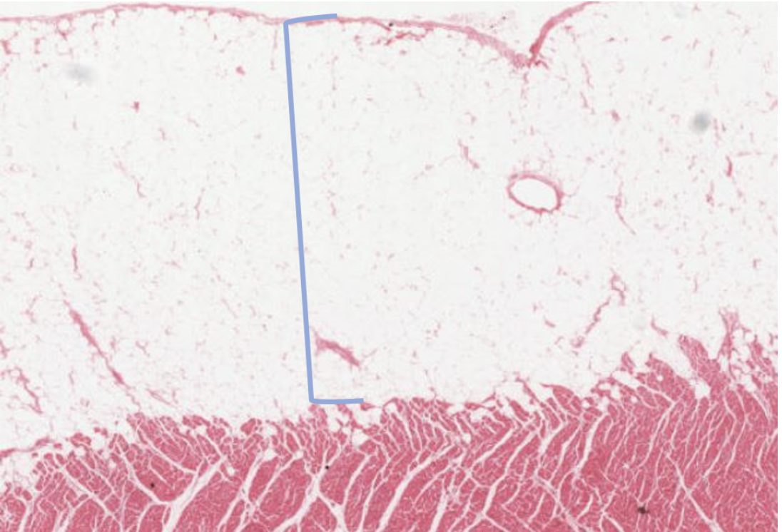
Epicardium
53
Identify the structures indicated by the arrows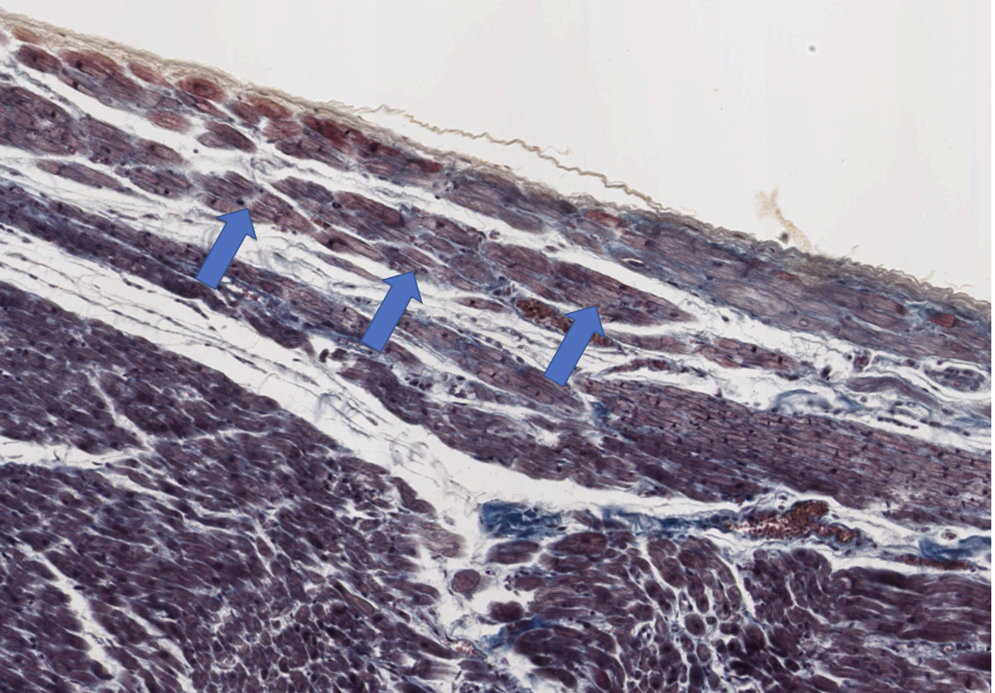
Purkinje fibers
54
Identify the euchromatic cell indicated by the pointer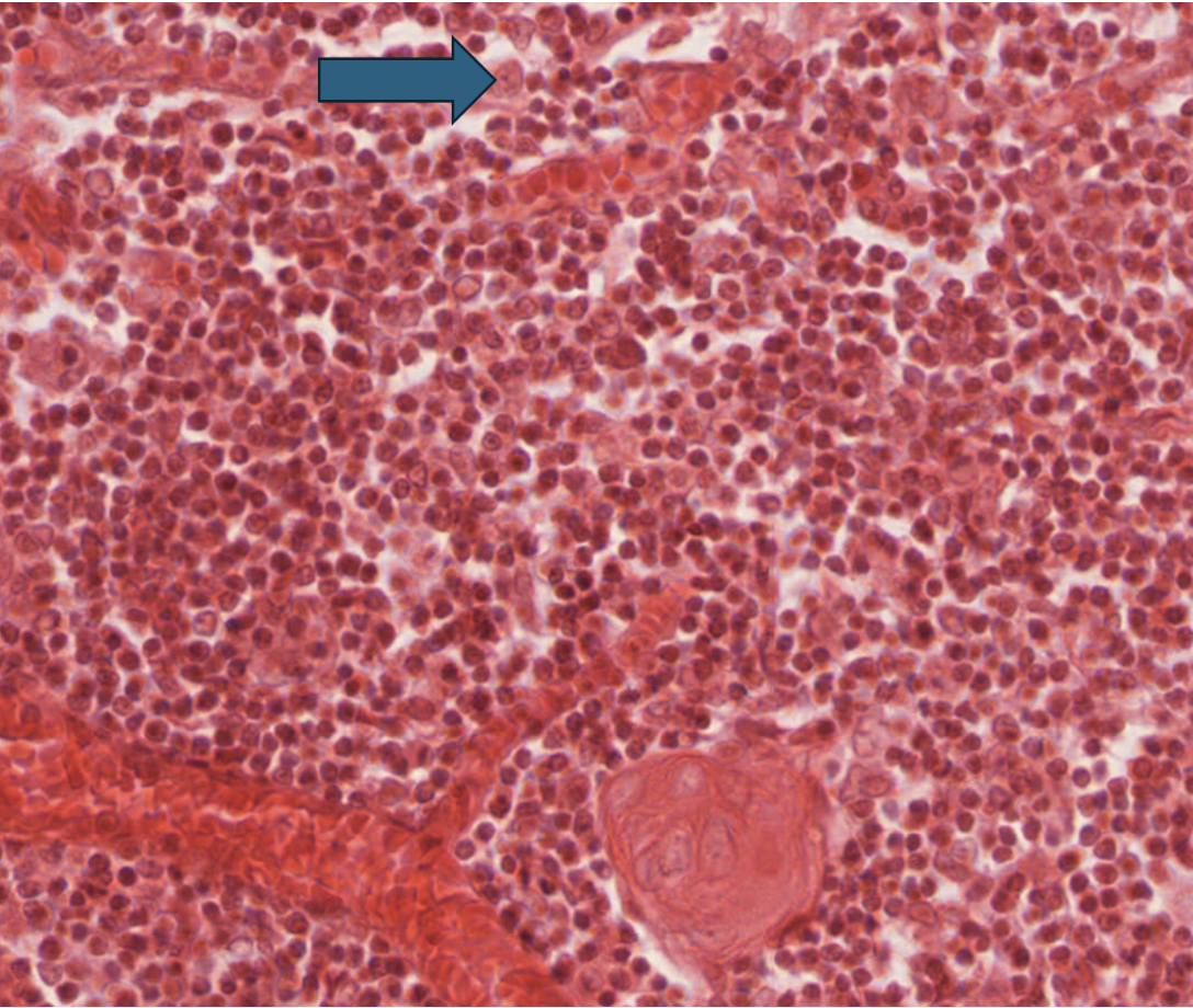
Thymic epithelial cell
55
The vessel at the bottom left is a __________________, the vessel at the top right is a __________________.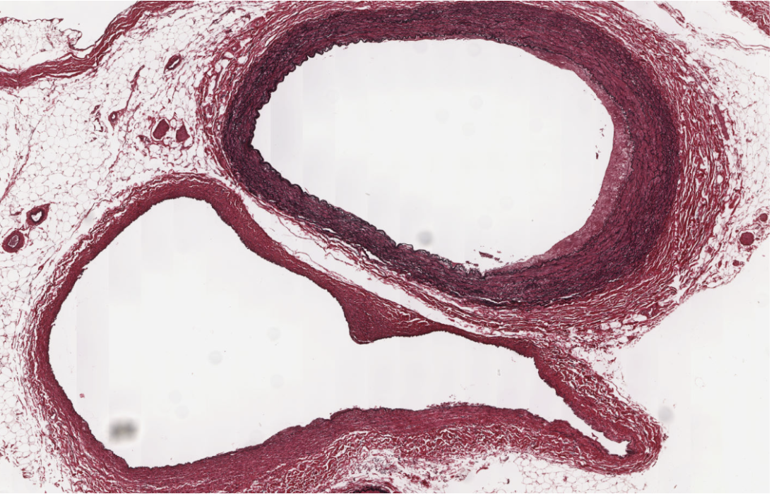
vein, artery
56
What is the name of the tissue layer lining the chamber indicated by the pointer?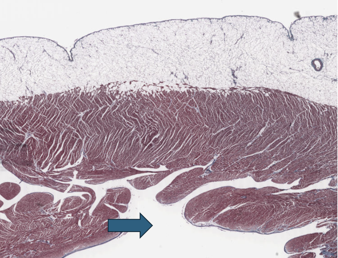
Endocardium