Chapter 3: Mammography Equipment Digital Mammography
Study these flashcardsFree · spaced repetition · iPhone, Android, Mac & web
All cards
1
Mammography is the gold standard for detecting
- To detect breast lesions
- To measure bone density
- To image abdominal organs
- To diagnose lung infections
2
Modern x‐ray mammography produce x‐rays that are
- high in quality but low in radiation dose.
- no longer considered effective
- higher doses to improve image clarity
3
Modern mammography systems are tightly monitored by the
- FDA Food Code
- Health Insurance Portability Act (HIPAA)
- Affordable Care Act (ACA)
- Mammography Quality Standard Act (MQSA)
4
The part of the mammography machine where the patient is positioned
- AWS (Acquisition WorkStation)
- Gantry
5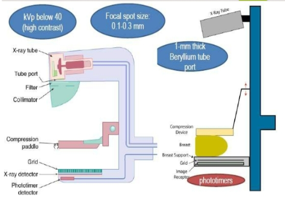
Image occlusion: name the hidden labels
6
Image occlusion: name the hidden labels
7
Image occlusion: name the hidden labels
8
Image occlusion: name the hidden labels
9
Image occlusion: name the hidden labels
10
Image occlusion: name the hidden labels
11
Image occlusion: name the hidden labels
12
Image occlusion: name the hidden labels
13
Image occlusion: name the hidden labels
14
Image occlusion: name the hidden labels
15
Image occlusion: name the hidden labels
16
Image occlusion: name the hidden labels
17
Image occlusion: name the hidden labels
18
Image occlusion: name the hidden labels
19
Image occlusion: name the hidden labels
20
Image occlusion: name the hidden labels
21
Image occlusion: name the hidden labels
22
Image occlusion: name the hidden labels
23
Image occlusion: name the hidden labels
24
The part of the mammography machine where the technologist has electronic control over the exam
- AWS (Acquisition WorkStation)
- Gantry
25
There are three principal parts to any x-ray imaging system:
- The operating console
The high-voltage generator
The x-ray tube - Image intensifier
26
X-ray production requires
high voltage
27
X-ray tube can only operate on
direct current (DC)
28
A rectifier is a solid-state device that allows
current flow in one direction only, thereby converting AC to DC
29
Timing circuit is used to
shuts off the high voltage
30
Mammography Tube Made of
Pyrex or metal electronic vacuum tube
31
Mammography Tube has two electrodes
Positive: Anode with target
Negative: Cathode with filament
Negative: Cathode with filament
32
The filament circuit is used to create
thermionic emission at the cathode
33
The filament circuit has 2 filaments
small for small focal spot and
large for routing images (large focal spot)
large for routing images (large focal spot)
34
Thermionic emission is the term used to describe
the boiling off of outer shell electrons of the filament atoms
35
Protective housing of the x-ray tube is
lead lined to reduce the levels of leakage radiation.
36
X-ray tube window or exit port is the
thin area of glass or metal enclosure
37
Xray production is ... heat/ ... x-ray
99%/1%
38
The tube in encased in an
oil bath that serves as a thermal cushion to dissipate heat and an insulator to protect against electric shock.
39
The atom consist of
central nucleus, which has protons, or positively charged particles, and neutrons, which have no charge.
40
Electrons are ... charged
negatively
41
The shell closest to the nucleus is called
the K shell
42
Types of X-ray Production
Characteristic radiation-characteristic of target material
Bremsstrahlung radiation-”braking”
Bremsstrahlung radiation-”braking”
43
The transition of the outer-shell electron to an inner shell results in the emission of
characteristic x-ray
44
The ejected electron in characteristic x-ray is called
the photoelectron
45
A photoelectric interaction cannot occur if the energy of the incident electron is
lower than the binding energy of the electron of the target
46
Bremsstrahlung Radiation produced when
electrons are slowed down in the atom
The incident electron interacts with the force
field of the nucleus
The incident electron interacts with the force
field of the nucleus
47
X-rays are dangerous to the human because
they ionize or interact with atoms in the human body
48
The most important molecule in the human body is
DNA, which contains the genetic information for each cell
49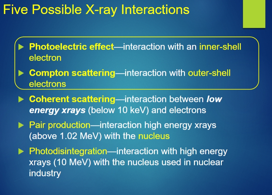
Image occlusion: name the hidden labels
50
Image occlusion: name the hidden labels
51
Image occlusion: name the hidden labels
52
Image occlusion: name the hidden labels
53
Image occlusion: name the hidden labels
54
Photoelectric Effect produces
characteristic radiation
55
Compton Interaction is X-ray interacts with an
outer-shell electron and ejects it from the atom
The ejected electron is called the Compton electron
The ejected electron is called the Compton electron
56
Negative Effects of Compton Scattering
Occupational radiation exposure
Reduced image contrast
backscatter
Reduced image contrast
backscatter
57
Coherent Scattering (also called classical scattering)
Incident photon interacts with electrons result in the incident x-ray changes direction without a change in energy
There is no energy transfer—and no ionization.
There is no energy transfer—and no ionization.
58
Pair Production
The incident electron has sufficient energy to come close to the nucleus of the atom and is influenced by the strong nuclear force field
The x-ray disappears and is replaced by two electrons - one positive (positron) and one negative
This process does not occur in x-ray imaging
The x-ray disappears and is replaced by two electrons - one positive (positron) and one negative
This process does not occur in x-ray imaging
59
Photodisintegration
Occurs at energies higher than 10MeV
The x-ray is completely absorbed by the nucleus
The nucleus becomes excited and emits a nucleon or another nuclear fragment
Photodisintegration does not occur in diagnostic radiology
The x-ray is completely absorbed by the nucleus
The nucleus becomes excited and emits a nucleon or another nuclear fragment
Photodisintegration does not occur in diagnostic radiology
60
Target (Anode) Material
Molybdenum (17–24 keV)
Rhodium (2–3 keV higher than molybdenum)
Rhodium (2–3 keV higher than molybdenum)
61
Digital mammography switches to ... target tubes
Tungsten
62
Tungsten Offers
wider dynamic range (more grays) that can be utilized for digital
Single tracks is more reliable and less expensive
Single-track tungsten tubes support 2–3 times the maximum anode load versus dual track
Single tracks is more reliable and less expensive
Single-track tungsten tubes support 2–3 times the maximum anode load versus dual track
63
The line-focus principle allows
a large area of focal spot heating while keeping the effective focal spot small
Decreasing target angle = decreasing effective focal spot size
Decreasing target angle = decreasing effective focal spot size
64
Filtration is defined by
HVL-half value layer
65
HVL-half value layer is define as
the thickness of a material required to reduce the ‘air kerma’of an x-ray or gamma ray to half its original value
66
2 Types of Filtration
Inherent
Added
Added
67
Inherent Filtration include
Oil bath
Glass or metal window of x-ray tube
Mirror assembly
Compression plate
Glass or metal window of x-ray tube
Mirror assembly
Compression plate
68
Added Filtration include
Rh/Ag/Al—Aluminum equivalent material
Mo
Mo
69
With molybdenum and rhodium targets, the filtration is generally the
same element as the tube target
70
With Tungsten tubes—targets, element of Filtration used for Fatty breast, Dense/large breast, and in digital tomosynthesis units are?
Fatty breast—rhodium (Rh)
Dense/large breast—silver (Ag)
Aluminum is used in digital tomosynthesis units and photon-
counting imaging systems
Dense/large breast—silver (Ag)
Aluminum is used in digital tomosynthesis units and photon-
counting imaging systems
71
Glass window of tube
Beryllium or borosilicate glass
Does not attenuate low-energy photons needed for mammography imaging
Does not attenuate low-energy photons needed for mammography imaging
72
Define Half-Field Geometry and the angle of anode and tube tilt
Central axis of beam parallel to chest wall and perpendicular to the image receptor
Typical 16-degree anode angle + 6-degree tube tilt
Typical 16-degree anode angle + 6-degree tube tilt
73
Focal Spot Size for routine work
0.4 mm or smaller
The most commonly used is 0.3 mm
The most commonly used is 0.3 mm
74
Focal Spot Size for magnification
0.15 mm or smaller
The most common focal spot size in magnification is 0.1 mm
The most common focal spot size in magnification is 0.1 mm
75
Anode Heel Effect
The tube is orientated to place the nipple at the anode to reduce the effect of anode heel effect
The strongest part of the beam is at the cathode side so the cathode is placed over the chest wall and anode over thinner tissue (to nipple)
The strongest part of the beam is at the cathode side so the cathode is placed over the chest wall and anode over thinner tissue (to nipple)
76
Exposure Time, mA, and mAs
mAs determines the number of x-rays produced
mAs must be kept short toreduce motion blur.
mAs will affect patient dose
mAs must be kept short toreduce motion blur.
mAs will affect patient dose
77
Compression Plate/Paddle is made of
Low attenuation material
78
AEC detectors
ensuring a uniform density of the final image
the chance of repeats and additional radiation dose to the patient is significantly reduced
system is easily able to respond tovery small or very large breast
the chance of repeats and additional radiation dose to the patient is significantly reduced
system is easily able to respond tovery small or very large breast
79
Backup Timers
avoid gross overexposure
600 mAs for grid techniques
Avoid backup timer by using higher kVp, not higher mAs
The backup time will reach when imaging implants using AEC-use manual technique!
600 mAs for grid techniques
Avoid backup timer by using higher kVp, not higher mAs
The backup time will reach when imaging implants using AEC-use manual technique!
80
Grids
Improve contrast
Decrease scattered radiation
Results in more radiation dose to
patient
Always used in routine imaging
(except DBT most of the time not
used)
Never used in magnification
mammography
Decrease scattered radiation
Results in more radiation dose to
patient
Always used in routine imaging
(except DBT most of the time not
used)
Never used in magnification
mammography
81
Linear or focused Grid
Grid ratio: 3:1 to 5:1—focused to the SID typical is 4:1
Grid interspace material
Carbon
Wood
Grid interspace material
Carbon
Wood
82
High Transmission Cellular (HTC) Grid (crossed grid)
Reduces scatter in 2 directions rather than 1
Copper as the lead strip
Air as the interspace material
Results in less radiation to patient
Copper as the lead strip
Air as the interspace material
Results in less radiation to patient
83
the exposure field is restricted to the size of the image plate/detector (entire image plate)
This is done to prevent extraneous light from compromising the perception of the fine detail when the image is viewed
under MQSA standards, the collimation should not extend beyond the chest wall edge of the image plate by more than 1% of the SID
This is done to prevent extraneous light from compromising the perception of the fine detail when the image is viewed
under MQSA standards, the collimation should not extend beyond the chest wall edge of the image plate by more than 1% of the SID
84
Recommended SID and OID
typical is 65-70cm
OID is as small as possible except in magnification
Typically with non mag imaging bring the AOI as close to the detector as possible
OID is as small as possible except in magnification
Typically with non mag imaging bring the AOI as close to the detector as possible
85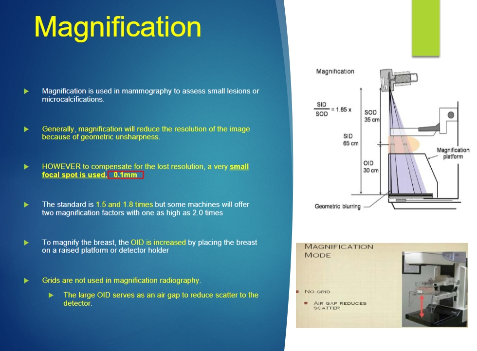
Image occlusion: name the hidden labels
86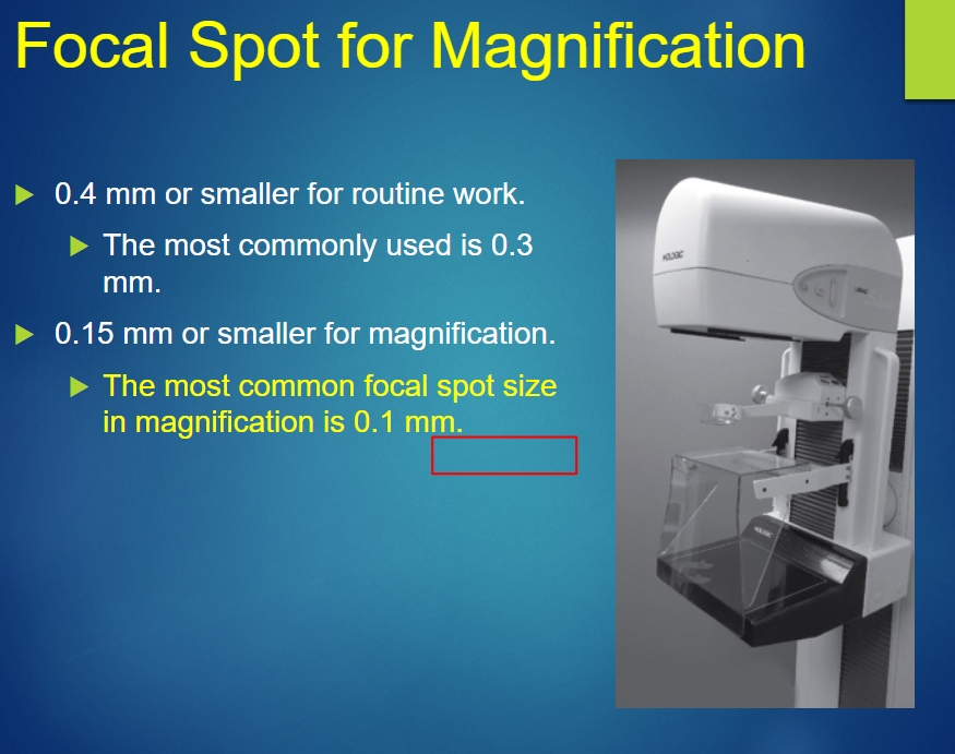
Image occlusion: name the hidden labels
87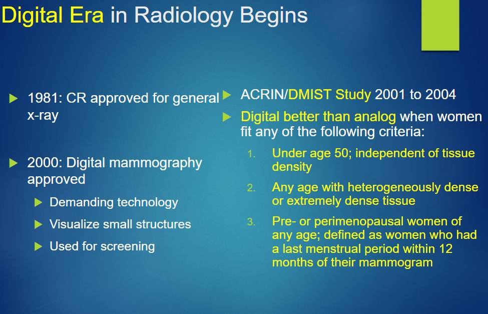
Image occlusion: name the hidden labels
88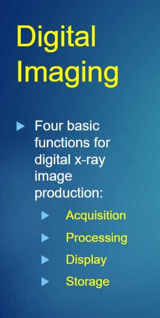
Image occlusion: name the hidden labels
89
FFDM stand for
Full Field Digital Mammography
90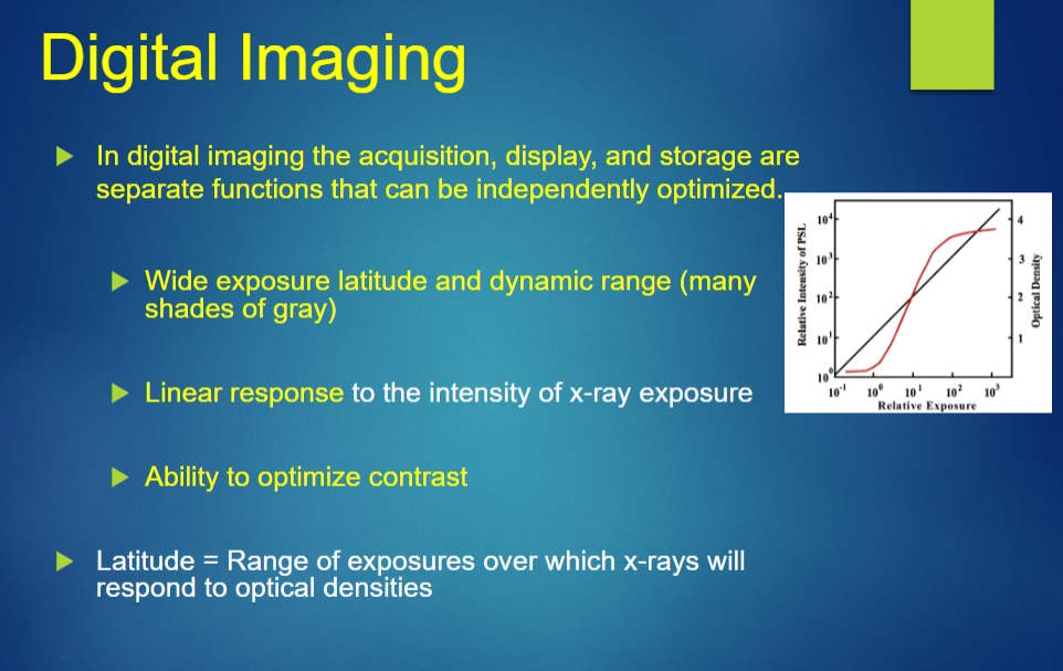
Image occlusion: name the hidden labels
91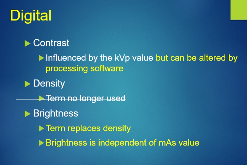
Image occlusion: name the hidden labels
92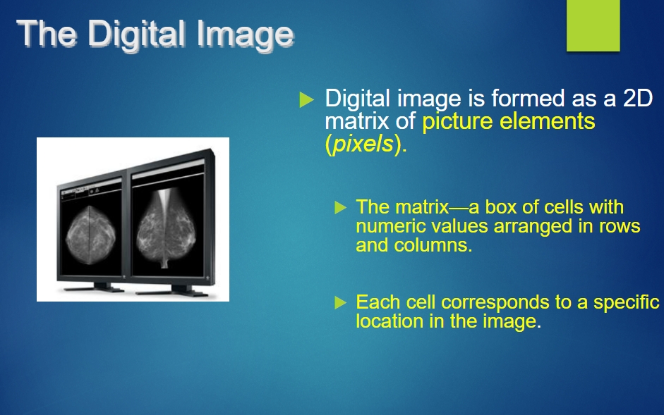
Image occlusion: name the hidden labels
93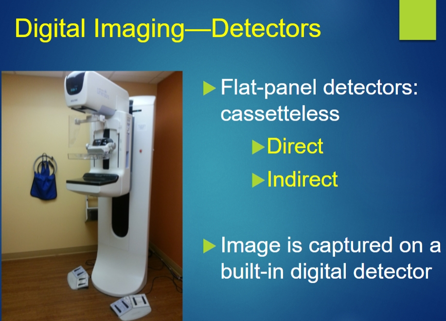
Image occlusion: name the hidden labels
94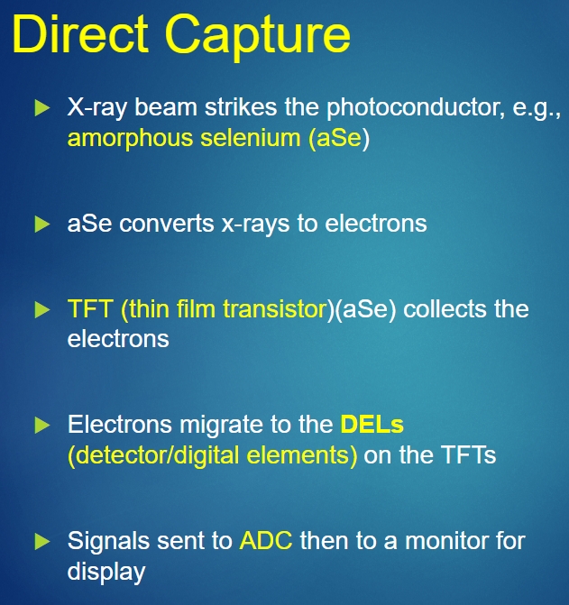
Image occlusion: name the hidden labels
95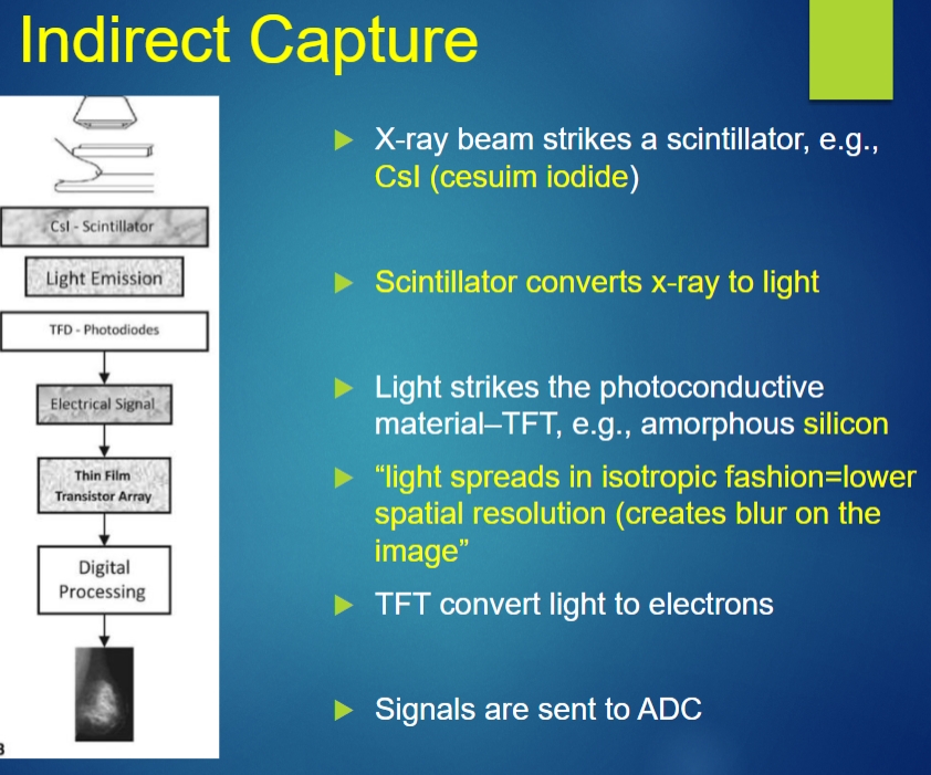
Image occlusion: name the hidden labels
96
formerly additional indirect used
PSP (photostimulated plates)=xray-light by psp-electrons
97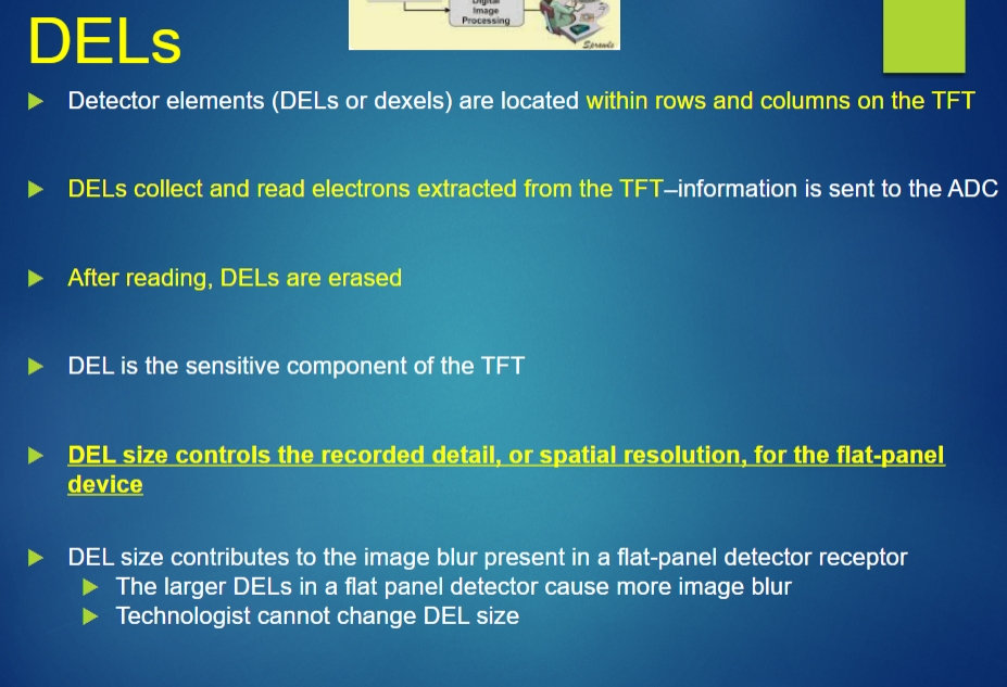
Image occlusion: name the hidden labels
98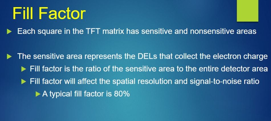
Image occlusion: name the hidden labels
99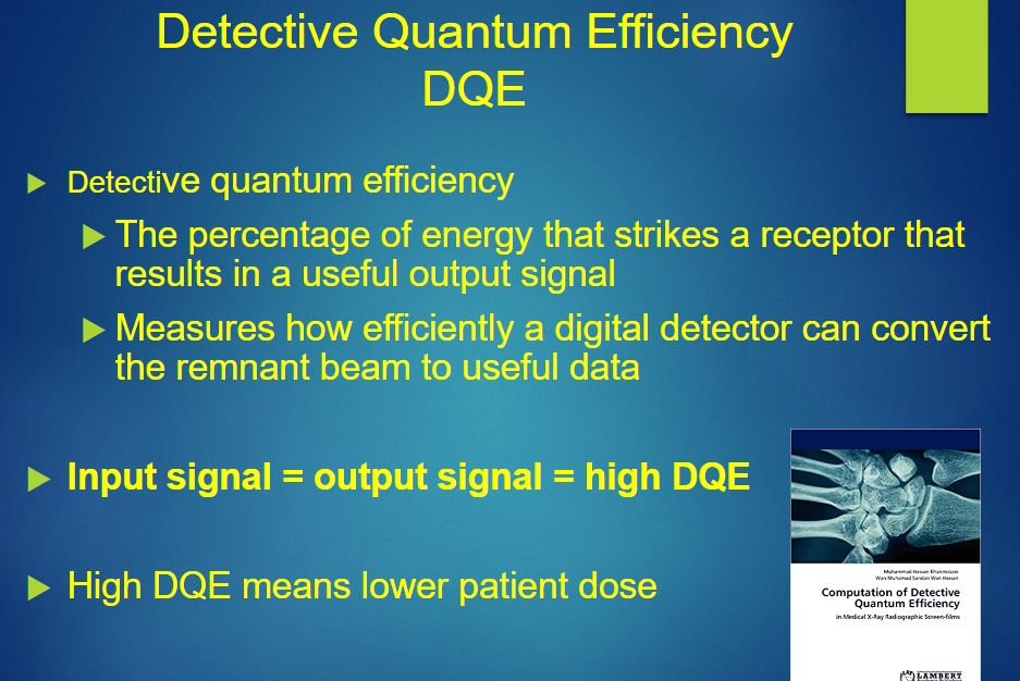
Image occlusion: name the hidden labels
100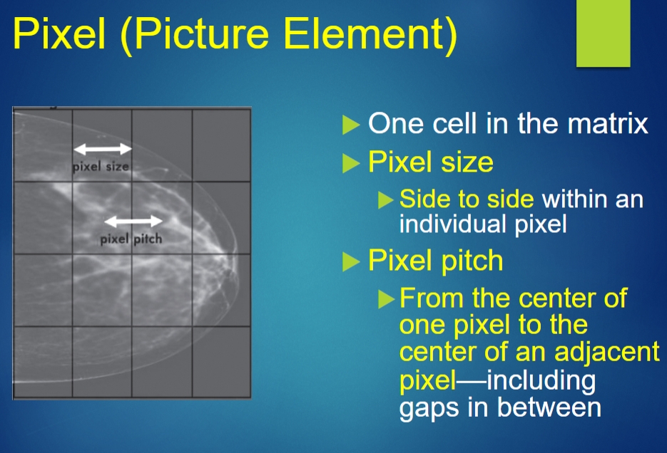
Image occlusion: name the hidden labels
101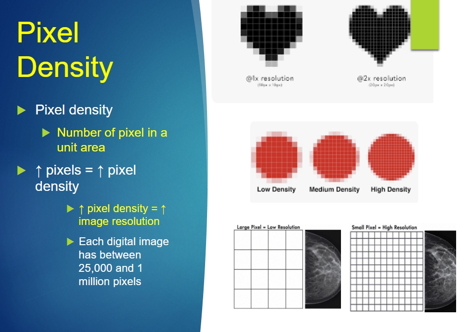
Image occlusion: name the hidden labels
102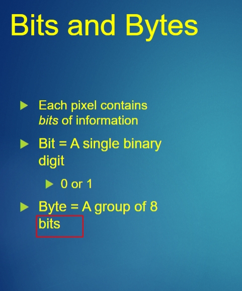
Image occlusion: name the hidden labels
103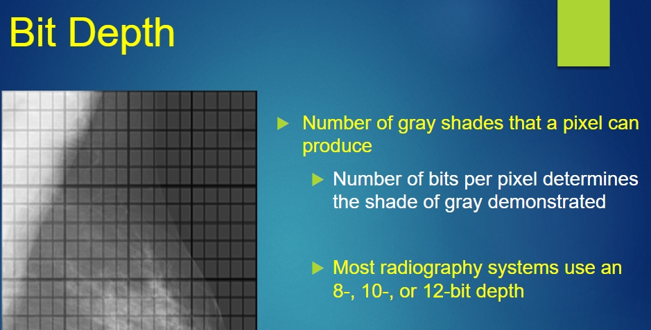
Image occlusion: name the hidden labels
104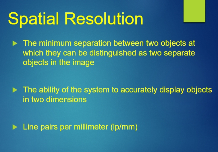
Image occlusion: name the hidden labels
105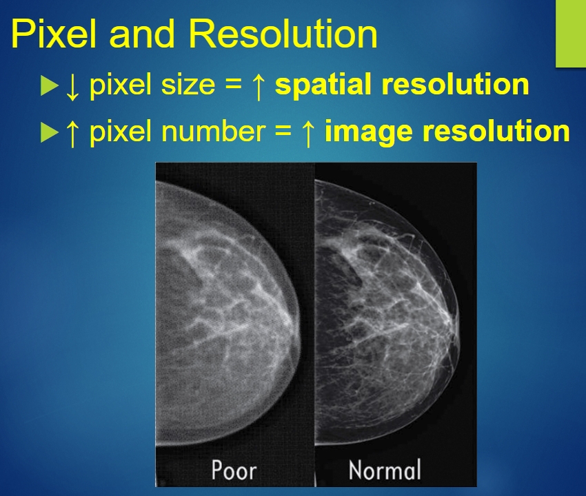
Image occlusion: name the hidden labels
106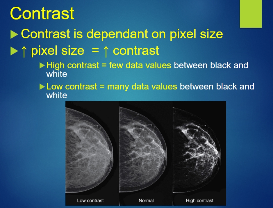
Image occlusion: name the hidden labels
107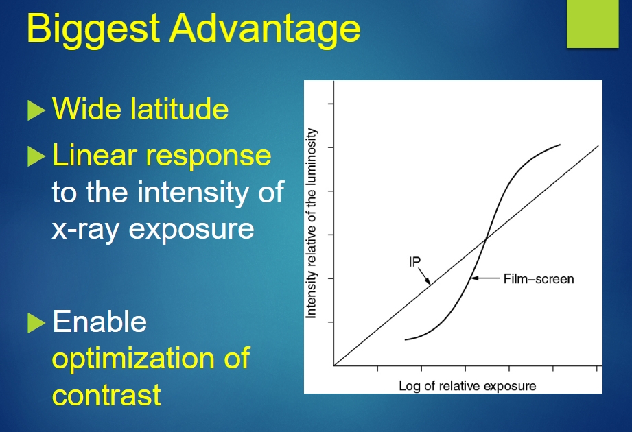
Image occlusion: name the hidden labels
108
Exposure Latitude of Digital receptors
50% underexposure to 400% overexposure
109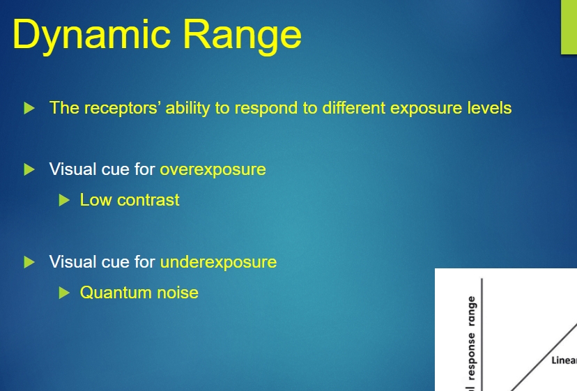
Image occlusion: name the hidden labels
110
Image occlusion: name the hidden labels
111
Image occlusion: name the hidden labels
112
Image occlusion: name the hidden labels
113
Image occlusion: name the hidden labels
114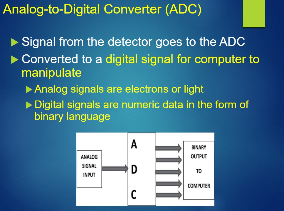
Image occlusion: name the hidden labels
115
Image occlusion: name the hidden labels
116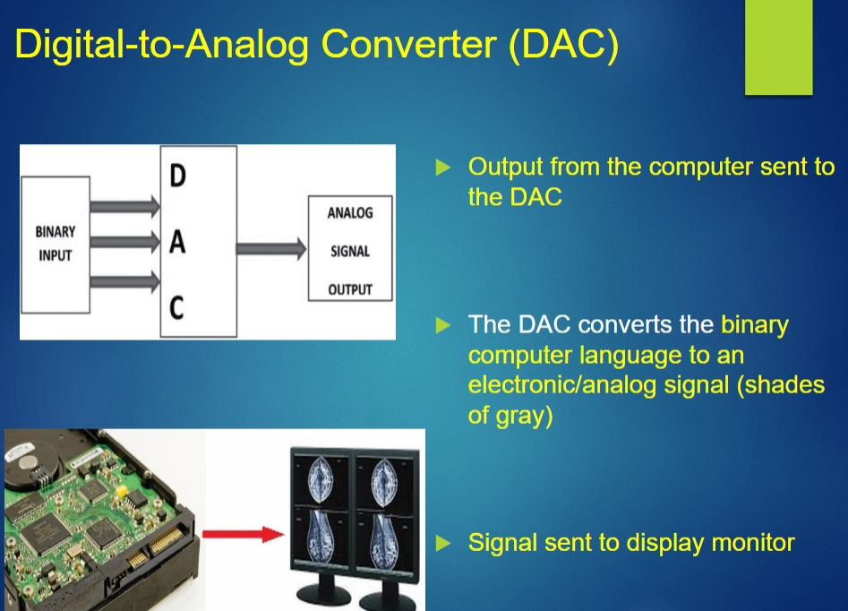
Image occlusion: name the hidden labels