Chapter 5 Breast Pathology Benign and Malignant Disease
Study these flashcardsFree · spaced repetition · iPhone, Android, Mac & web
All cards
1
Main Location of Breast Cancers
TDLU (carcinoma)
2
Other Location of Breast Cancers
Fibrous tissue
Connective tissue
Larger ducts
(sarcoma)
Connective tissue
Larger ducts
(sarcoma)
3
4 Changes Leading to Malignancy
Epithelial hyperplasia
Atypical (epithelial) hyperplasia
Carcinoma in situ
Metastases
Atypical (epithelial) hyperplasia
Carcinoma in situ
Metastases
4
2 Type of Augmented Breast
Silicon
Saline
Saline
5
2 Method of placement of Augmented Breast
Subglandular or retromammary placement
Subpectoral or retropectoral placement
Subpectoral or retropectoral placement
6
Silicone Leakage is called ... sign
Linguine
7
What is Reduction Mammoplasty
Elevation of nipple
Removal of glandular
tissue
Removal of glandular
tissue
8
Shapes of Benign Breast Lesion(masses)
Round
Oval
Lobulated
Oval
Lobulated
9
Shapes of Malignant Breast Lesion(masses)
Lobulated
Irregular
Spiculated
Irregular
Spiculated
10
Margins of Benign Breast Lesions
Circumscribed
Obscured
Microlobulated
Obscured
Microlobulated
11
Margins of Malignant Breast Lesions
Microlobulated
Indistinct
Spiculated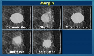
Indistinct
Spiculated
12
2 types of Density of a Mass
Low density: Tends to indicate
benign lesion
High density: Tends to indicate
malignant lesions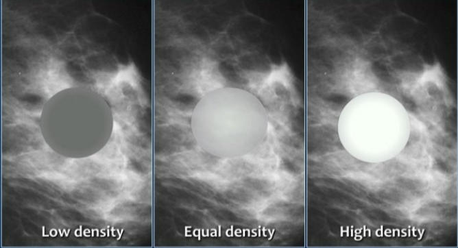
benign lesion
High density: Tends to indicate
malignant lesions
13
3 Mammographic Characteristics of Benign Circular or Oval lesions
Radiolucent
Radiolucent and radiopaque combined
Low-density radiopaque
Radiolucent and radiopaque combined
Low-density radiopaque
14
Examples of High-density radiopaque circular/oval lesion
Carcinoma
Abscess
Galactocele—calcified
Hematoma—calcified
Skin cyst
Abscess
Galactocele—calcified
Hematoma—calcified
Skin cyst
15
Define Halo Sign
Thin capsule—curved
radiolucent line(ring) around
the lesion
radiolucent line(ring) around
the lesion
16
Does The halo always represent a benign lesion?
often (not always)
17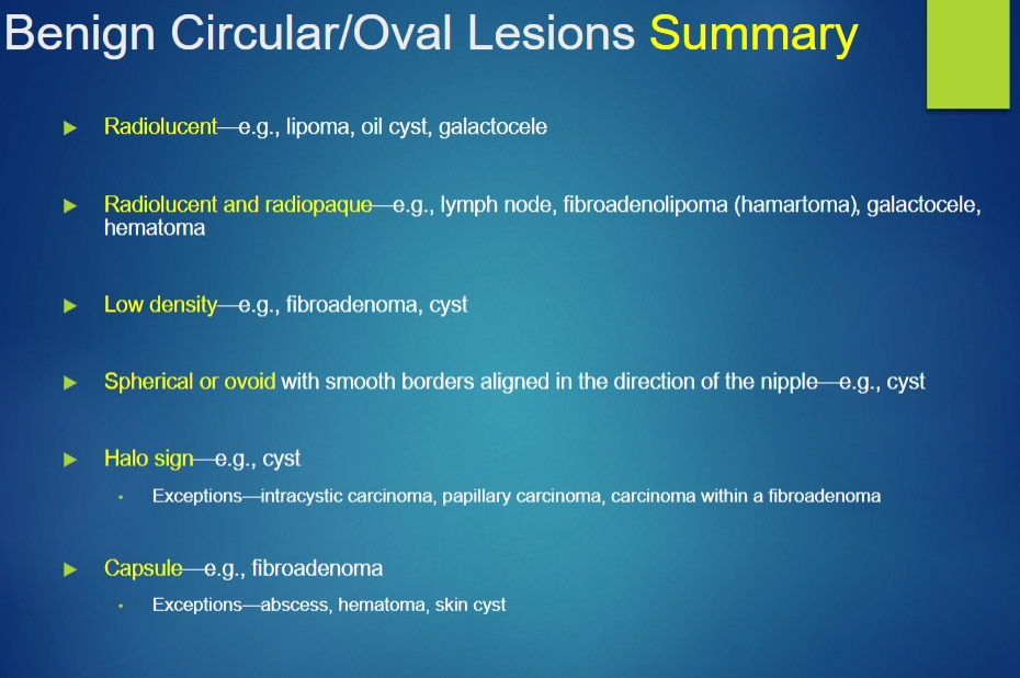
Image occlusion: name the hidden labels
18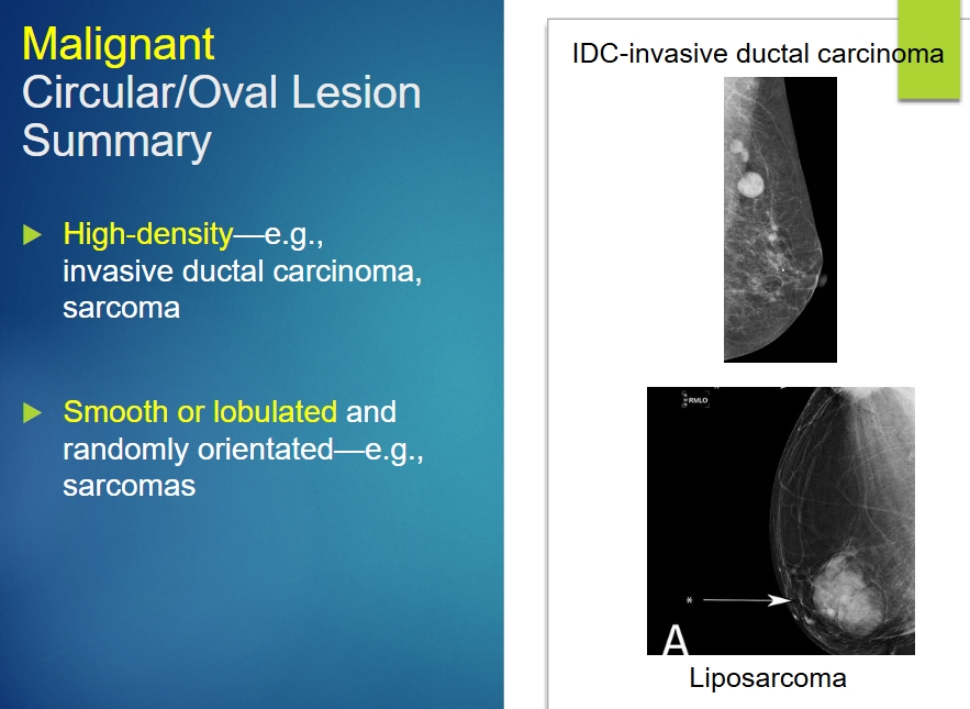
Image occlusion: name the hidden labels
19
Charateristic of Malignant Spiculated/Stellate Lesions
Central tumor (Distinct central mass) withradiating structures
Spicules reaching the skin or muscle may cause localized skin thickening or skin dimpling, e.g., IDC
Spicules reaching the skin or muscle may cause localized skin thickening or skin dimpling, e.g., IDC
20
Charateristic of Benign Spiculated/Stellate Lesion
No solid, dense, or distinct central mass
radial scar-was considered “high risk” often now considered benign but often still excised
radial scar-was considered “high risk” often now considered benign but often still excised
21
Mammographic Characteristics of Benign Circular or Oval lesions
Radiolucent
Radiolucent and radiopaque combined
Low-density radiopaque
Radiolucent and radiopaque combined
Low-density radiopaque
22
High-density radiopaque lesion example
Carcinoma
Abscess
Galactocele—calcified
Hematoma—calcified
Skin cyst
Abscess
Galactocele—calcified
Hematoma—calcified
Skin cyst
23
Halo Sign
Thin capsule—curved radiolucent line(ring) around the lesion
often (not always) represent a benign lesion
often (not always) represent a benign lesion
24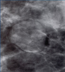
Halo Sign
25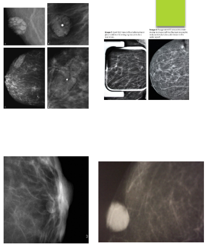
Skin Lesions
26
Benign Circular/Oval Lesions Sign include:
Radiolucent
Radiolucent and radiopaque
Low density
Spherical or ovoid
Halo sign
Capsule
Radiolucent and radiopaque
Low density
Spherical or ovoid
Halo sign
Capsule
27
Malignant
Circular/Oval Lesion Sign include:
Circular/Oval Lesion Sign include:
High-density
Smooth or lobulated
Smooth or lobulated
28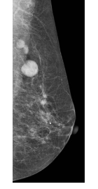
IDC-invasive ductal carcinoma
29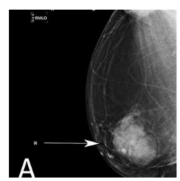
Liposarcoma
30
Spiculated/Stellate Lesions sign
Central tumor with radiating structures
31
Malignant Spiculated/Stellate Lesions Sign
Distinct central mass
Spicules reaching the skin or muscle may cause localized skin thickening or skin dimpling
Spicules reaching the skin or muscle may cause localized skin thickening or skin dimpling
32
Benign Spiculated/Stellate Lesion sign
No solid, dense, or distinct central mass
radial scar-was considered “high risk” often now considered benign but often still excised”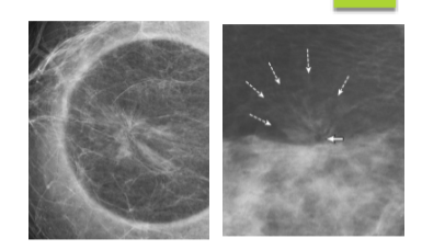
radial scar-was considered “high risk” often now considered benign but often still excised”
33
What is Architectural Distortion
the indenting or
tenting of the breast parenchyma
tenting of the breast parenchyma
34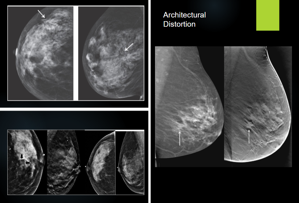
Image occlusion: name the hidden labels
35
what is Asymmetry
glandular tissue in one portion of the breast that is not present in a similar position in the contralateral breast
36
What is focal asymmetric lesion
asymmetric fibroglandular breast tissue seen on both the CC and MLO views
37
Global asymmetry
cover a
larger portion of the breast, typically more than one quadrant.
larger portion of the breast, typically more than one quadrant.
38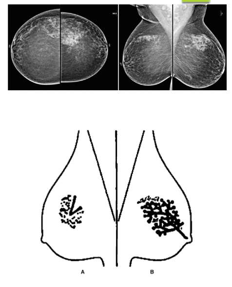
Asymmetry
39
Main Causes of Breast Calcifications
Cystic changes
Dermal calcification
Calcified fibroadenoma
Sutural
Dermal calcification
Calcified fibroadenoma
Sutural
40
Evaluation/Classification of Micro calcifications
Density
Distribution
Change over time
Number
Morphology
Distribution
Change over time
Number
Morphology
41
Calcification Morphology - Benign
Skin calcifications
Vascular calcifications
Coarse calcifications—“pop-corn like”
Linear rod-like
Round calcifications - punctuate if they are smaller than 0.5 mm in diameter
Large rod-like calcifications
Rim calcifications/”eggshell”
Dystrophic calcifications
Milk of calcium
Sutural calcifications
Vascular calcifications
Coarse calcifications—“pop-corn like”
Linear rod-like
Round calcifications - punctuate if they are smaller than 0.5 mm in diameter
Large rod-like calcifications
Rim calcifications/”eggshell”
Dystrophic calcifications
Milk of calcium
Sutural calcifications
42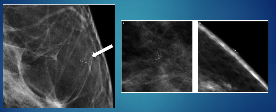
Skin Calcifications
43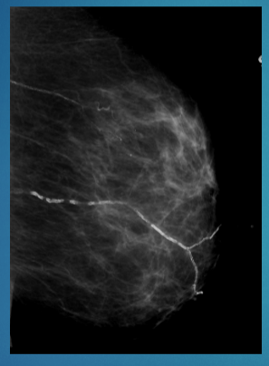
Vascular Calcifications
distinctive parallel lines
distinctive parallel lines
44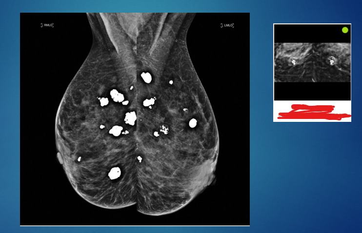
Coarse Calcifications
Popcorn like
Popcorn like
45
Linear rod-like calcifications
46
Round, puntuate calcifications
are more likely to be malignant
are more likely to be malignant
47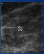
Rim calcifications/”eggshell”
48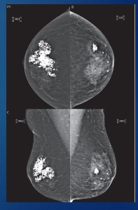
Dystrophic calcifications
often associated with surgery
often associated with surgery
49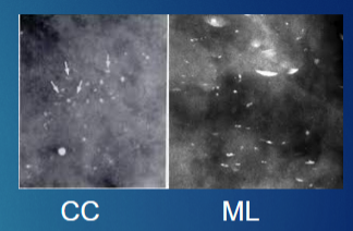
Milk of calcium
50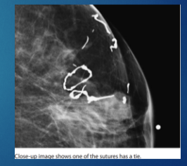
Sutural calcifications
51
Calcification Morphology - Intermediate Concern
Coarse heterogeneous
Amorphous or indistinct
Amorphous or indistinct
52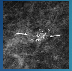
Coarse heterogeneous
they can also be associated with malignancy
they can also be associated with malignancy
53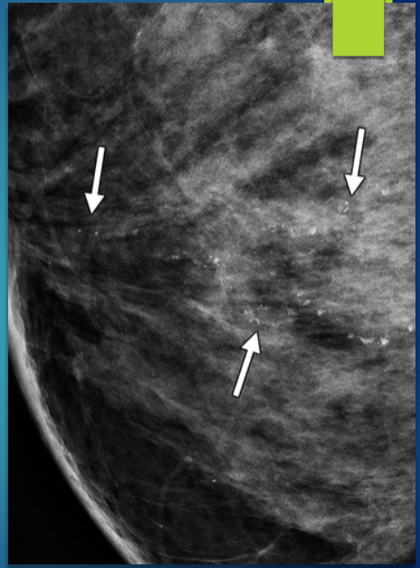
Amorphous calcifications are of
intermediate concern and can be
malignant or benign
intermediate concern and can be
malignant or benign
54
Malignant Calcifications
Fine linear or branching - casting- type calcifications
Pleomorphic
Pleomorphic
55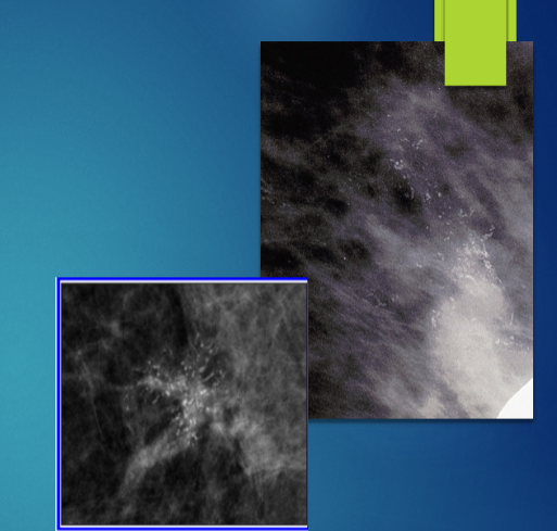
Fine linear or branching calcifications
casting- type calcifications, forming a “cast”
around the ducts.
casting- type calcifications, forming a “cast”
around the ducts.
56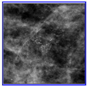
Pleomorphic calcifications
very fine calcifications
appear similar to shattered glass or crushed stones
very fine calcifications
appear similar to shattered glass or crushed stones
57
What is typically malignant calcifications (e.g., DCIS).
Pleomorphic calcifications
58
Suspicious Calcifications include:
Fine linear or branching calcification
Amorphous or indistinct calcifications
Pleomorphic calcifications
Coarse heterogenous or granular calcifications
Amorphous or indistinct calcifications
Pleomorphic calcifications
Coarse heterogenous or granular calcifications
59
Benign Masses (Lesions)
Simple Cyst
Galactocele
Fibroadenoma
Lipoma
Hamartoma - Fibroadenolipoma
Papilloma
Duct Ectasia
Hematoma
Abscess
Fat Necrosis/Oil cyst
Lymph Nodes
Gynecomastia-male
Radial Scar-no central mass
Galactocele
Fibroadenoma
Lipoma
Hamartoma - Fibroadenolipoma
Papilloma
Duct Ectasia
Hematoma
Abscess
Fat Necrosis/Oil cyst
Lymph Nodes
Gynecomastia-male
Radial Scar-no central mass
60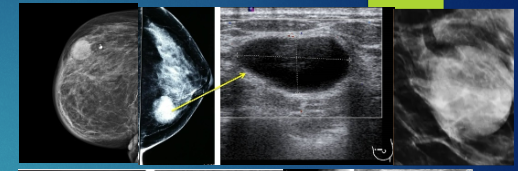
Simple Cyst
61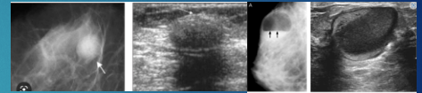
Galactocele
62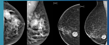
Fibroadenoma
63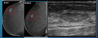
Lipoma
64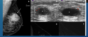
Abscess
65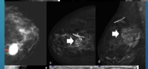
Hematoma
66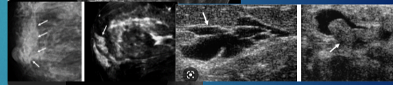
Duct Ectasia-green, brown discharge
67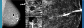
Papilloma
68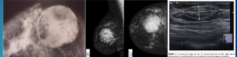
Hamartoma
69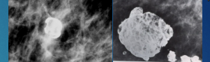
Fat Necrosis/Oil cyst
70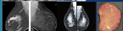
Lymph Nodes
71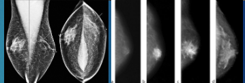
Gynecomastia-male
72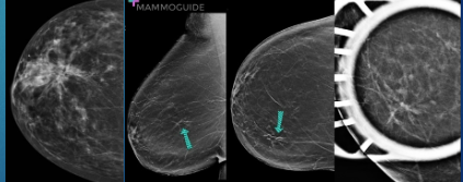
Radial Scar
73
Fibrocystic changes result in
calcifications that can vary in size from 0.5 to 1 mm
74
Sclerosing Adenosis/Ductal epithelia hyperplasia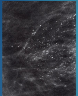
Calcifications as a result of increased cellular activity
75
Discharge can be green,brown,yellowish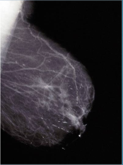
Ductal Ectasia
76
Vascular tumors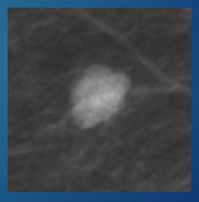
Hemangioma
77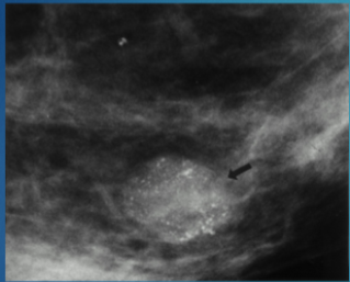
Intraductal Papilloma - Small wart-like
bloody/serous nipple discharge
bloody/serous nipple discharge
78
What is Histological analysis?
Microscopic analysis of organ and body tissues
79
What is Cytological analysis?
Analysis of cells
80
Thickened Skin Syndrome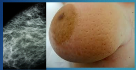
Entire breast or localized
Peau d’orange appearance
Associated with increased density
Caused by benign or malignant
changes
Normal skin is 0.5–2 mm thick
Peau d’orange appearance
Associated with increased density
Caused by benign or malignant
changes
Normal skin is 0.5–2 mm thick
81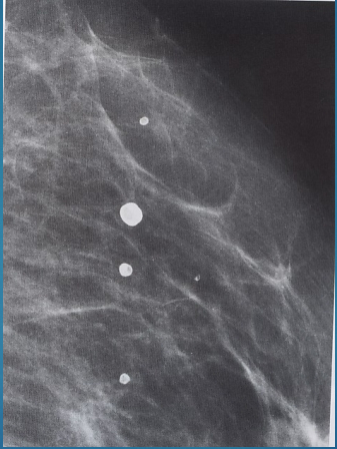
Microhematoma
82
Benign Skin Thickening treatment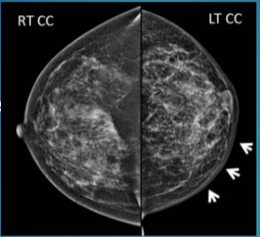
Radiation treatment
83
Malignant Skin Thickening Signs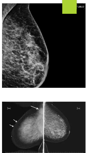
Inflammatory carcinoma
Obstruction of the lymphatic
Obstruction of the lymphatic
84
Most common types of Breast Cancer
Ductal—90% of all breast cancers
Lobular—5%–10% of all breast cancers
Lobular—5%–10% of all breast cancers
85
Example of Non-Invasive Breast Cancer-In Situ
DCIS-DUCTAL CARCINOMA IN SITU
86
DCIS-DUCTAL CARCINOMA IN SITU
confined to the ducts
Most common non-invasive breast cancer
contain microcalcifications
Best detected with mammography
Most common non-invasive breast cancer
contain microcalcifications
Best detected with mammography
87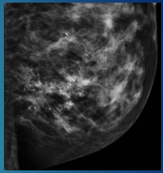
Ductal Carcinoma In Situ (DCIS)
88
Not considered a cancer but marks an increased risk for future breast cancer
LCIS- Lobular Carcinoma In Situ
Incidental finding on biopsy
Incidental finding on biopsy
89
High Risk Breast Pathology include:
LCIS- Lobular carcinoma In-situ (incidental finding)
ALH- Atypical Lobular Hyperplasia
ADH- Atypical Ductal Hyperplasia
Papilloma with Atypia
Radial Scar- more recently considered benign
Phyllodes Tumor
ALH- Atypical Lobular Hyperplasia
ADH- Atypical Ductal Hyperplasia
Papilloma with Atypia
Radial Scar- more recently considered benign
Phyllodes Tumor
90
Invasive or Infiltrating Ductal Carcinoma (IDC)
tissue (broken through the basement membrane)
Most common invasive breast cancer-75%
Most common invasive breast cancer-75%
91
Invasive Lobular Carcinoma (ILC)
Most frequently missed breast cancer
Often multifocal, multicentric, bilateral
Difficult to detect on imaging
Single file pattern
Often multifocal, multicentric, bilateral
Difficult to detect on imaging
Single file pattern
92
Other Breast Carcinomas
Inflammatory
Infiltrating medullary
Colloid comedo
Tubular
Mucinous
Papillary carcinoma
Paget’s disease
Infiltrating medullary
Colloid comedo
Tubular
Mucinous
Papillary carcinoma
Paget’s disease
93
Medullary Carcinoma
Most common in younger women, Japanese and African Americans
<35yrs of age
<35yrs of age
94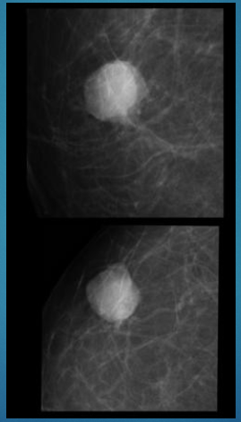
Colloid (mucinous) Carcinoma
95
Papillary Carcinoma
in older women
Nipple discharge
Nipple discharge
96
Phyllodes (sarcoma) tumor
does not arise from TDLU, but from stroma
leaf-like
leaf-like
97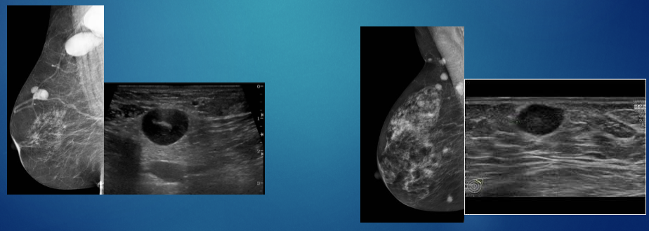
Lymphoma
Metastases to the Breast
Metastases to the Breast
98
Inflammatory Processes
Mastitis
Seroma, Hematoma
Abscess
Inflammatory Carcinoma
Paget’s Disease-
Seroma, Hematoma
Abscess
Inflammatory Carcinoma
Paget’s Disease-
99
2 types of Mastitis
Puerperal(Lactating)Mastitis
Plasma Cell Mastitis
Plasma Cell Mastitis
100
Puerperal(Lactating)Mastitis
Most common type
Bacterial (staphylococcus)due to cracked nipple entry,inflamed ducts;milk stasis
Bacterial (staphylococcus)due to cracked nipple entry,inflamed ducts;milk stasis
101
Plasma Cell Mastitis
Cellular not bacterial
102
Paget Disease of the Breast underlying of ....
IDC