Module 3
Study these flashcardsFree · spaced repetition · iPhone, Android, Mac & web
All cards
1
functions of the pulmonary system
Exchange of Co2 and O2
temperature
metabolizes toxic substances
temperature
metabolizes toxic substances
2
Issues of the pulmonary system
airway obstruction
alveoli obstruction/ restriction
Decreased compliance: thoracic cavity and interstitial lung tissue
alveoli obstruction/ restriction
Decreased compliance: thoracic cavity and interstitial lung tissue
3
Obstructive Diseases
Asthma
COPD
Cystic Fibrosis
COPD
Cystic Fibrosis
4
anatomy affected in obstructive diseases
Airways
5
breathing phase in obstructive diseases
expiratory
6
pathophysiology in obstructive diseases
increased airway resistance
7
PFT's for obstructive disease
Fev1/FVC ratio, flow rates
8
restrictive diseases
atelectasis
Infections
Acute Respiratory Disorder (ARDS)
Intersitital Lung Disease
Infections
Acute Respiratory Disorder (ARDS)
Intersitital Lung Disease
9
anatomy affected in restrictive diseases
thoracic cage, lung tissue
10
breathing phase restrictive diseases
inspiratory
11
pathophysiology restrictive diseases
decreased lung or thoracic compliance
12
PFT's restrictive lung diseases
Volumes, capacities
13
Lung Volumes
Tidal Volume
Expiratory Reserve Volume (ERV)
Inspiratory Reserve Volume (IRV)
Residual Volume (RV)
Expiratory Reserve Volume (ERV)
Inspiratory Reserve Volume (IRV)
Residual Volume (RV)
14
Tidal Volume
regular amount of air inhaled/exhaled
around 0.5L
around 0.5L
15
ERV
amount of air that can be forcefully exhaled after a passive exhalation
around 1.1 L
around 1.1 L
16
IRV
amount of air you can breathe in at the end of the tidal volume
around 3L
around 3L
17
RV
air left over after forced exhalation
you need some so your lungs don't collapse!
you need some so your lungs don't collapse!
18
Lung capacities
Total Lung Capacity (TLC)
Inspiratory Capacity (IC)
Functional Residual Capacity (FRC)
Vital Capacity (VC)
Inspiratory Capacity (IC)
Functional Residual Capacity (FRC)
Vital Capacity (VC)
19
TLC
max amount lungs can hold
TLC = VC + RV
around 5.8L
increases with emphysema
decreases with chest wall restrictions
TLC = VC + RV
around 5.8L
increases with emphysema
decreases with chest wall restrictions
20
IC
amount of air you can breathe in after normally breathing out
IC = V(t) (tidal volume) + IRV
around 3.5L
decreases with restrictive lung disease
IC = V(t) (tidal volume) + IRV
around 3.5L
decreases with restrictive lung disease
21
FRC
air left after normal expiration
FRC = ERV + RV
around 2.3L
decreases with restrictive lung disorder
FRC = ERV + RV
around 2.3L
decreases with restrictive lung disorder
22
VC
max air out after max air in
VC = IRV + ERV + Vt
ability to breathe deeply and cough; VC = 3x > Vt to make effective cough
insp/exp muscle strength
decreases w/obstructive (sometimes) lung disorders + restrictive (always)
VC = IRV + ERV + Vt
ability to breathe deeply and cough; VC = 3x > Vt to make effective cough
insp/exp muscle strength
decreases w/obstructive (sometimes) lung disorders + restrictive (always)
23
Asthma
Chronic Inflamatory Disorder of airway that leads to airway obstruction
Episodic hyper reactivity of airway to certain irritants/stimulants -> inflammation -> obstruction
Episodic hyper reactivity of airway to certain irritants/stimulants -> inflammation -> obstruction
24
asthma pathophysiology
genetics
low birthweight
maternal smoking
second hand smoking
allergens
irritant -> inflammation
bronchiole smooth muscle contracts => bronchoconstriction
low birthweight
maternal smoking
second hand smoking
allergens
irritant -> inflammation
bronchiole smooth muscle contracts => bronchoconstriction
25
asthma signs/symptoms
wheezing
dyspnea
chest tightness
couging
using accessory breathing muscles
dyspnea
chest tightness
couging
using accessory breathing muscles
26
PFTs for Asthma
Between attacks: reduced FVC
During/ right after: reduced PEF, FVC, FEV1
During/ right after: reduced PEF, FVC, FEV1
27
Asthma risks
people can "outgrow" asthma
remodeling of airways, even in absence of symptoms
bronchial biopses => inflammatory factors, thickening of airways
remodeling of airways, even in absence of symptoms
bronchial biopses => inflammatory factors, thickening of airways
28
asthma diagnosis
FEV 1 -> improvement of >12% or increase of >200mL post bronchodilator
reversible -> unlike other obstructive diseases
reversible -> unlike other obstructive diseases
29
asthma medical interventions
Goal: prevent repeated damage -> airway scarring + permanent obstruction
identifying "triggers", minimize exacerbations
Meds: bronchodilators, anti-inflammatory agents
identifying "triggers", minimize exacerbations
Meds: bronchodilators, anti-inflammatory agents
30
asthma PT interventions
Impaired Ventilation
-breathing exercises like controlled breathing techniques, posture
Ventilatory Muscle Weakness
-breathing exercises
-inspiratory muscle training
Aerobic Capacity
-energy conservation techniques -> paced breathing
-exercise
-breathing exercises like controlled breathing techniques, posture
Ventilatory Muscle Weakness
-breathing exercises
-inspiratory muscle training
Aerobic Capacity
-energy conservation techniques -> paced breathing
-exercise
31
Asthma PT Implications
asthma vs laryngeal spasms!
differential diagnosis
refer to SLP
accurate diagnosis -> good outcomes
differential diagnosis
refer to SLP
accurate diagnosis -> good outcomes
32
FEV 1 - forced expiratory volume in 1 sec
volume of air that is forcibly exhaled after full inspiration in the first second
>80% predicted value
>80% predicted value
33
Forced Vital Capacity (FVC)
volume of air forcible exhaled after full inspiration
>80% predicted value
4.75-5 men
3.25-3.75 women
>80% predicted value
4.75-5 men
3.25-3.75 women
34
FEV1/FVC
identifies obstructive or restrictive ventilatory defects
FEV1/FVC <70% aka FEV1<FVC => obstructive
FEV1/FVC >70% aka FVC reduced more => restrictive
FEV1/FVC <70% aka FEV1<FVC => obstructive
FEV1/FVC >70% aka FVC reduced more => restrictive
35
Peak Expiratory Flow (PEF)
maximal flow that can be exhaled when blowing out at a steady state
speed of air, 400-700 L/m or 5-6 L/s
speed of air, 400-700 L/m or 5-6 L/s
36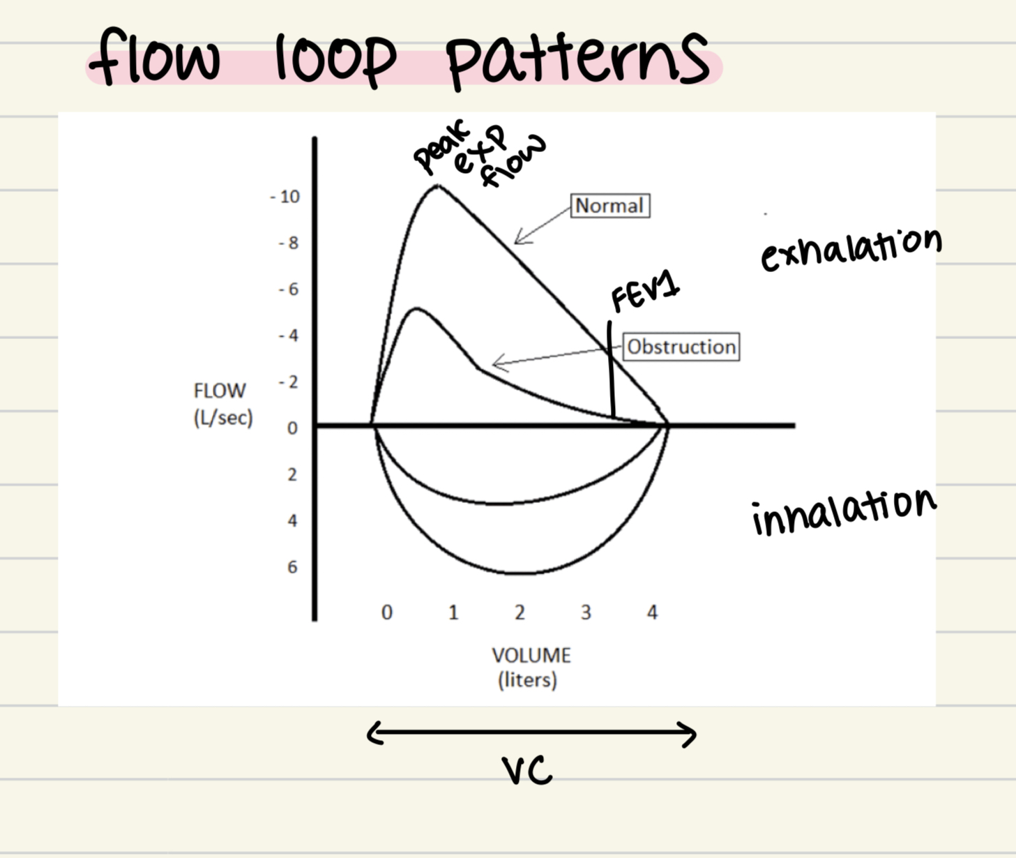
Image occlusion: name the hidden labels
37
Image occlusion: name the hidden labels
38
Image occlusion: name the hidden labels
39
Image occlusion: name the hidden labels
40
Image occlusion: name the hidden labels
41
obstructive disease flow loop patterns
slower
similar VC
decreased PEF
similar VC
decreased PEF
42
bronchodilator how it works
increased bronchomotor tone in airways -> smooth muscle constriction, increased mucous production, inflammatory edema
43
Types of bronchodilators
Beta Adrenergic Agonist
Xanthine Derivatives
Anticholinergics
Xanthine Derivatives
Anticholinergics
44
beta adrenergic agonist
stimulates beta-2 receptors => smooth muscle dilation
beta 1 - heart, beta 2 - lungs
Non selective - Beta 1 and 2
-epinephrine, isoproterenol
Selective - Beta 2
-albuterol, metaproterenol, formoterol, salmeterol, terabutaline
Fast onset!
beta 1 - heart, beta 2 - lungs
Non selective - Beta 1 and 2
-epinephrine, isoproterenol
Selective - Beta 2
-albuterol, metaproterenol, formoterol, salmeterol, terabutaline
Fast onset!
45
beta adrenergic agonist side effects
CNS: nervousness, anxiety, restlessness
CV: tachycardia
GI: none
Other: bronchial irritation, bronchial constriction
CV: tachycardia
GI: none
Other: bronchial irritation, bronchial constriction
46
xanthine dervatives
initiates smooth muscle dilation
theophylline, aminophylline
theophylline, aminophylline
47
xanthine derivatives side effects
CNS: confusion, irritability, restlessness, seizures
CV: arrhythmia
GI: nausea
Other: theophylline toxicity
CV: arrhythmia
GI: nausea
Other: theophylline toxicity
48
anticholinergics
block acetylcholine receptors => bronchial smooth muscle relaxation
ipatropium, tiotropium, revefenacin, aclidinium, umeclidinium
ipatropium, tiotropium, revefenacin, aclidinium, umeclidinium
49
anticholinergics side effects
CNS: confusion
CV: tachycardia
GI: constipation
Other: dry mouth
CV: tachycardia
GI: constipation
Other: dry mouth
50
corticosteroid
reduce inflammatory response + decrease mucosal swelling due to bronchial irritation/constriction
IV administration in acute situations
systemic (oral) administration
newer development of inhaled agents for long term preventative use
IV administration in acute situations
systemic (oral) administration
newer development of inhaled agents for long term preventative use
51
Corticosteroid common meds
Oral/IV: prednisone, prenisolone, methyl-predisone
Inhaled: beclamethasone, budesonide, fluticasone
Inhaled: beclamethasone, budesonide, fluticasone
52
corticosteroid indications
asthma: acute and long term management of airway inflammation
COPD: don't use IV/oral to prevent, only acute
CF: acute management of airway inflammation
COPD: don't use IV/oral to prevent, only acute
CF: acute management of airway inflammation
53
corticosteroid side effects
CNS: irritability, emotional liability
CV: none
GI: General upset
Other: muscle weakness/atrophy, osteoporosis, immunosuppression, hyperglycemia
side effects usually with higher doses
CV: none
GI: General upset
Other: muscle weakness/atrophy, osteoporosis, immunosuppression, hyperglycemia
side effects usually with higher doses
54
antihistamines
used to treat histamine-mediated coughing, sneezing, irritation by blocking H receptor
decrease effects of histamine on upper respiratory tract
used in the treatment of asthma to decrease mucosal congestion, irritation caused by allergens
also used for seasonal allergies, colds: reduced coughing + sneezing
decrease effects of histamine on upper respiratory tract
used in the treatment of asthma to decrease mucosal congestion, irritation caused by allergens
also used for seasonal allergies, colds: reduced coughing + sneezing
55
antihistamines side effects
CNS: sedation, fatigue, loss of coordination, blurred vision
CV: none
GI: general GI upset
Other: none
CV: none
GI: general GI upset
Other: none
56
antihistamines PT implications
sedative effects: risk of falls
dry out respiratory tract: limit productive cough
dry out respiratory tract: limit productive cough
57
supplemental O2
increases FiO2 to reduce hypoxemia
methods of delivery: nasal cannula mask, tent/hood, direct - endotrachral tube or tracheostomy
methods of delivery: nasal cannula mask, tent/hood, direct - endotrachral tube or tracheostomy
58
Supplemental O2: Fraction of Inspired oxygen (FiO2)
% of O2 in the atmosphere being breathed in
Room air (RA) - 21%, FiO2 of RA = 0.21
supplemental O2 increases FiO2
Low Flow O2: prescribed 1L/min - 5L/min: for every 1L/min, FiO2 increased by 0.03 (3%)
High Flow O2: prescribed by the FiO2 %
Room air (RA) - 21%, FiO2 of RA = 0.21
supplemental O2 increases FiO2
Low Flow O2: prescribed 1L/min - 5L/min: for every 1L/min, FiO2 increased by 0.03 (3%)
High Flow O2: prescribed by the FiO2 %
59
supplemental oxygen PT implications
understand how to change oxygen
how much O2 is in tank
know prescribed dosages
must have orders to change/titrate during exercise
how much O2 is in tank
know prescribed dosages
must have orders to change/titrate during exercise
60
supplemental oxygen: signs of O2 toxicity
visual changes
dizziness
tinnitis
nausea
muscle twitching
behavior changes: irritability, confusion, anxiety
dizziness
tinnitis
nausea
muscle twitching
behavior changes: irritability, confusion, anxiety
61
supplemental oxygen: risks
Oxygen toxicity
-high concentration >24 hrs = interstitial lung disease
-24-72 hrs = alveoli edema, hemorrhage, atelectasis
-lung fibrosis
-blindness
-high concentration >24 hrs = interstitial lung disease
-24-72 hrs = alveoli edema, hemorrhage, atelectasis
-lung fibrosis
-blindness
62
obstructive diseases limit airflow by
decreasing bronchial lumen size
increasing mucous production
damaging protective actions of airways (cilia)
damaging alveolar elasticity
increasing mucous production
damaging protective actions of airways (cilia)
damaging alveolar elasticity
63
obstructive diseases limiting airflow leads to
bronchial constriction
weakened airway walls
retained secretions
inflammation of mucosal lining of airways
alveolar sac destruction + over inflation
lung hyperinflation due to air trapping
weakened airway walls
retained secretions
inflammation of mucosal lining of airways
alveolar sac destruction + over inflation
lung hyperinflation due to air trapping
64
COPD MS changes
Diaphragm
-flattens: overinflation of lungs
-loses elasticity + exhalation becomes active
Increase in intrabdominal pressure
-increases stress on pelvic floor muscles: incontinence
Difficulty with Inspiration
-regular use of accessory muscles: hypertrophied and shortened muscles
-affects posture: forward head, increased thoracic kyphosis, rounded shoulders
General Skeletal Muscle Weakness
-flattens: overinflation of lungs
-loses elasticity + exhalation becomes active
Increase in intrabdominal pressure
-increases stress on pelvic floor muscles: incontinence
Difficulty with Inspiration
-regular use of accessory muscles: hypertrophied and shortened muscles
-affects posture: forward head, increased thoracic kyphosis, rounded shoulders
General Skeletal Muscle Weakness
65
COPD Risk Factors
Primary:
smoking (active and secondhand)
-destroys lung parenchyma (tissue)
-destroys alveoli
Environmental Exposures
-pollution
-occupational exposures to dust, chemicals, fumes
Other:
Socioeconomic factors
-linked to environmental exposures and access to healthcare
Frequent respiratory infections as a child and asthma
Age
Genetics (emphysema)
-alpha 1 antitrypsin deficiency
smoking (active and secondhand)
-destroys lung parenchyma (tissue)
-destroys alveoli
Environmental Exposures
-pollution
-occupational exposures to dust, chemicals, fumes
Other:
Socioeconomic factors
-linked to environmental exposures and access to healthcare
Frequent respiratory infections as a child and asthma
Age
Genetics (emphysema)
-alpha 1 antitrypsin deficiency
66
COPD Pathophysiology
Risks:
smoking and environment
bronchi and alveoli inflammation
impairs and destroys cilia function, hyperplasia of mucous secreting cells
hypersecretion of mucous and mucous plugging
edema of airway, reactive airways, destruction of terminal airways + alveoli
-hyperinflation of lungs from trapping air in alveoli
-loss of elastic recoil of airways/alveoli
-reduced gas exchange leading to hypoxemia + hypercapnea
smoking and environment
bronchi and alveoli inflammation
impairs and destroys cilia function, hyperplasia of mucous secreting cells
hypersecretion of mucous and mucous plugging
edema of airway, reactive airways, destruction of terminal airways + alveoli
-hyperinflation of lungs from trapping air in alveoli
-loss of elastic recoil of airways/alveoli
-reduced gas exchange leading to hypoxemia + hypercapnea
67
COPD PT Interventions
aerobic endurance training
-strength training: IMT, general
-Patient education: disease process, energy conservation, preventing/managin dyspnea
Ventilatory Interventions
-acapella, flutter, theraPEP
-airway clearance techniques: postural drainage
-controlled breathing: pursed lip + paced breath
-strength training: IMT, general
-Patient education: disease process, energy conservation, preventing/managin dyspnea
Ventilatory Interventions
-acapella, flutter, theraPEP
-airway clearance techniques: postural drainage
-controlled breathing: pursed lip + paced breath
68
COPD PT considerations
coordination w/meds
manage anxiety
energy conservation and paced breathing
manage anxiety
energy conservation and paced breathing
69
Exercise for different grades of COPD
Grade 1+2: 70-80% HR max
Grade 3+4: RPE 4-6/10
Walking: 80% average walking speed
Grade 3+4: RPE 4-6/10
Walking: 80% average walking speed
70
Types of COPD
Chronic bronchitis
Emphysema
Emphysema
71
chronic bronchitis
malfunction of the mucociliary transport
-buildup of mucous in airways that narrows lumen of airways
hypersecretion of mucous in large airways 1st, smaller later in disease
thickening of airway walls => inflammatory response
diagnosis: productive cough 3months/yr, 2 consecutive years and not due to any other infection
Increased mucous and damaged airways, normal compliance
-buildup of mucous in airways that narrows lumen of airways
hypersecretion of mucous in large airways 1st, smaller later in disease
thickening of airway walls => inflammatory response
diagnosis: productive cough 3months/yr, 2 consecutive years and not due to any other infection
Increased mucous and damaged airways, normal compliance
72
chronic bronchitis symptoms
persistent progressive dyspnea + DOE (dyspnea on exertion)
chronic productive cough w sputum
cyanosis: lack of O2 => whitereylue tint to skin
fluid retention (central) : enlarged abdomen
chronic productive cough w sputum
cyanosis: lack of O2 => whitereylue tint to skin
fluid retention (central) : enlarged abdomen
73
chronic bronchitis ABG
hypoxemia: PaO2 decreased w/severity
PaCO2 elevated, increased with progression - hypercapnia
PaCO2 elevated, increased with progression - hypercapnia
74
chronic bronchitis PFT
decreased FEV1 + FVC
Decreased FEV1/FVC: <70% normal TLC, maybe increased: air not that trapped
increased RV
Decreased FEV1/FVC: <70% normal TLC, maybe increased: air not that trapped
increased RV
75
emphysema
destruction of the alveolar walls and enlargement of: respiratory bronchiles, alveolar ducts, alveoli
weakening of the airways: airway lumen collapses during exhalation
loss of recoil of lung tissues + alveoli
All result in air trapping + hyperinflation of lungs: alveoli are stretched out so air isn't exiting passively as it should, pt has to force air out => collapsed airway
weakening of the airways: airway lumen collapses during exhalation
loss of recoil of lung tissues + alveoli
All result in air trapping + hyperinflation of lungs: alveoli are stretched out so air isn't exiting passively as it should, pt has to force air out => collapsed airway
76
emphysema symptoms
persistent, progressive dyspnea, esp during exertion (DOE)
hypertrophied SCM, scalenes, other accessory muscles due to chronic use
pursed lip breathing
thin frail appearance
increased A-P dimension thorax + elevated shoulder "Barrel Chest"
peripheral edema + R-side HF
pulm HTN
hypertrophied SCM, scalenes, other accessory muscles due to chronic use
pursed lip breathing
thin frail appearance
increased A-P dimension thorax + elevated shoulder "Barrel Chest"
peripheral edema + R-side HF
pulm HTN
77
emphysema ABG
hypoxeia: PaO2 decreased with severity
PaCO2: decreased early and increased as prog hypercapnia
PaCO2: decreased early and increased as prog hypercapnia
78
emphysema PFT
decreased FVC + FEV1
decreased FEV1/FVC
Increased TLC
Increased RV
decreased FEV1/FVC
Increased TLC
Increased RV
79
Imaging results emphysema
lung hyperinflation, horizontal ribs, low flat diaphragm, barrel chest
80
Acidemia
pH < 7.35
81
Alkalemia
pH>7.45
82
Acidosis
causes acidemia
83
alkalosis
causes alkalemia
84
metabolic alkalosis symptoms
confusion
coma
tingling/numb
muscle weakness
coma
tingling/numb
muscle weakness
85
metabolic acidosis symptoms
tachycardia
nausea
vomiting
dizziness
nausea
vomiting
dizziness
86
respiratory alkalosis symptoms
syncope
tremors
dyspnea
hyperventilation
tremors
dyspnea
hyperventilation
87
respiratory acidosis symptoms
SOB
confusion
fatigue
seizures
muscle weakness
confusion
fatigue
seizures
muscle weakness
88
Respiratory System ABG
Minutes, 2nd line of defense maintains pH by controlling CO2 => carbonic acid
CO2 is acidic
alterations in RR can effect pH by breathing off or retaining CO2 which will increase or decrease acid: don't hold your breath
CO2 + H2O = H2CO3
PaCO2 decrease, decrease acid avail => resp alkalosis (<35)
PaCO2 increase, increase acid avail => resp acidosis (>45)
CO2 is acidic
alterations in RR can effect pH by breathing off or retaining CO2 which will increase or decrease acid: don't hold your breath
CO2 + H2O = H2CO3
PaCO2 decrease, decrease acid avail => resp alkalosis (<35)
PaCO2 increase, increase acid avail => resp acidosis (>45)
89
Blood Buffer
maintain pH
works within seconds
proteins, hemoglobin, phosphate
works within seconds
proteins, hemoglobin, phosphate
90
Renal Systems ABG
several hrs -> days
bicarbonate: HCO3- (reabsorb or produce)
Excretes H+
Increase in HCO3- => more base => metabolic alkalosis (>24)
Decrease in HCO2- => less base => metabolic acidosis (<24)
bicarbonate: HCO3- (reabsorb or produce)
Excretes H+
Increase in HCO3- => more base => metabolic alkalosis (>24)
Decrease in HCO2- => less base => metabolic acidosis (<24)
91
Normative values: pH, PaO2, PaCO2, HCO3-, SaO2, Base Excess
pH: 7.40
PaO2: 75-100 mmHg
PaCO2: 35-45 mmHg
HCO3-: 22-26 mEq/L
SaO2: 100%
Base Excess: -2-2 mmol/2
PaO2: 75-100 mmHg
PaCO2: 35-45 mmHg
HCO3-: 22-26 mEq/L
SaO2: 100%
Base Excess: -2-2 mmol/2
92
Compensation
only when there is both one acidosis and one alkalosis
if pH normal, compensation full
if pH abnormal, compensation partial
acute: resp osis = pH emia but kidney hasn't compensated yet
>24 hrs for kidneys => chronic
if pH normal, compensation full
if pH abnormal, compensation partial
acute: resp osis = pH emia but kidney hasn't compensated yet
>24 hrs for kidneys => chronic
93
mild, moderate, severe hypoxemia
mild hypoxemia: 65- age norm
moderate: 40-64
Severe: <40
moderate: 40-64
Severe: <40
94
ABG Eitiology
V/Q mismatch
R->L shunt
diffusion impairment
hypoventilation
low inspired PO2
R->L shunt
diffusion impairment
hypoventilation
low inspired PO2
95
Cystic Fibrosis
genetic disorder that causes excessive production of thick, sticky mucous secretions in various organ systems
historically a pediatric disease
historically a pediatric disease
96
CF pathophysiology
progressive, multisystem genetic disorder
causes excessive production, thick, tough, viscous secretions in the lungs, pancreas and other organs
mutation in CF transmembrane conductance regulator (CTFR) gene which causes CTFR protein to become dysfunctional
this causes the inability to move the chloride component of the salt to the cell surface and therefore the cell does not attract water
the mucous becomes thick and sticky in various organs
causes excessive production, thick, tough, viscous secretions in the lungs, pancreas and other organs
mutation in CF transmembrane conductance regulator (CTFR) gene which causes CTFR protein to become dysfunctional
this causes the inability to move the chloride component of the salt to the cell surface and therefore the cell does not attract water
the mucous becomes thick and sticky in various organs
97
CF Lungs
upper respiratory system: sinus infections
lower respiratory systems: thick viscous mucous plugs form in airways cause inflammation, obstruction + chronic infections leading up to respiratory failure
lower respiratory systems: thick viscous mucous plugs form in airways cause inflammation, obstruction + chronic infections leading up to respiratory failure
98
CF Pancreas
build up of mucous prevents the release of digestive enzymes that help the body absorb nutrients
malnutrition, poor growth
malnutrition, poor growth
99
CF Liver
thick mucous bile duct leading to liver disease
100
CF Males
infertility
101
Normal airway
mucous secreted by cells in the respiratory system
up to 100 mL of mucous per day is produced in the normal lung
mucociliary clearance/transport
-defense mechanism to protect to lower airways
-cough, clear throat, etc to clear mucous
up to 100 mL of mucous per day is produced in the normal lung
mucociliary clearance/transport
-defense mechanism to protect to lower airways
-cough, clear throat, etc to clear mucous
102
CF airways
mucous is thicker than normal + accumulation occurs in the airways
mucociliary function also impaired making it difficult to mobilize secretions
chronic inflammation stretches airways, destorys cartilage that supports airways
=> bronchiectasis
mucociliary function also impaired making it difficult to mobilize secretions
chronic inflammation stretches airways, destorys cartilage that supports airways
=> bronchiectasis
103
msk changs in thorax during CF
structural changes in chest wall
changes in breathing mechanics
accessory muscle use
increased A-P dimensions - barrel chest
changes in breathing mechanics
accessory muscle use
increased A-P dimensions - barrel chest
104
CF signs and symptoms
Initial presentation in infants
wheezing/SOB
frequent lung infections
slow weight gain/poor growth
frequent, greasy stool
salty "taste" to skin
wheezing/SOB
frequent lung infections
slow weight gain/poor growth
frequent, greasy stool
salty "taste" to skin
105
CF Diagnosis
newborn screening: heel stick and blood analysis
infants with + family history for CF
-DNA analysis and genetic testing in utero
Sweat Test!
-gold standard for diagnosis of CF
-required for final diagnosis
-elevated level of Cl+
<29 mmol/L: CF unlikely
30-59 mmol/L: CF is possible + more testing needed
>60 mmol/L: CF highly likely
infants with + family history for CF
-DNA analysis and genetic testing in utero
Sweat Test!
-gold standard for diagnosis of CF
-required for final diagnosis
-elevated level of Cl+
<29 mmol/L: CF unlikely
30-59 mmol/L: CF is possible + more testing needed
>60 mmol/L: CF highly likely
106
CF PFT
Children >6 yo
younger children - disease evaluated by ABGs only
Obstructive Pattern: increase in TLC, RV early in disease
As disease progresses, decrease in FEV1/FV ratio
Decrease FEV1 + FVC
younger children - disease evaluated by ABGs only
Obstructive Pattern: increase in TLC, RV early in disease
As disease progresses, decrease in FEV1/FV ratio
Decrease FEV1 + FVC
107
CF ABGs
early in disease: mild decrease in PaO2
as disease progresses: PaO2 decrease, PaCO2 increases
as disease progresses: PaO2 decrease, PaCO2 increases
108
Bronchiectasis
anatomical abnormality that results from chronic inflammation of the mucous membranes of the bronchi
-irreversible dilation of 1+ bronchi
-distortion of the conducting airways: thickening, herniation, dilation
long violent bouts of coughing + hemoptysis, dyspnea, increased sputum production, recurrent chest infections
can also devlop in patients with COPD: chronic bronchitis + people with chronic pulm infections
-irreversible dilation of 1+ bronchi
-distortion of the conducting airways: thickening, herniation, dilation
long violent bouts of coughing + hemoptysis, dyspnea, increased sputum production, recurrent chest infections
can also devlop in patients with COPD: chronic bronchitis + people with chronic pulm infections
109
CF observations
chronic productive cough w/frequent hemoptysis
dyspnea
hypertrophy of accessory muscles from excessive use
malnourished
digital clubbing: fingers nails huge
dyspnea
hypertrophy of accessory muscles from excessive use
malnourished
digital clubbing: fingers nails huge
110
CF auscultations
breath sounds: increased RR, diminshed, prolonged expiratory phase
adventitious sounds: crackles, wheezes
adventitious sounds: crackles, wheezes
111
CF Pulmonary risks
frequent infections
hemoptysis
bronchiectasis
spontaneous pneumothorax
-air in the pleural space, pressure increases in the lungs
pulmonary HTN
respiratory failure
hemoptysis
bronchiectasis
spontaneous pneumothorax
-air in the pleural space, pressure increases in the lungs
pulmonary HTN
respiratory failure
112
CF cardiac risks
RV enlargement
R sided HF
malnutrition
R sided HF
malnutrition
113
CF msk risks
osteopenia
osteoporosis
osteoporosis
114
CF digestive/endocrine risks
diabetes
115
CF reproductive risks
infertility
116
CF medical interventions
Controlling infections: antibiotics, flu/covid vaccine
Nutritional Support: supplement, tube feeding
In adoescents/YA, controlling: diabetes, bone diseases, depression
promoting mucous clearance
-meds to thin mucous and help with mobilize secretions
-nebulizers/inhalers (bronchodilators)
-encourage airway clearance either independently or through PT intervention, vest system, family assistance, or combo
Nutritional Support: supplement, tube feeding
In adoescents/YA, controlling: diabetes, bone diseases, depression
promoting mucous clearance
-meds to thin mucous and help with mobilize secretions
-nebulizers/inhalers (bronchodilators)
-encourage airway clearance either independently or through PT intervention, vest system, family assistance, or combo
117
CF mucoactive agents
promote mobilization and removal mucous from resp system
118
CF mucolytics
disrupt chemical bond in mucous and purulent secretions to decrease thickness of secretions
beneficial prior to airway clearance techniques
-postural drainage, percussion, shaking, vibration, acapella, flutter
common meds: administered by inhalation, acetylcyteine not used much, pulmozyme common
Side effects:
-CNS + CV none
-GI nausea, vomiting
Other: irritation of mouth
beneficial prior to airway clearance techniques
-postural drainage, percussion, shaking, vibration, acapella, flutter
common meds: administered by inhalation, acetylcyteine not used much, pulmozyme common
Side effects:
-CNS + CV none
-GI nausea, vomiting
Other: irritation of mouth
119
CF expectorants
facilitate removal of secretions by increasing hydration of the airway and/or secretions
common meds:
-guaifenesin - minimal effects
-warm saline mist: hydration is best, meds often given by nebulizer to add hydration
side effects:
CNS + CV: none
GI: nausea, vomiting
Other: irritation of mouth
common meds:
-guaifenesin - minimal effects
-warm saline mist: hydration is best, meds often given by nebulizer to add hydration
side effects:
CNS + CV: none
GI: nausea, vomiting
Other: irritation of mouth
120
CF Surgery
lung transplant survival 9.5 years => disease still present and malnutrition present
median sternotomy, same precautions as heart transplant
median sternotomy, same precautions as heart transplant
121
PT Intervention CF
Airway Clearance techniques
address ventilationespiration, ventilatory pattern, aerobic capacity and endurance
aerobic exercise
address ventilationespiration, ventilatory pattern, aerobic capacity and endurance
aerobic exercise
122
CF airway clearance techniques
postural drainage
percussion/shaking/vibration
active cycle of breathing technique (ACBT)
acapella
TheraPEP
High-frequency chest wall oscillation (vest)
coughing/huffing
percussion/shaking/vibration
active cycle of breathing technique (ACBT)
acapella
TheraPEP
High-frequency chest wall oscillation (vest)
coughing/huffing
123
CF address ventilationespiration, ventilatory pattern, aerobic capacity and endurance
Breathing techniques
-pursed lip breathing
-deep breathing/diaphragmatic breathing
-IMT
-paced breathing
-lateral costal expansion
Exercise Program
-aerobic exercise + strengthesistive exercise
Other:
-mindfulness, meditation, relaxation + positioning
-pursed lip breathing
-deep breathing/diaphragmatic breathing
-IMT
-paced breathing
-lateral costal expansion
Exercise Program
-aerobic exercise + strengthesistive exercise
Other:
-mindfulness, meditation, relaxation + positioning
124
CF aerobic exercise
maintains overall strength and endurance to preserve functional ability
assists with airway clearance
measure: 6MWT, shuttle walk tests (pacer test)
assists with airway clearance
measure: 6MWT, shuttle walk tests (pacer test)
125
CF Foundation recs
Airway clearance 3-4x per day: acapella, vest, TheraPEP, nebulizer
Exercise: aerobic and strengthening
Exercise: aerobic and strengthening
126
CF PT considerations
timing airway clearance with mucolutic meds
encourage pts to stay well hydrated
watch for signs and symptoms of acute pulm exacerbation
-increased cough, sputum, temp, RR
-changes in lung sounds during auscultation
-decrease in apetite/activity
PT education
encourage pts to stay well hydrated
watch for signs and symptoms of acute pulm exacerbation
-increased cough, sputum, temp, RR
-changes in lung sounds during auscultation
-decrease in apetite/activity
PT education