Cardiovascular System
Study these flashcardsFree · spaced repetition · iPhone, Android, Mac & web
All cards
1
functions of blood
transportation
regulation
protection
regulation
protection
2
what does blood transport
oxygen
carbon dioxide
nutrients
waste
hormones
carbon dioxide
nutrients
waste
hormones
3
what does blood regulate
body temperature by carrying heat
pH
fluid balance throughout the body
pH
fluid balance throughout the body
4
how does blood protect
white blood cells and antibodies in the blood protect against infection
5
the heart is a pump at the center of 2 intertwining circulatory systems:
pulmonary circulation
systemic circulation
systemic circulation
6
pulmonary circulation
delivers blood to the lungs where it gets oxygenated
7
systemic circulation
brings blood to all the tissues in the body, thus bringing oxygen to all the cells of the body and removing carbon dioxide
8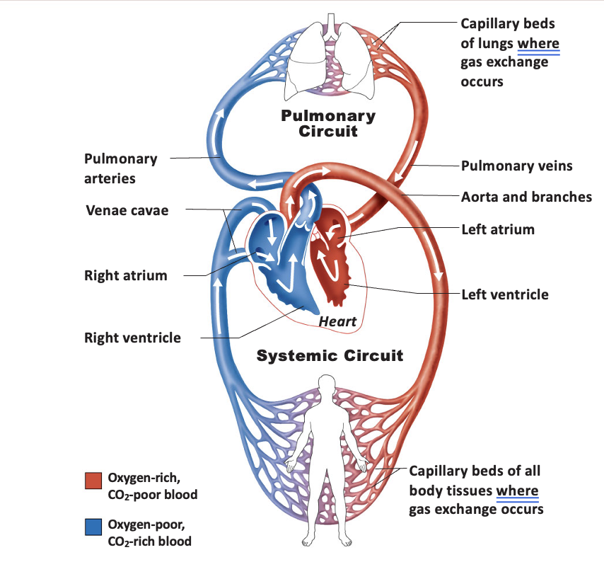
Image occlusion: name the hidden labels
9
Image occlusion: name the hidden labels
10
Image occlusion: name the hidden labels
11
Image occlusion: name the hidden labels
12
Image occlusion: name the hidden labels
13
Image occlusion: name the hidden labels
14
Image occlusion: name the hidden labels
15
Image occlusion: name the hidden labels
16
Image occlusion: name the hidden labels
17
Image occlusion: name the hidden labels
18
position of the heart
thoracic cavity
left of midline
posterior to sternum
left of midline
posterior to sternum
19
__ chambered heart
4
20
inferior pointed end of the heart is called
apex
21
posterior superior region of heart is flat and called
base
22
heart is contained within a fibrous sac called the
pericardium
23
functions of pericardium
restricts heart's movement within thorax
prevents overflow
prevents overflow
24
pericardium is attached
superiorly to the large vessels exiting the heart
inferiorly to the diaphragm
inferiorly to the diaphragm
25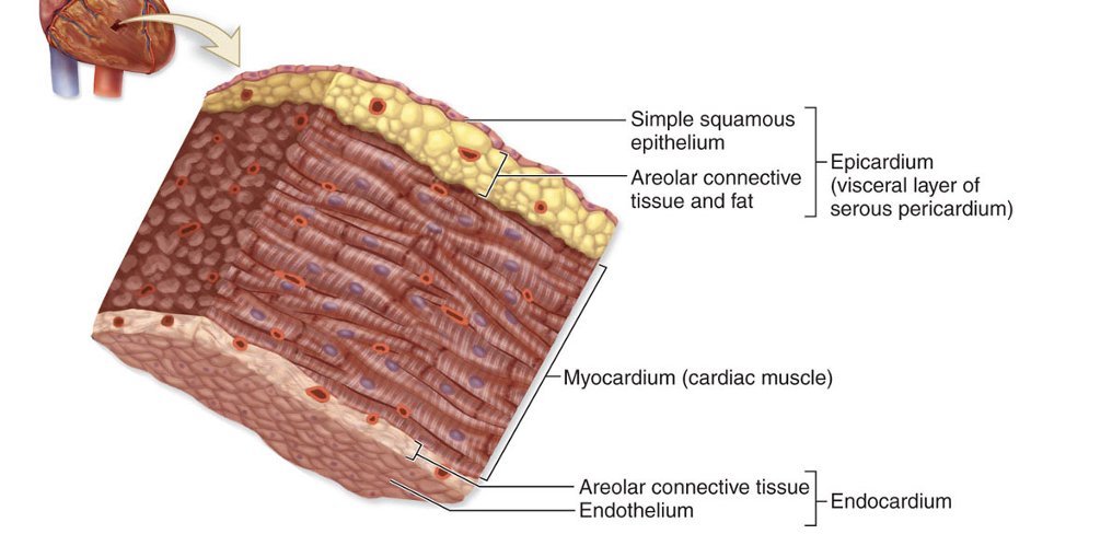
Image occlusion: name the hidden labels
26
Image occlusion: name the hidden labels
27
Image occlusion: name the hidden labels
28
Image occlusion: name the hidden labels
29
Image occlusion: name the hidden labels
30
Image occlusion: name the hidden labels
31
Image occlusion: name the hidden labels
32
each atrium has an anterior flap-like extension called an _____ that
auricle
expands their volume
expands their volume
33
the external surface of the heart has several
large grooves (sulci)
34
________ ______ rest in the sulci
coronary vessels
35
the coronary sulcus -
separates the atria from the ventricles
36
the interventricular sulci -
separate the ventricles from each other (posterior and anterior)
37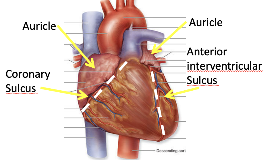
Image occlusion: name the hidden labels
38
Image occlusion: name the hidden labels
39
Image occlusion: name the hidden labels
40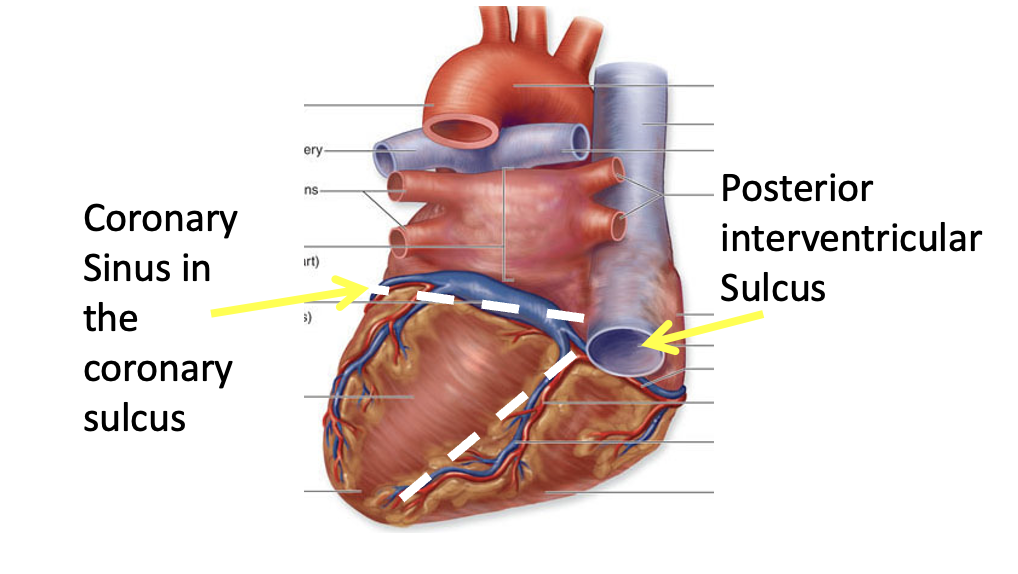
Image occlusion: name the hidden labels
41
Image occlusion: name the hidden labels
42
blood is received from the ______ _______ circulation by the ______ _______
venous systemic
right atrium
right atrium
43
3 vessels empty into the right atria:
superior vena cava
inferior vena cava
coronary sinus
inferior vena cava
coronary sinus
44
the intratrial septum is
the thin wall between the atria
has a depression called fossa ovalis
has a depression called fossa ovalis
45
fossa ovalis
hole from the fetal period that closed in the atria
46
blood leaves the right atria through _____ _____
atrioventricular (AV) opening when the AV valve opens
47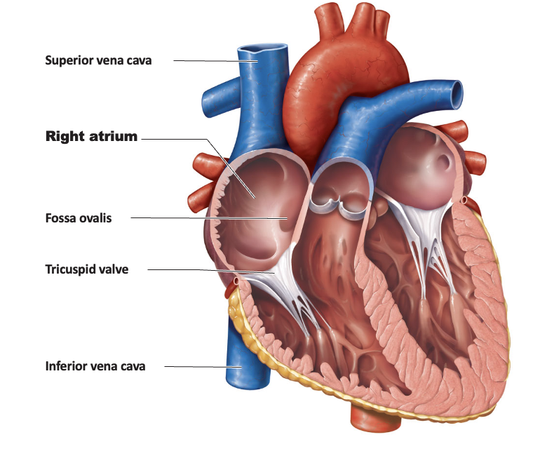
Image occlusion: name the hidden labels
48
Image occlusion: name the hidden labels
49
Image occlusion: name the hidden labels
50
Image occlusion: name the hidden labels
51
Image occlusion: name the hidden labels
52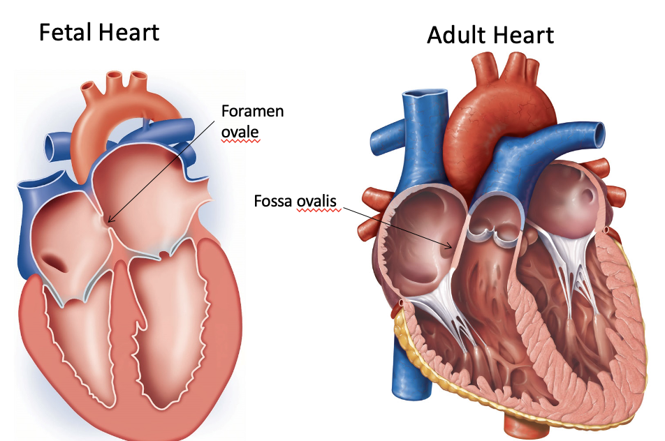
Image occlusion: name the hidden labels
53
Image occlusion: name the hidden labels
54
the right ventricle recieves blood from
the right atrium
55
the AV valve is anchored to the ventricle walls by
fibrous cords called chordae tendinae
56
chordae tendinae serve to
keep the av valve from flipping backwards when the ventricle opens
57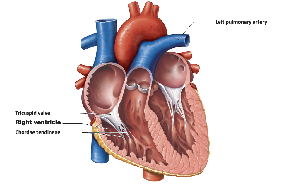
Image occlusion: name the hidden labels
58
Image occlusion: name the hidden labels
59
Image occlusion: name the hidden labels
60
Image occlusion: name the hidden labels
61
when the right ventricle contracts, it forces the blood upwards through the ______ _________ ______ into the ____________ _____
pulmonary semilunar valve
pulmonary trunk
pulmonary trunk
62
the pulmonary trunk splits into ________ ______ that
pulmonary arteries
bring deoxygenated blood to the lungs for gas exchange
bring deoxygenated blood to the lungs for gas exchange
63
the pulmonary veins carry oxygenated blood into
the left atrium
64
the left atrium also has an
auricle
65
the blood flows anterio-inferiorly from the left atrium to the ventricle through the
atrioventricular when the AV valve opens
66
The AV valve on the left is sometimes called the
bicuspid or mitral valve
67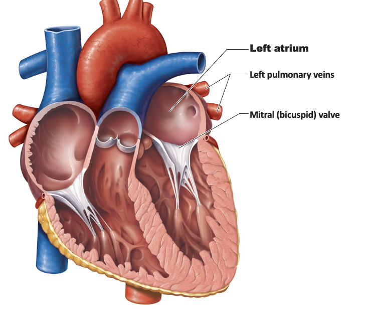
Image occlusion: name the hidden labels
68
Image occlusion: name the hidden labels
69
Image occlusion: name the hidden labels
70
the left ventricular wall is ____ than the right
thicker
71
when the left ventricle contracts, it forces blood through the
aortic semilunar valve into the aorta
72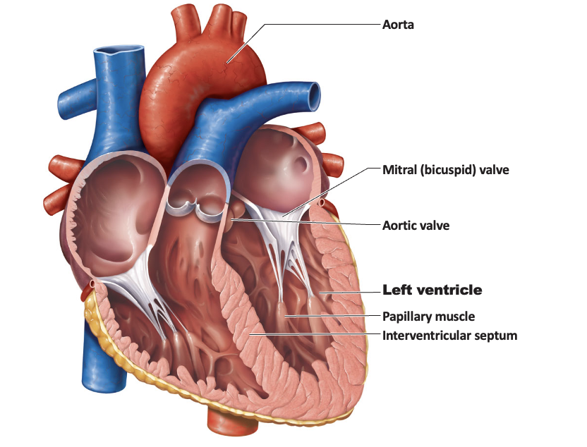
Image occlusion: name the hidden labels
73
Image occlusion: name the hidden labels
74
Image occlusion: name the hidden labels
75
Image occlusion: name the hidden labels
76
Image occlusion: name the hidden labels
77
Image occlusion: name the hidden labels
78
the left and right coronary arteries exit the aorta and feed
the cardiac muscle
79
coronary arteries branch into
progressively smaller blood vessels
80
the venous return for the heart occurs through the
cardiac veins
81
cardiac veins merge into the
coronary sinus
82
blood flows in the coronary vessels during the periods
between heart contractions
83
blood flow to the heart is impaired when the heart beats
excessively
tachycardia
tachycardia
84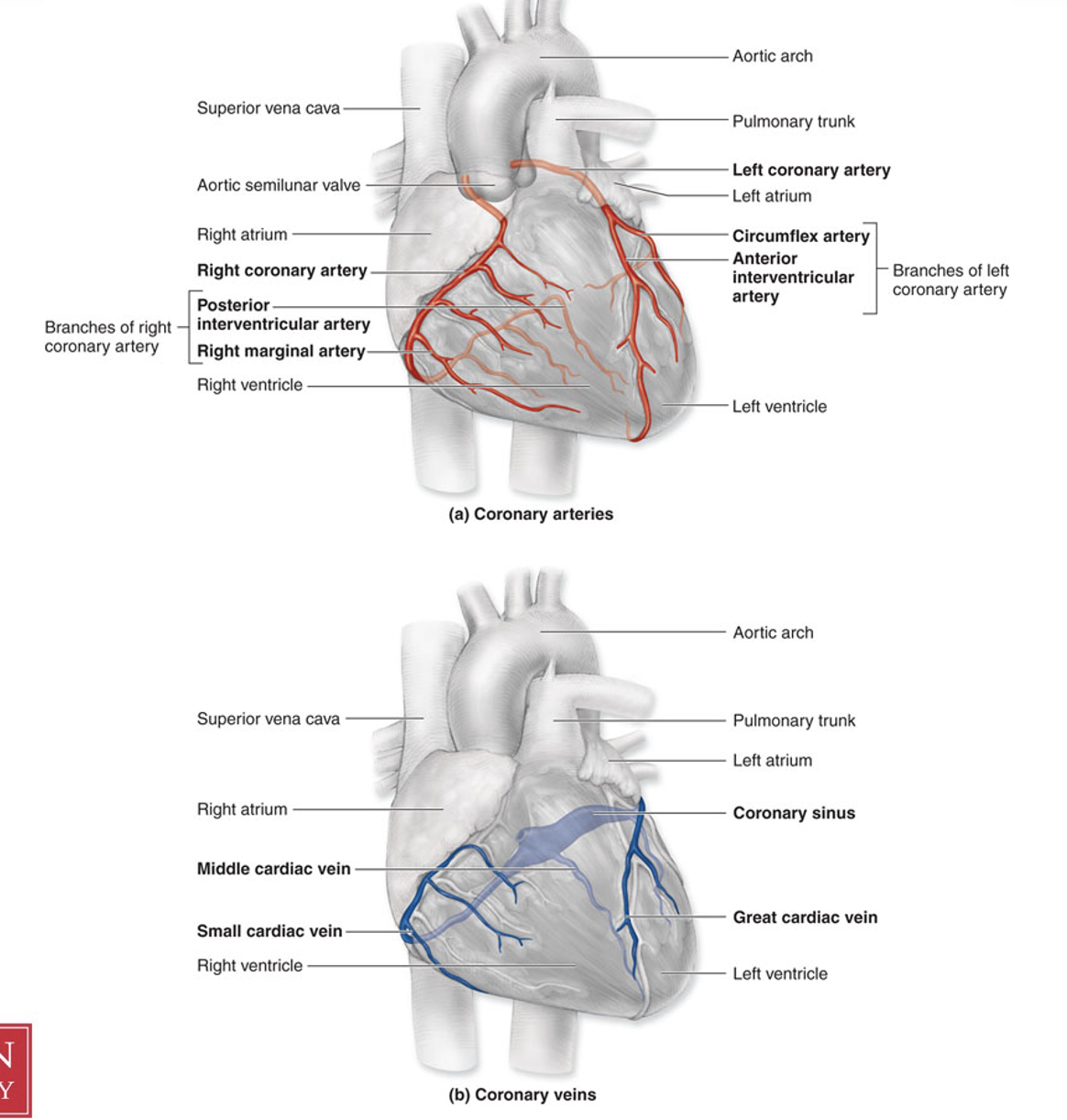
Image occlusion: name the hidden labels
85
Image occlusion: name the hidden labels
86
Image occlusion: name the hidden labels
87
Image occlusion: name the hidden labels
88
Image occlusion: name the hidden labels
89
Image occlusion: name the hidden labels
90
Image occlusion: name the hidden labels
91
Image occlusion: name the hidden labels
92
Image occlusion: name the hidden labels
93
Image occlusion: name the hidden labels
94
Image occlusion: name the hidden labels
95
Image occlusion: name the hidden labels
96
Image occlusion: name the hidden labels
97
Image occlusion: name the hidden labels
98
Image occlusion: name the hidden labels
99
three main types of blood vessels
arteries
capillaries
veins
capillaries
veins
100
progression of blood vessels away from the heart
elastic arteries
muscular arteries
arterioles
capillaries
muscular arteries
arterioles
capillaries
101
elastic capillaries
due to fluctuations in pressure between heart contractions and rest, these arteries have an extremely high proportion of elastin in their walls
102
muscular arteries
increased proportion of muscle for vasodilation and constriction
103
arterioles and capillaries
vessels continue to branch, increasing surface area covered while each individual vessel decreases in diameter
104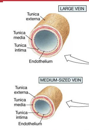
Image occlusion: name the hidden labels
105
Image occlusion: name the hidden labels
106
Image occlusion: name the hidden labels
107
Image occlusion: name the hidden labels
108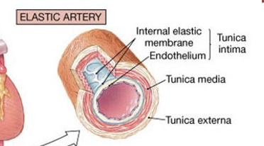
Image occlusion: name the hidden labels
109
Image occlusion: name the hidden labels
110
Image occlusion: name the hidden labels
111
Image occlusion: name the hidden labels
112
Image occlusion: name the hidden labels
113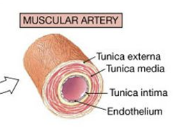
Image occlusion: name the hidden labels
114
Image occlusion: name the hidden labels
115
Image occlusion: name the hidden labels
116
Image occlusion: name the hidden labels
117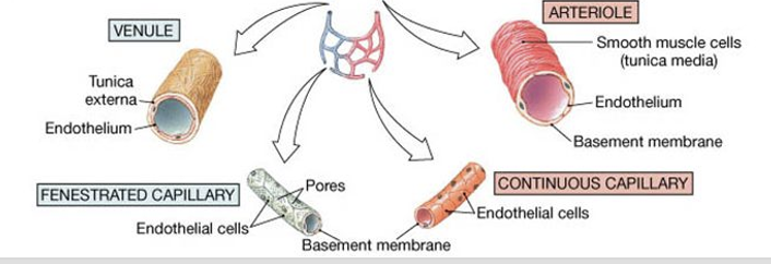
Image occlusion: name the hidden labels
118
Image occlusion: name the hidden labels
119
Image occlusion: name the hidden labels
120
Image occlusion: name the hidden labels
121
Image occlusion: name the hidden labels
122
Image occlusion: name the hidden labels
123
progression of blood vesse;s toward the heart
venules -> veins
vessels merge together, decreasing surface area covered and increasing in diameter
no significant changes to structure of vessel walls
vessels merge together, decreasing surface area covered and increasing in diameter
no significant changes to structure of vessel walls
124
precapillary sphincter-ring of muscle that
controls bloodflow
125
sphincter opens when the tissue is ______ and close when _____
low in O2
sufficient O2
5-10 minute cycles
sufficient O2
5-10 minute cycles
126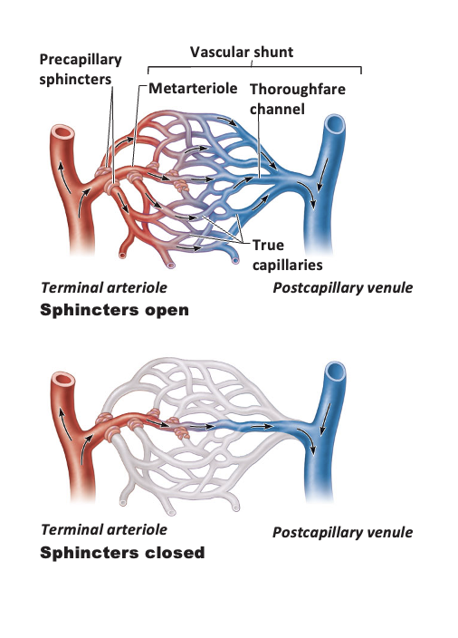
Image occlusion: name the hidden labels
127
Image occlusion: name the hidden labels
128
Image occlusion: name the hidden labels
129
Image occlusion: name the hidden labels
130
Image occlusion: name the hidden labels
131
Image occlusion: name the hidden labels
132
Image occlusion: name the hidden labels
133
portal systems
capillary beds connected without blood returning to the heart
134
anastomoses
many parts of the body receive blood from branches of more than 1 artery
places where these branches connect are called anastomoses
places where these branches connect are called anastomoses
135
veins drain ______ and return
venules
deoxygenated blood to the heart
deoxygenated blood to the heart
136
compared to arteries, vein walls are ____ and lumens are ______
thinner, and lumens are larger
137
blood pressure in veins is greatly ____ so there is ____ blood in veins than arteries
reduced
more (travels slower)
more (travels slower)
138
blood pressure in veins is too low to
overcome gravity
139
2 adaptations for veins
valves
muscle pumps
muscle pumps
140
valves in veins
prevent backflow down the vein
141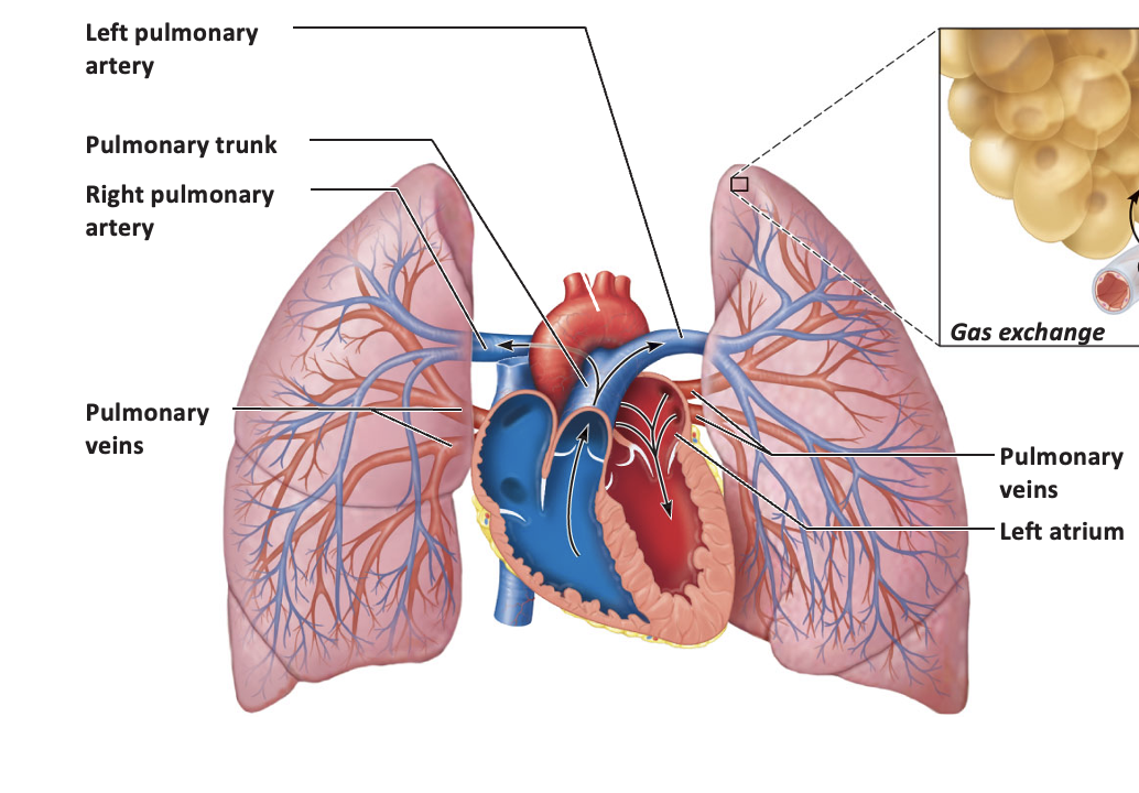
Image occlusion: name the hidden labels
142
Image occlusion: name the hidden labels
143
Image occlusion: name the hidden labels
144
Image occlusion: name the hidden labels
145
Image occlusion: name the hidden labels
146
Image occlusion: name the hidden labels
147
the coronary arteries branch immediately after the
SL valve
148
the aorta curves and becomes the
aortic arch
149
the aortic arch turns downward and is called the
descending aorta
150
the descending aorta continues and becomes
the descending abdominal aorta
151
oxygenated blood leaving the left ventricle needs to go
up to the head
down to the abdomen and beyond
out to the arms
down to the abdomen and beyond
out to the arms
152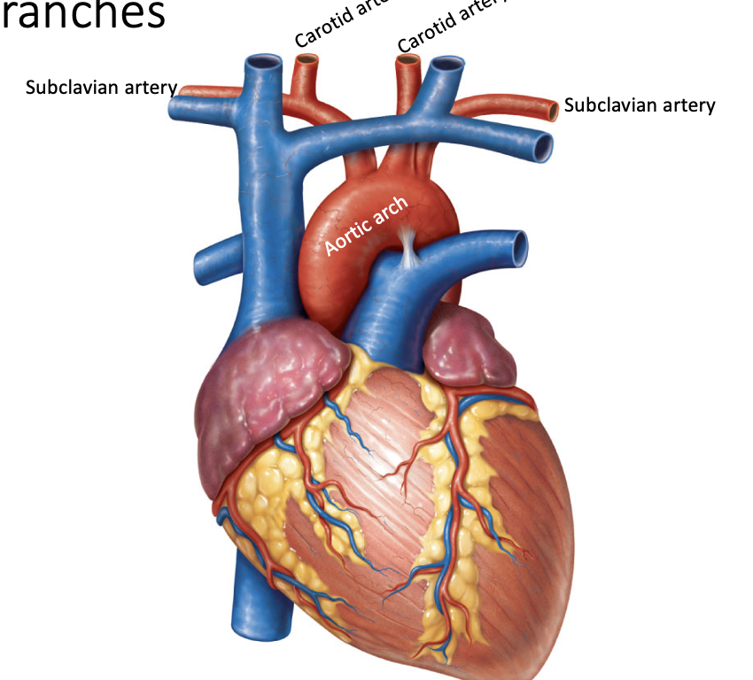
Image occlusion: name the hidden labels
153
Image occlusion: name the hidden labels
154
Image occlusion: name the hidden labels
155
Image occlusion: name the hidden labels
156
Image occlusion: name the hidden labels
157
carotid arteries bring blood
up to the brain and face
158
subclavian arteries pass under the _____ and bring blood
the clavicle and bring blood toward the arm
159
a short primary vessel before it divides into two is called a
trunk
160
leftmost branch (rightmost anatomically) off the aorta is called
brachiocephalic trunk
161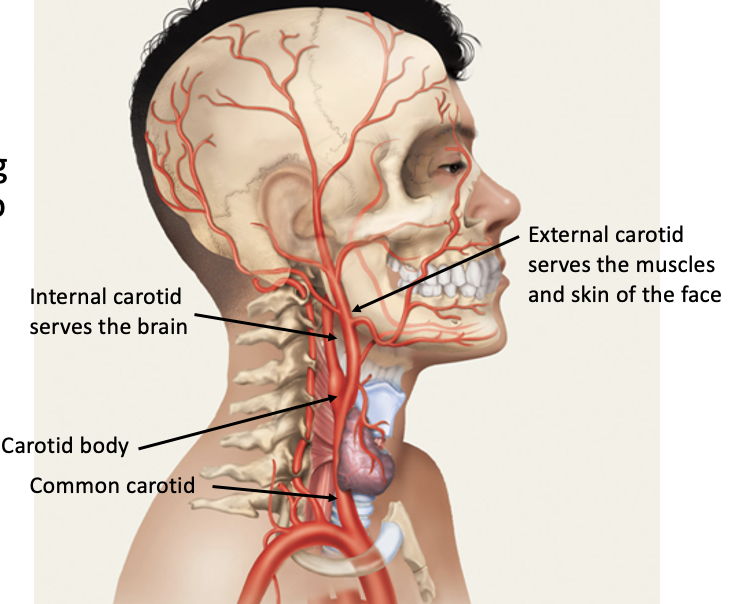
Image occlusion: name the hidden labels
162
Image occlusion: name the hidden labels
163
Image occlusion: name the hidden labels
164
Image occlusion: name the hidden labels
165
external carotid serves the
muscles and skin of the face
166
internal carotid serves
the brain
167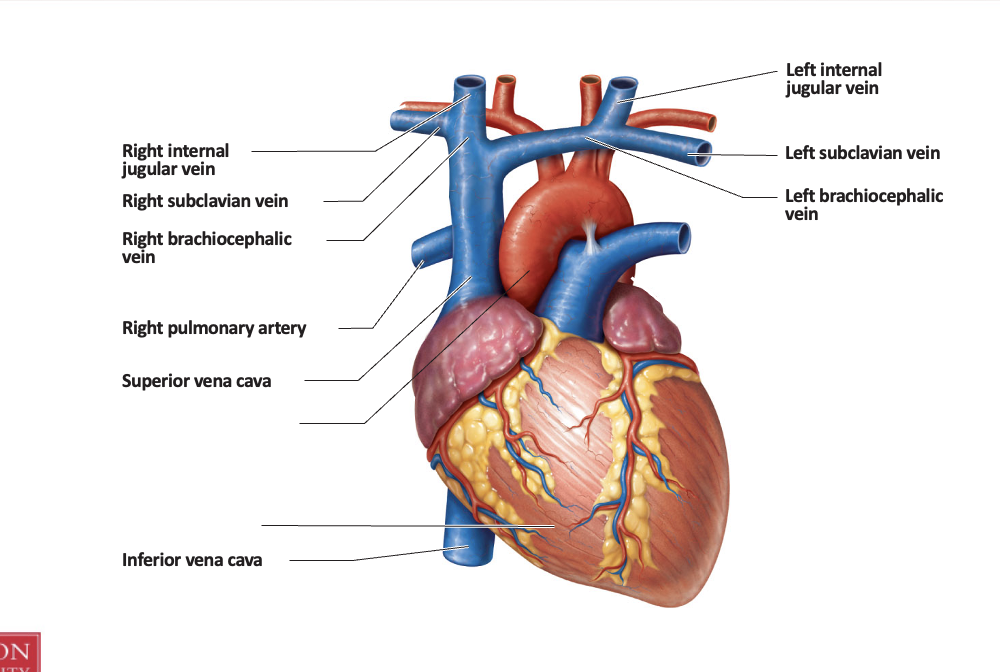
Image occlusion: name the hidden labels
168
Image occlusion: name the hidden labels
169
Image occlusion: name the hidden labels
170
Image occlusion: name the hidden labels
171
Image occlusion: name the hidden labels
172
Image occlusion: name the hidden labels
173
Image occlusion: name the hidden labels
174
Image occlusion: name the hidden labels
175
Image occlusion: name the hidden labels
176
R internal jugular and subclavian veins merge to become
R brachiocephalic vein
177
L internal jugular and subclavian veins merge to become
L brachiocephalic vein
178
external jugular vein drains the
face and muscles
179
blood from the brain descends in the
internal jugular vein
180
independently the external and internal jugular veins join the _____ vein which joins the
subclavian
superior vena cava
superior vena cava
181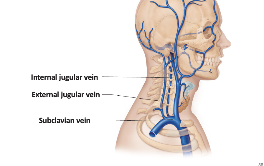
Image occlusion: name the hidden labels
182
Image occlusion: name the hidden labels
183
Image occlusion: name the hidden labels
184
thoracic aorta becomes the
abdominal aorta
185
branches off the subclavian artery supply the
head and inferior neck
186
as subclavian arteries approach the axilla, the artery is now called the
axillary artery
187
as axillary reaches the arm, its is called the
brachial artery
188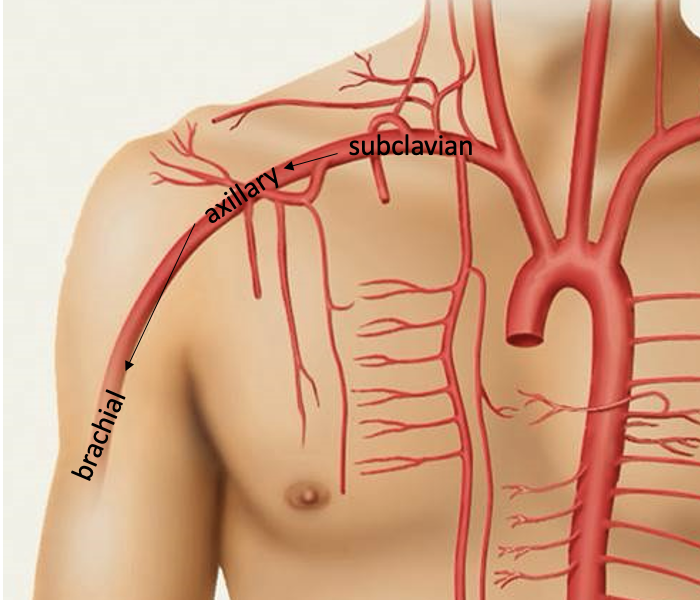
Image occlusion: name the hidden labels
189
Image occlusion: name the hidden labels
190
Image occlusion: name the hidden labels
191
as the thoracic aorta descends through the thorax, the aorta divides into
visceral and parietal branches
192
visceral branches of the thoracic aorta serve
thoracic organs
193
parietal branches of the thoracic aoirta serve the
body wall
194
the thoracic aorta becomes the abdominal aorta as it passes through the
hiatus of the diaphragm
195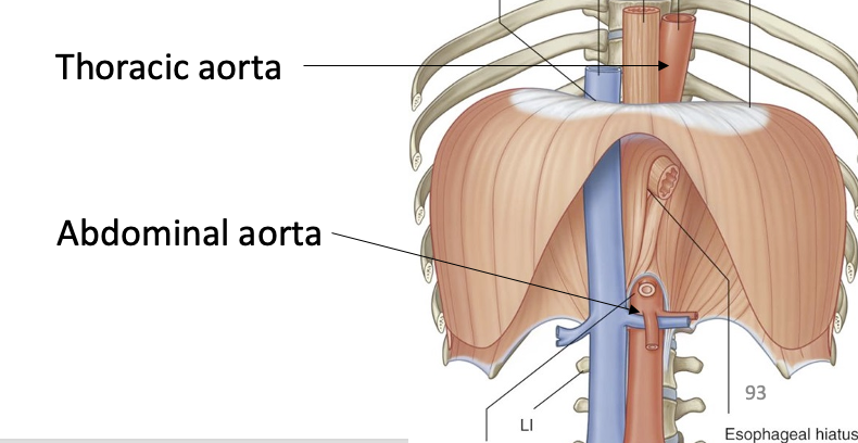
Image occlusion: name the hidden labels
196
Image occlusion: name the hidden labels