Lecture 4 - Autoimmunity Pt2
Study these flashcardsFree · spaced repetition · iPhone, Android, Mac & web
All cards
1
What is self tolerance?
Ability of immune system to not mount immune response to self antigens
2
How does the immune system prevent an attack on self?
By establishing self tolerance so the immune system does not respond to self antigens
3
What are the two types of tolerance to self?
Central Tolerance and Peripheral Tolerance
4
Where is central tolerance established?
Bone Marrow and Thymus
5
Where is peripheral tolerance established?
Peripheral Tissues
6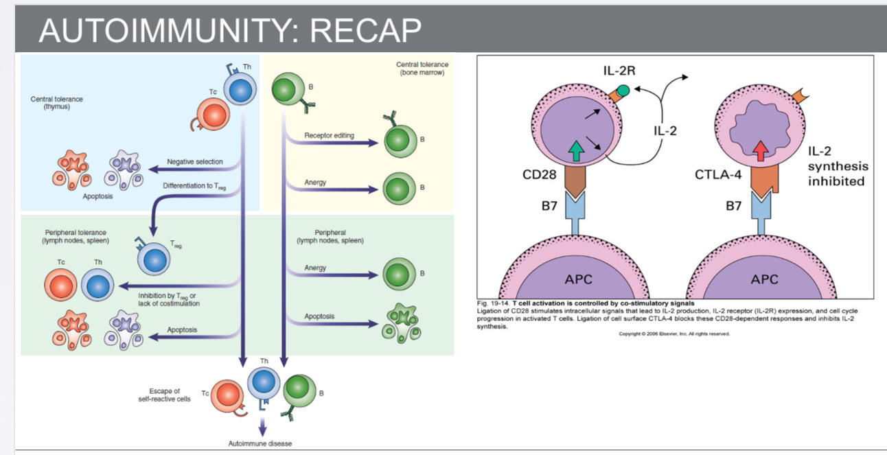
7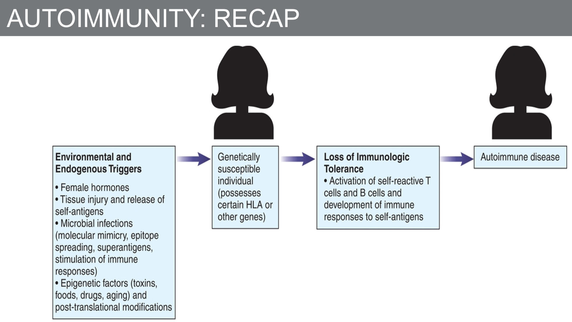
8
know this 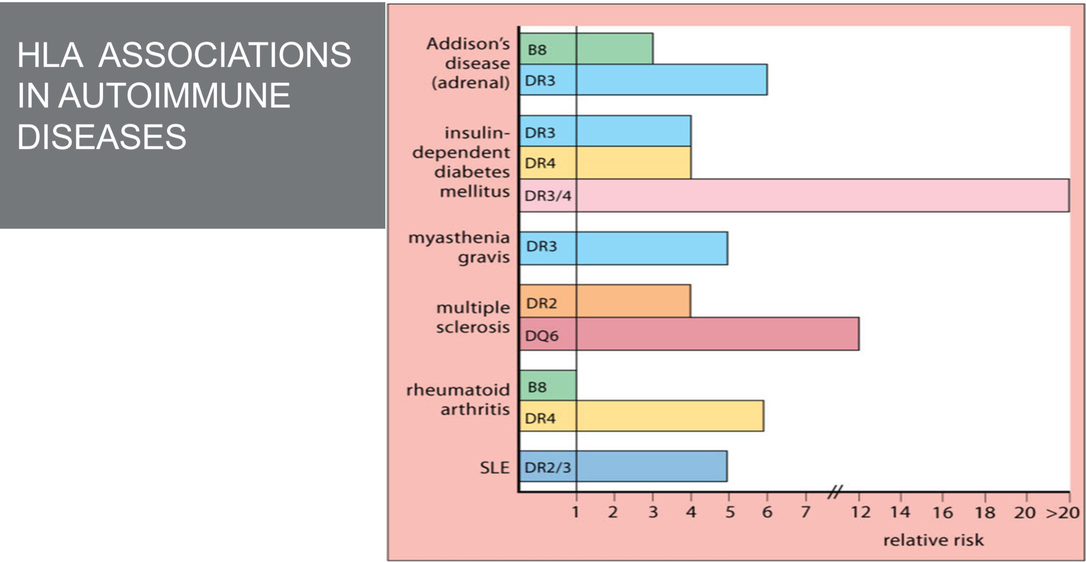
9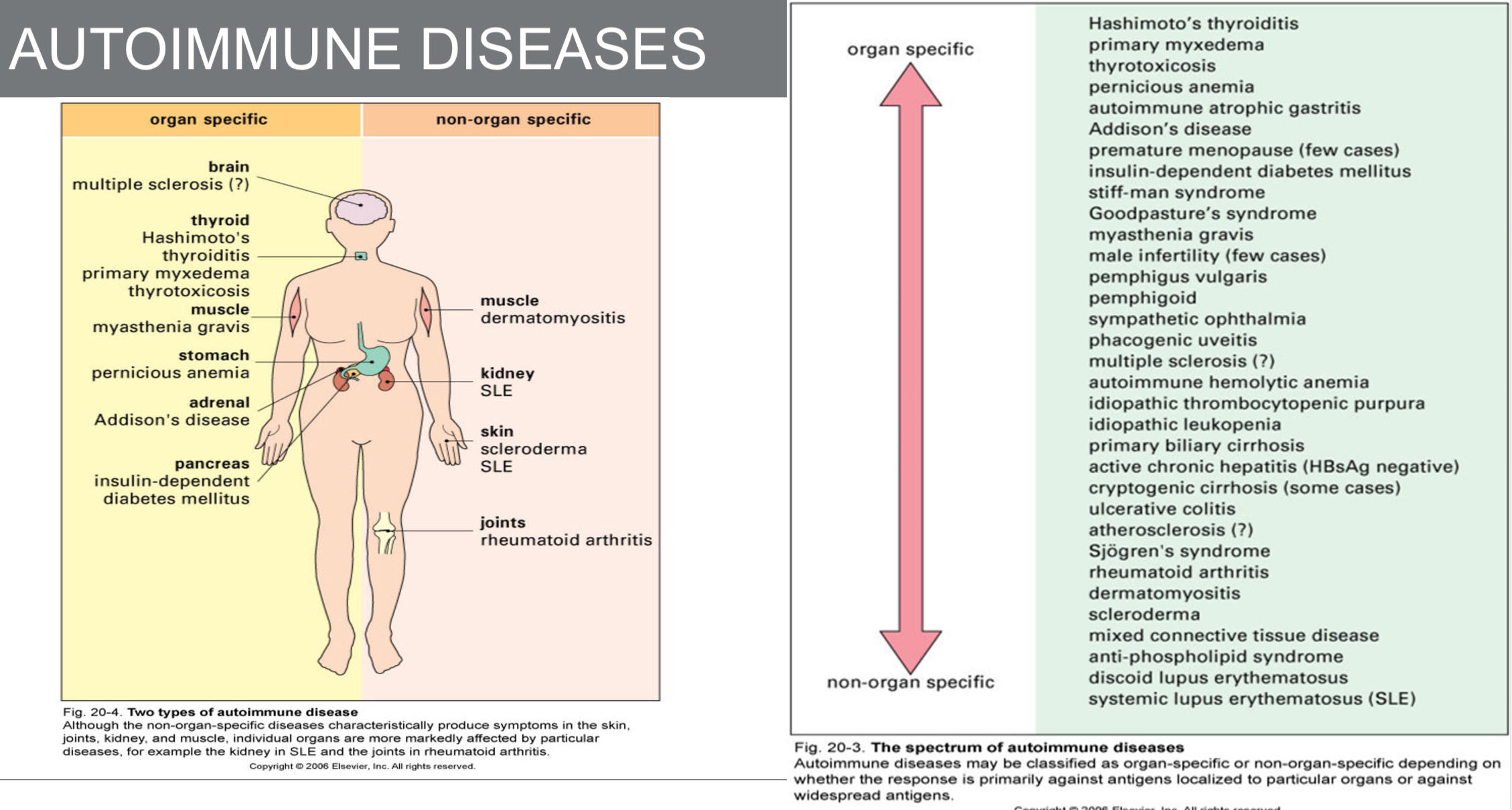
10
What is organ-specific autoimmunity?
Immune response to self antigens of a specific organ
11
What does organ-specific autoimmunity cause?
Damage to organ structure and function
12
What is non-organ specific autoimmunity?
Systemic–auto Ag found on any type of cell throughout body (DNA)
13
What does non-organ specific autoimmunity involve?
Widespread attack of self antigens
14
What damage occurs in non-organ specific autoimmunity?
Damage to numerous structures
15
How is autoimmune activity distributed in non-organ specific autoimmunity?
Autoimmune activity is widely spread throughout the body
16
What do systemic diseases affect and how many major groups are included?
They affect multiple organs and tissues and include two important groups; Systemic autoimmune rheumatic diseases (SARDs) and Anti-neutrophil cytoplasmic antibodies (ANCA).
17
What are systemic autoimmune rheumatic diseases (SARDs)?
Diseases that involve inflammation of the joints and their associated structures
18
What are Anti-neutrophil cytoplasmic antibodies (ANCA)?
Associated vasculitides, which are characterized by inflammation of the blood vessels
19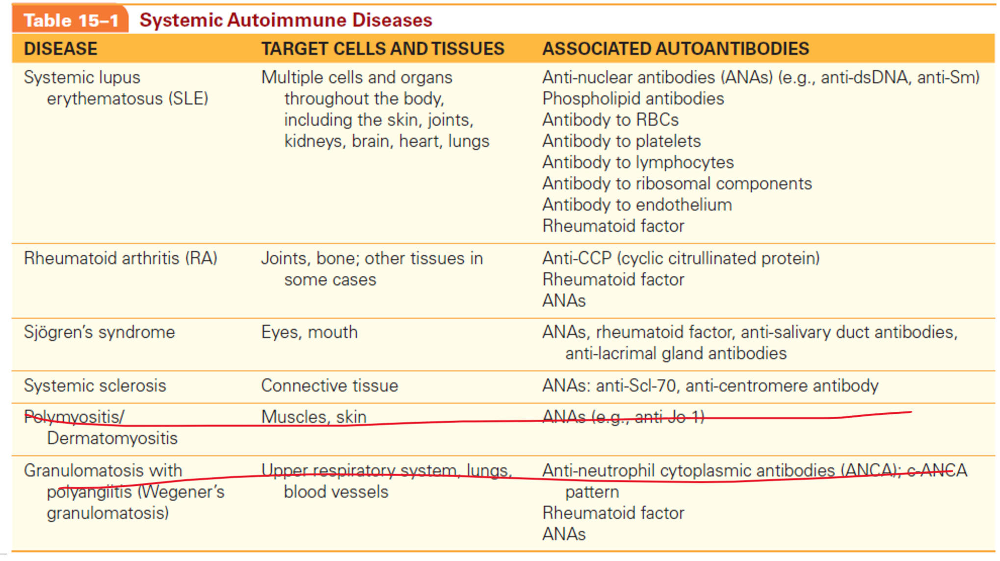
20
How is Systemic Lupus Erythematosous (SLE) described?
A chronic inflammatory disease, relapsing, flares correlates with disease activity
21
What type of autoimmune condition is SLE?
A multisystem autoimmune disease
22
Who is most affected by SLE?
Women more than men (9:1 ratio), with 90% of cases being women
23
Which racial group is more affected by SLE?
African Americans more than Whites
24
When does SLE usually onset?
Between ages 15 and 45 years
25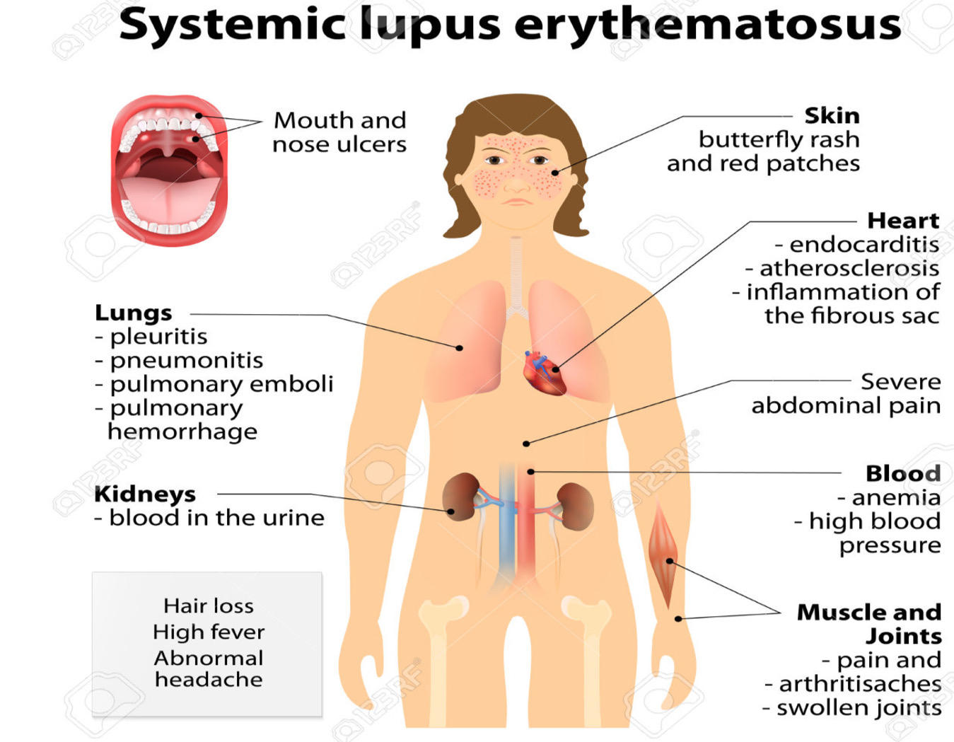
26
What interactions contribute to SLE etiology?
Environmental factors, genetic susceptibility, and abnormalities within the immune system
27
What environmental factors are linked to SLE?
Some drugs (procainamide, isoniazid, hydralazine), UV exposure, infections, hormones (estrogen-containing contraceptives)
28
What genetic makeup, findings are associated with SLE?
>60 genetic loci associated with lupus in humans have been discovered including HLA-A1, B8, and DR3
29
What immune system abnormalities contribute to SLE?
Defects in CRs on phagocytic cells, defects in FcRs, and deficiencies in C1q, C2, or C4; polymorphisms in cytokine genes, etc
30
What autoantibodies occur in SLE?
Over 100 autoantibodies (dsDNA, histones, nucleoli, lymphocytes, erythrocytes, platelets, phospholipids, ribosomal components, and endothelium)
31
What is typical regarding autoantibodies in SLE patients?
The typical patient has several circulating autoantibodies, which can precede disease onset by several years (critical role of B cells and Abs)
32
What is the significance of anti-dsDNA?
Found in >70% of patients and highly specific
33
What immune and cellular targets are affected in SLE?
Impaired immune complex clearance (IC deposit on tissues) and autoantibodies against RBCs, PLTs, and endothelial cells
34
What pregnancy-related and coagulation issues are linked to SLE?
Anti-phospholipid antibodies (associated with increased miscarriage, stillbirth, and preterm delivery in pregnant women with lupus) and lupus anticoagulant
35
What conditions are included in the SLE diagnostic classification (at least 4 conditions present)?
Butterfly rash; discoid lupus of head and neck with scarring and disfigurement; photosensitivity with UV-induced DNA damage; oral ulcers; arthritis; serositis; neurologic; hematologic; renal involvement; autoantibodies; cardiopulmonary
36
What renal and immunologic findings are SLE criteria?
Renal involvement is present in about half of lupus patients; nephritis is a major cause of illness and death
37
What are the most common causes of death in lupus patients?
Renal failure and infection, followed by heart disease
38
What is included in the initial evaluation for SLE?
CBC, platelet count, creatinine levels, and urinalysis to assess renal function; it may show leukopenia, possible thrombocytopenia and anemia; ESR may also be high, but CRP may be low or normal
39
What is the most important screening test if SLE is suspected?
ANA will be done which is the most important screening test
40
What should be done after ANA screening is positive?
Further tests should be done to differentiate the type of ANA
41
What antibodies are highly specific for SLE?
Antibodies to dsDNA and antibodies to the Sm nuclear antigen are highly specific for SLE
42
43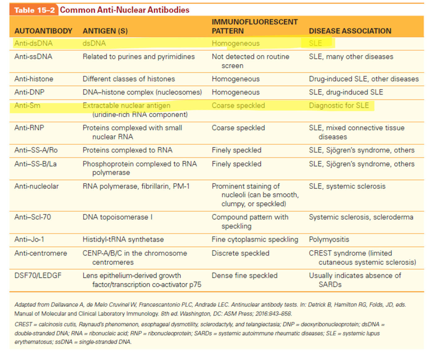
44
What is the gold standard ANA detection method?
Indirect Immunofluorescence (IIF)
45
What is FANA testing and why is it widely used and accepted?
Fluorescent anti-nuclear antibody (FANA) testing has been the most widely used and accepted test because it is highly sensitive, detects a wide range of antibodies, and is inexpensive and easy to perform
46
How is the ANA test performed using the indirect immunofluorescence method?
A commercially prepared slide with fixed HEp2 cell line; incubation with patient’s serum (serial dilutions, starting with 1:80 or 1:160), ANA will bind to HEp2 nucleus; wash to remove unbound Ab molecules; add anti-human Ig labeled with a fluorescent dye and incubate; wash again to remove unbound Ab; put a cover slip, mount the slide and view it under microscope (400× magnification); titers above 160 are clinically significant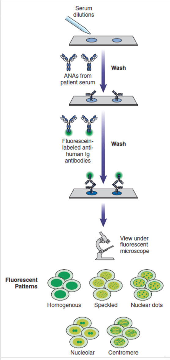
47
What is the homogeneous ANA fluorescence pattern and what is it associated with?
Homogeneous—This pattern is characterized by uniform staining of the entire nucleus in interphase (nondividing) cells and of the condensed chromosomal region in metaphase cells. It is associated with antibodies to dsDNA, histones, and nucleosomes. The homogeneous pattern is found in patients with SLE, drug-induced lupus, chronic autoimmune hepatitis (AIH), and Juvenile idiopathic arthritis.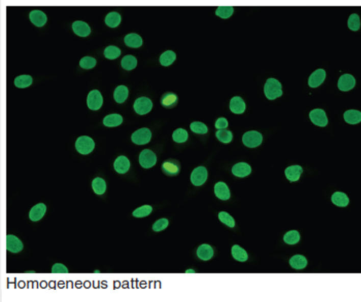
48
What describes the speckled ANA fluorescence pattern and its associations?
Speckled—These patterns are characterized by discrete, fluorescent specks throughout the nuclei of interphase cells. The speckled group is divided into three subgroups depending on whether the specks are dense fine, tiny/fine, or large/coarse. The fine and coarse speckled patterns are associated with antibodies to ENAs and can be found in patients with SLE, Sjögren’s syndrome, systemic sclerosis (SSc), and other SARDs.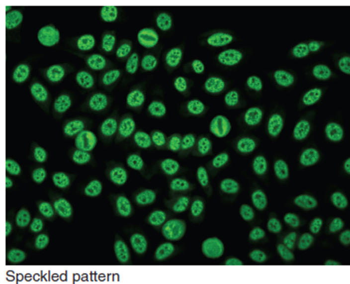
49
What describes the nucleolar ANA fluorescence pattern and what antibodies cause it?
Nucleolar—Prominent staining of the nucleoli within the nuclei of interphase cells is seen in this pattern. The size, shape, and number of the nucleoli per cell are variable, and staining can be smooth, clumpy, or speckled, depending on the type of antibody present. Staining may or may not be present in the dividing cells. The nucleolar patterns are primarily caused by antibodies to RNA and RNP and are seen mainly in patients with SSc but can also be present in patients with other SARDs.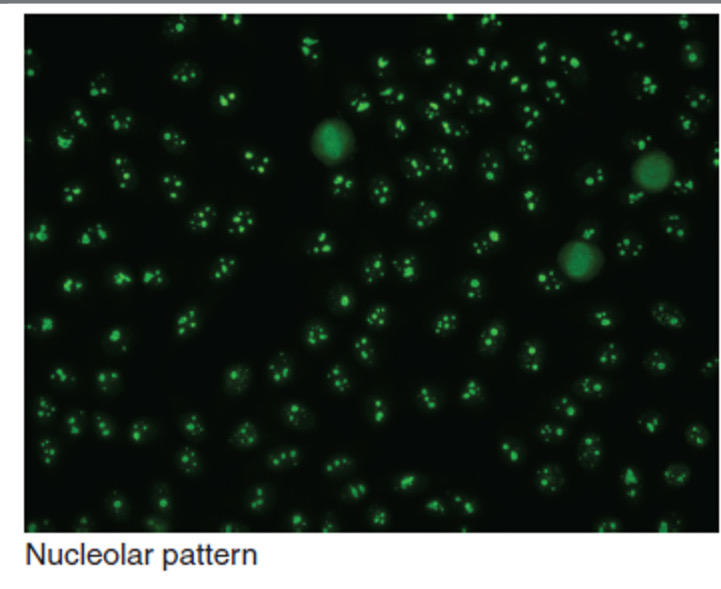
50
What are the solid-phase immunoassay methods used for ANA detection?
ELISA, chemiluminescence immunoassay (CLIA), and microsphere multiplex immunoassay [MIA].
51
What materials are used and what is the first step in these assays?
Polystyrene microtiter plates, nitrocellulose strips, or synthetic beads coated with auto-Ags; incubation with patient’s serum.
52
How are antibodies detected in these solid-phase ANA methods?
Detection by enzyme/fluo/chemilum tagged AHG.
53
What antibodies can these methods identify?
Antibodies to a range of Ags can be identified: dsDNA, Sm, U1-RNP, SS-A/Ro, SS-B/La, Jo-1, Scl-70, PM/Scl, Mi-2, CENP-B, and histones.
54
What are the advantages and disadvantages of these solid-phase methods?
Advantage: automation, ease of performance, and yielding objective results. Disadvantage: sensitivity lower than IIF, false positive results.
55
What ANA detection method uses Crithidia luciliae and what does it detect?
Immunofluorescence Using Crithidia luciliae. This IIF assay is used to detect antibodies to dsDNA.
56
What organism is used as the substrate in this method?
A hemoflagellate organism called Crithidia luciliae as the substrate.
57
What unique structure does this organism contain and what is it composed of?
This trypanosome has a circular organelle called a kinetoplast that is composed mainly of dsDNA.
58
How is the Crithidia luciliae test performed?
In this test, patient serum is incubated on a microscope slide coated with C. luciliae organisms, and binding is detected with a fluorescent-labeled anti-Ig conjugate; washing of the slide is performed after each step to remove unbound antibody.
59
What indicates a positive test in this method?
A positive test is indicated by a brightly stained kinetoplast.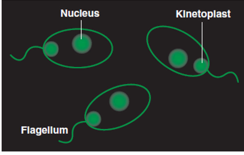
60
What is the Ouchterlony test used for in ANA detection?
Detection of ANAs by immunodiffusion in agarose gel.
61
How can the Ouchterlony test be applied clinically?
Can be used to determine the immunologic specificity of a positive FANA test, particularly when a speckled pattern is observed.
62
What does the Ouchterlony test detect?
It detects antibody to several ENAs including antibodies to Sm, RNP, SS-A/Ro, SS-B/La, Scl-70 (DNA topoisomerase I), and Jo-1.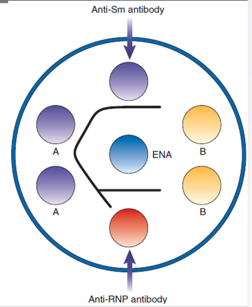
63
What is serological identity?
If the antigens are identical, they will react with the same antibody, and the precipitate line forms a continuous arc.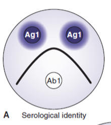
64
What is nonidentity?
If the antigens share no identical determinants, they will react with different antibodies, and two crossed lines are formed.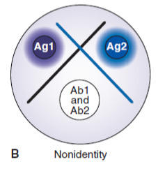
65
What is partial identity?
Partial identity. Antibodies react with two similar antigens, forming two lines and a spur that points to the simpler antigen (the antigen that reacts least with the antibody).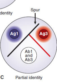
66
Give an example of Partial Identity.
For example, suppose antigen 3 has an epitope in common with antigen 1, and there are two antibodies in the center well (anti-1 and anti-3). One of the antibodies reacts with the epitope common to both antigens. The other antibody reacts with a different epitope on antigen 1 (that is absent on antigen 3). Two precipitin lines form, with a spur pointing toward antigen 3.
67
What genetic associations are linked to rheumatoid arthritis (RA)?
Association with >100 genetic regions; the strongest associations with specific HLA-DRB1 alleles or PTPN22 gene polymorphisms. These patients are positive for rheumatoid factor (RF) or antibodies to cyclic citrullinated peptides (CCP).
68
What epigenetic factors contribute to RA?
Epigenetic factors, such as altered DNA methylation affecting the transcription of critical genes, are also believed to play a role in the pathogenesis of RA.
69
What is the strongest environmental risk factor for RA?
The strongest environmental risk factor for RA is believed to be cigarette smoking, which doubles the risk of developing the disease.
70
What is the immunopathology of rheumatoid arthritis (RA) caused by?
Caused by an inflammatory process that results in the destruction of bone and cartilage in the joints.
71
What does infiltration of the inflamed synovium indicate in RA?
Infiltration of the inflamed synovium with T and B lymphocytes, plasma cells, dendritic cells, mast cells, and granulocytes is evidence of immunologic activity within the joint.
72
What cytokine imbalance contributes to RA inflammation?
The balance between proinflammatory and anti-inflammatory cytokines in RA appears to be tipped toward continual inflammation: IL-1, IL-6, IL-17 and TNF-α (TNF-α plays the key role).
73
What do these cytokines trigger in RA?
These cytokines trigger the release of matrix metalloproteinases from fibroblasts and macrophages; these enzymes degrade important structural proteins in the cartilage.
74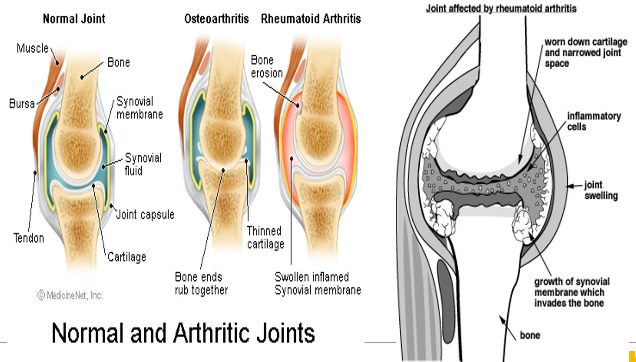
75
What immunopathology finding is noted in RA?
Bone lesions due to hyperactivation of osteoclasts.
76
What are the two key antibodies in RA?
Rheumatoid factor (RF), an IgM against FC portion of IgG, enhances activity of macrophages and Ag presentation to APCs; Anti-cyclic citrullinated peptide antibody [anti-CCP or ACPA].
77
What are citrullinated proteins and why are they important in RA?
Citrullinated proteins released from dead granulocytes or macrophages; overexpression of these antigens may provoke an immune response in individuals with certain HLA-DRB1 alleles.
78
What is the initial lab test for RA and what are key points about RF results?
Initial test is RF, positive in 70–90%; negative RF does not exclude RA; positive RF is not specific to RA; RF is also positive in about 5% of healthy individuals and in 10% to 25% of those over the age of 65.
79
Where else can RF be found outside of RA?
RF can be found in patients with other SARDs, such as SLE, Sjögren’s syndrome, scleroderma, as well as in people with some chronic infections.
80
How is RF testing performed and what does it detect?
RF is done using latex agglutination on a slide, detects only IgM isotype.
81
What methods are more sensitive and specific than latex RF testing and what do they detect?
ELISA and nephelometry are more sensitive and precise, they detect IgG isotype (more specific).
82
What role does anti-CCP play in RA diagnosis?
Anti-CCP is done using ELISA, great test for early diagnosis where RF may be negative; a large number of RF-negative RA patients are anti-CCP positive.
83
What is Sjögren’s syndrome and what symptoms are typical?
Chronic inflammation of the exocrine glands (eyes, saliva); most individuals with Sjögren syndrome present with sicca symptoms, such as xerophthalmia (dry eyes), xerostomia (dry mouth), and parotid gland enlargement.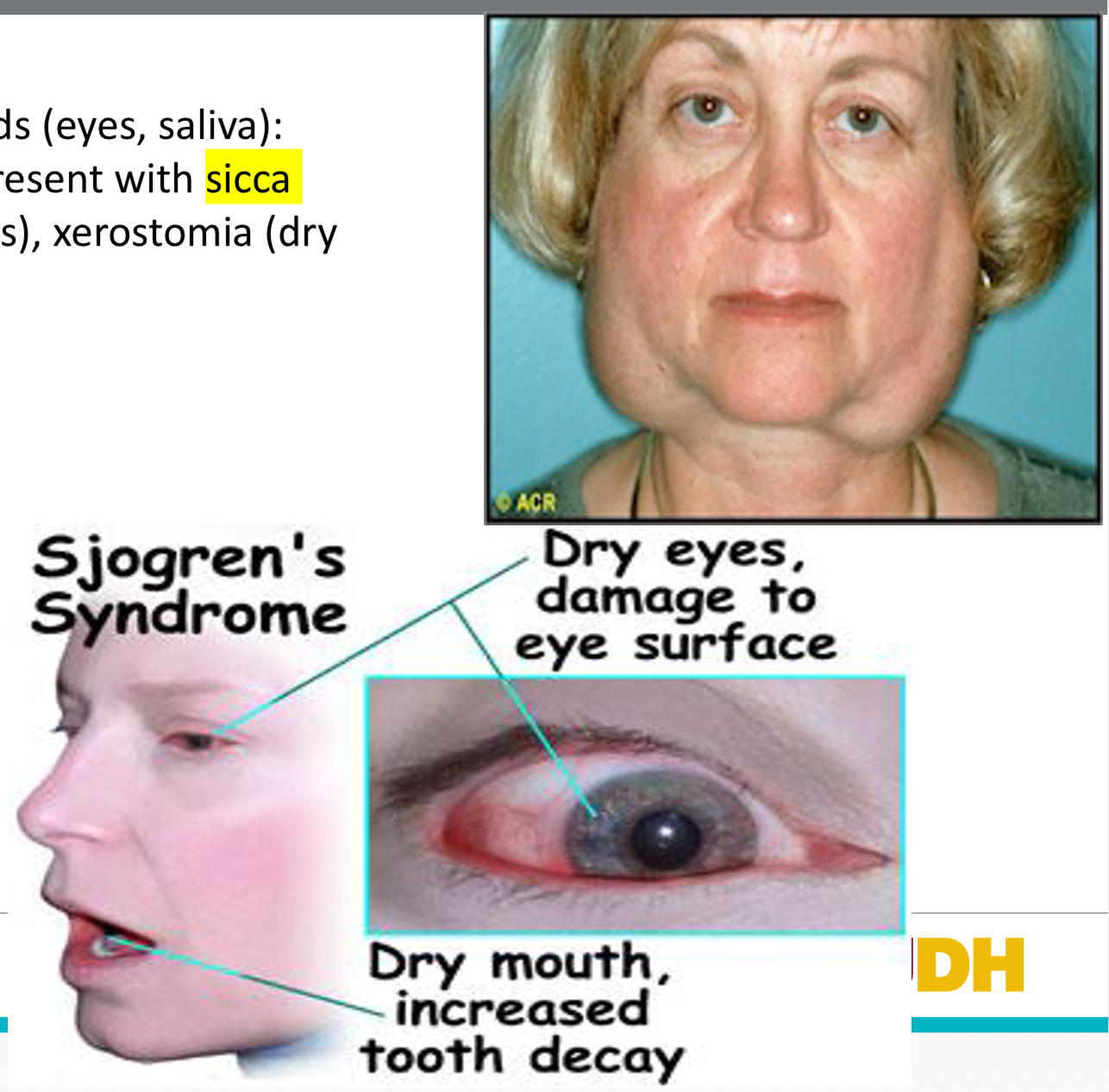
84
What is primary Sjögren’s syndrome?
Primary Sjögren’s syndrome occurs in the absence of other diseases.
85
What is secondary Sjögren’s syndrome?
Secondary Sjögren’s syndrome occurs in patients who also have other diseases, most commonly SLE or RA.
86
What serologic findings are common in primary Sjögren’s syndrome?
The majority of patients with the primary form of the disease test positive for RF and ANAs, exhibiting a fine speckled pattern of fluorescence that is indicative of anti–SS-A/Ro and/or anti–SS-B/La.
87
What is SSc (scleroderma) and how does it present?
SSc, also known as scleroderma, is a rare SARD: excessive fibrosis and vascular abnormalities that affect the skin and joints and progress over time to involve internal organs.
88
What symptoms are associated with SSc?
Symptoms: hardening of the skin, musculoskeletal pain, Raynaud’s phenomenon, and heartburn.
89
What clinical combination defines CREST syndrome?
The combination of calcinosis cutis, Raynaud’s phenomenon, esophageal dysmotility, sclerodactyly, and telangiectasia has been referred to as the CREST syndrome.
90
What is the major consequence of internal organ involvement in SSc?
Complications involving the internal organs often result in functional impairment, leading to death.
91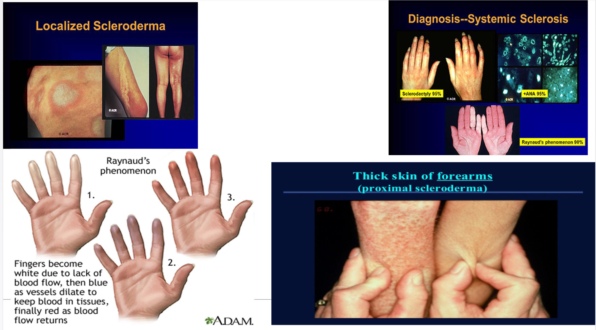
92
What does the slide say about the number of ANAs in SSc and which are useful?
At least 10 different types of ANAs have been identified in patients with SSc. Three of these are useful to determine the diagnosis, subgrouping, and prognosis.
93
What do anti-centromere antibodies indicate?
Anti-centromere antibodies are found only in patients with limited cutaneous SSc and correlate with a better survival rate.
94
What do antibodies to topoisomerase I (Scl-70) indicate?
Antibodies to topoisomerase I (also known as Scl-70) are associated with diffuse cutaneous SSc and a poor prognosis.
95
What do antibodies to RNA polymerase III indicate?
Antibodies to RNA polymerase III are associated with the diffuse form of the disease and rapid thickening of the skin but have a better prognosis than those with anti-Scl-70.
96
What autoantibody finding characterizes Mixed Connective Tissue Disease?
High titer of autoantibodies to U1-RNP.
97
What autoantibody is associated with Polymyositis?
Anti JO-1.
98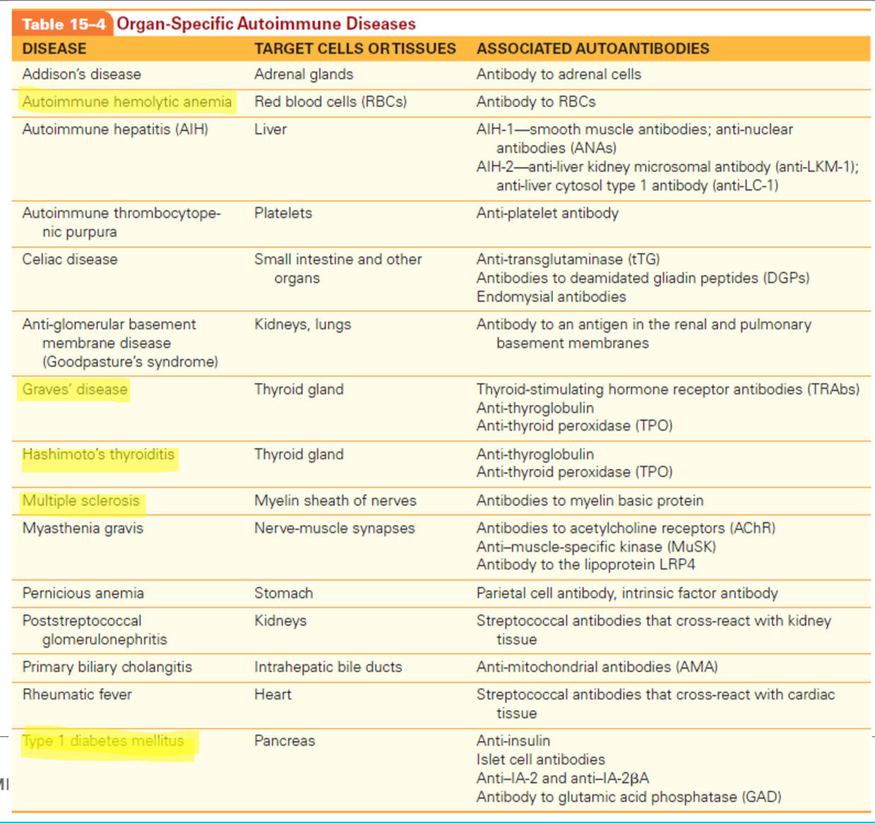
99
What is Hashimoto’s thyroiditis?
A common autoimmune disease, affecting about 8 out of every 1,000 individuals.
100
Who most often develops Hashimoto’s thyroiditis?
The disease is most often seen in middle-aged women; in addition, women are 5 to 10 times more likely to develop the disease than men.
101
What thyroid change occurs in Hashimoto’s thyroiditis?
Patients develop an enlarged thyroid called a goiter, which is irregular and rubbery.
102
What immune response causes Hashimoto’s thyroiditis?
Patients also produce thyroid-specific autoantibodies and cytotoxic T cells leading to destruction of the thyroid gland, which results in hypothyroidism.
103
What are the symptoms of hypothyroidism in Hashimoto’s thyroiditis?
Dry skin, decreased sweating, puffy face with edematous eyelids, pallor with a yellow tinge, weight gain, fatigue, and dry and brittle hair.
104
What autoantibodies do many patients with Hashimoto’s produce?
Antibodies to Tg and TPO.
105
What characterizes Graves’ disease in contrast to Hashimoto’s thyroiditis?
Graves’ disease is characterized by hyperthyroidism, a state of excessive thyroid function.
106
How common is Graves’ disease?
Graves’ disease is, in fact, one of the most frequently occurring autoimmune diseases and the most common cause of hyperthyroidism.
107
Who is more susceptible to Graves’ disease?
Women exhibit greater susceptibility.
108
What is thyrotoxicosis in Graves’ disease?
Thyrotoxicosis: an excess of thyroid hormones, with a diffusely enlarged goiter that is firm instead of rubbery
109
What are the clinical symptoms of Graves’ disease?
Nervousness, insomnia, depression, weight loss, heat intolerance, sweating, rapid heartbeat, palpitations, breathlessness, fatigue, cardiac dysrhythmias, and restlessness.
110
What causes exophthalmos in Graves’ disease?
Hypertrophy of the eye muscles and increased connective tissue in the orbit cause the eyeball to bulge out so that the patient has a large-eyed, staring expression.
111
What are the main antibodies involved in the pathogenesis of Graves’ disease?
The main antibodies involved in the pathogenesis of Graves’ disease are the TSH receptor antibodies (TRAbs).
112
What causes the hyperthyroidism seen in Graves’ disease?
The type responsible for the hyperthyroidism seen in Graves’ disease are called thyroid-stimulating immunoglobulins (TSIs). When TSIs bind to the TSH receptor, they mimic the action of TSH, resulting in uncontrolled receptor stimulation with excessive release of thyroid hormones.
113
What is the best indicator of Hashimoto’s thyroiditis and why?
For patients with Hashimoto’s thyroiditis, antibodies to TPO are the best indicator of the disease because they are found in up to 95% of patients but in only 10% to 15% of the general population.
114
What makes TRAbs highly indicative of Graves’ disease?
TRAbs, on the other hand, are highly indicative of Graves’ disease because they are present in 98% to 100% of patients; TRAbs.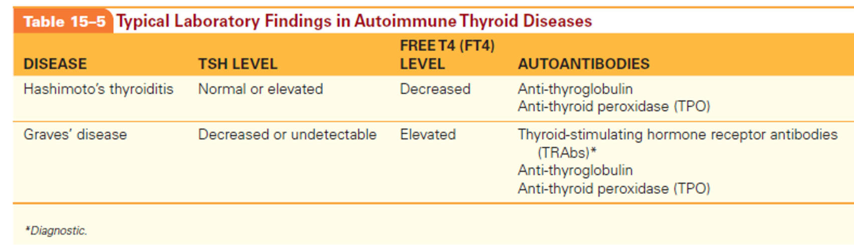
115
What percentage of DM patients have a complete or nearly complete deficiency in insulin, and what portion of them have an immune-mediated form?
About 5% to 10% of patients with DM (a complete or nearly complete deficiency in insulin). Of these patients, 90% have an immune-mediated form of the disease known as type 1A diabetes.
116
What is Type 1 diabetes mellitus (T1D) and what pancreatic cells are affected?
T1D is a chronic autoimmune disease that involves selective destruction of the beta cells of the pancreas.
117
What problems result from insufficient insulin in T1D?
Insufficient insulin production, hyperglycemia, and toxic effects on the body.
118
What long-term effects are associated with T1D?
Long-term effects include cardiovascular complications, renal disease, nerve damage, blindness, and infections of the lower extremities, which can lead to amputation.
119
What treatment is required for T1D to control glucose and reduce complications?
Patients require lifelong insulin injections to control glucose levels and lower the risk of these complications.
120
What role does genetics play in T1D?
Family history (role of genetics, multiple genes involved).
121
What HLA association increases risk for T1D?
Association with inheritance of HLA-DR3 or DR4 (higher risk if inherited together).
122
What does progressive inflammation cause in the islets of Langerhans in T1D?
Progressive inflammation of the islets of Langerhans in the pancreas leads to fibrosis and destruction of most beta cells.
123
What immune cells accumulate in T1D immunopathology?
Accumulation of CD8+ lymphocytes, plasma cells and macrophages.
124
How long before clinically evident T1D does autoantibody production begin?
Autoantibody production precedes the development of clinically evident T1D by several years.
125
According to the ADA, what is the criteria a person needs to meet to considered diabetic?
According to the ADA, a person is considered to have diabetes if he or she meets one of four criteria:
1. A fasting glucose greater than 126 mg/dL on more than one occasion (normal value is lower than 100 mg/dL)
2. A random plasma glucose level of 200 mg/dL or more with classic symptoms of diabetes
3. An oral glucose tolerance test of 200 mg/dL or more in a 2-hour sample with a 75 g glucose load
4. Or a hemoglobin A1c value (HbA1c) greater than 6.5%.
1. A fasting glucose greater than 126 mg/dL on more than one occasion (normal value is lower than 100 mg/dL)
2. A random plasma glucose level of 200 mg/dL or more with classic symptoms of diabetes
3. An oral glucose tolerance test of 200 mg/dL or more in a 2-hour sample with a 75 g glucose load
4. Or a hemoglobin A1c value (HbA1c) greater than 6.5%.
126
How is T1D differentiated from type 2 DM?
T1D needs to be differentiated from type 2 DM by serologic tests: ICA detection in serum.
127
What tests confirm T1D if suspected?
If T1D suspected: tests for detection of antibodies to GAD and IA-2A confirm diagnosis.
128
What is multiple sclerosis (MS)?
An autoimmune disorder involving inflammation and destruction of the CNS.
129
How many people does MS affect?
It affects more than 900,000 Americans and millions of individuals worldwide.
130
What ages are most commonly affected by MS?
Common between the ages of 20 and 50 and is twice as common in women as in men.
131
What genetic factor is most closely associated with MS?
Multiple genes may contribute to the development of MS, but the disease is most closely associated with inheritance of the HLA allele, DRB1*1501.
132
What environmental factors contribute to MS?
Exposure to sunlight, vitamin D deficiency, cigarette smoking, and infection with EBV after early childhood.
133
What forms in the white matter of the brain and spinal cord in MS?
Formation of plaques in the white matter of the brain and spinal cord.
134
What cells predominate within MS plaques?
T cells and macrophages predominate within the plaques and orchestrate progressive destruction of the myelin sheath of axons (demyelination).
135
What does the antibody bind to in MS?
Antibody binds to the myelin membrane and may stimulate specialized phagocytes called microglial cells.
136
MS diagnosis is primarily based upon what?
Clinical picture and detection of lesions in the brain using MRI.
137
What is detected in supportive lab tests for MS that is not found in serum?
Oligoclonal bands in CSF electrophoresis.
138
What is the IgG index?
A calculated ratio of cerebral spinal fluid (CSF) IgG/albumin ÷ serum IgG/albumin.
139
What happens to the IgG index in MS?
It will be increased in MS.
140
What autoantibody is detected in the majority of MS patients?
Detection of autoantibody against myelin basic protein (positive in majority of patients)