Heme Dry
Study these flashcardsFree · spaced repetition · iPhone, Android, Mac & web
All cards
1
Q: What causes electrical resistance, or impedance, in the current during cell counting?
A: Impedance occurs as the cells pass through the sensing aperture, causing voltage pulses that are measurable.
2
Q: In impedance counting, what is the relationship between the number of pulses and the number of cells?
A: The number of pulses is proportional to the number of cells counted.
3
Q: In impedance counting, what does the height of the voltage pulse represent?
A: The height of the voltage pulse is directly proportional to the volume of the cell.
4
Q: How are pulses collected and sorted in impedance cell counting?
A: Pulses are collected and sorted (channelized) according to their amplitude by pulse height analyzers.
5
Q: What standard modules are included in XN series?
A: XN-CBC, XN-DIFF, XN-BF, nRBCs, RET-He (reticulocyte hemoglobin), and IRF (immature reticulocyte fraction).
6
Q: What unique platelet analysis method is available on the XN series?
A: The platelet analysis on the XN also uses a fluorescent count, in addition to the impedance count and optical count, called the PLT-F, performed by optical measurement.
7
Q: What dye does the PLT-F method use?
A: A fluorocell fluorescent dye (oxazine) combined with an extended PLT counting volume and time.
8
Q: How are platelets differentiated in PLT-F analysis and how can PLT-F be set up?
A: Based on differences in intensity of the fluorescence combined with forward scattered light. PLT-F is performed on each specimen or as a reflex test based on laboratory PLT criteria.
9
Q: How many hydraulic subsystems are used for determining the hemogram and what is measured directly?
A: The WBC, RBC, platelet counts, hemoglobin, and HCT are considered to be measured directly. Three hydraulic subsystems: WBC channel, RBC/platelet channel, and a separate hemoglobin channel.
10
Q: What method is used in WBC and RBC transducer chambers for counting and sizing cells?
A: In the WBC and RBC transducer chambers, diluted WBC and RBC samples are aspirated through the different apertures and counted using the impedance (DC detection) method for counting and volumetrically sizing cells.
11
Q: What two features enhance impedance technology in Sysmex analyzers?
A: Two unique features enhance the impedance technology: In the RBC/platelet channel, a sheathed stream with hydrodynamic focusing is used to direct cells through the aperture, which reduces coincident passage, particle volume distortion, and recirculation of blood cells around the aperture; and in the WBC and RBC/platelet channels, floating thresholds are used to discriminate each cell population.
12
Q: What happens as cells pass through the apertures?
A: Signals are transmitted in sequence to the analog circuit and particle volume distribution analysis circuits for conversion to cumulative cell volume distribution data. Particle volume distribution curves are constructed, and optimal position of the autodiscrimination level (i.e., threshold) is set by the microprocessor for each cell population.
13
Q: What are the platelet threshold volume ranges?
A: The lower platelet threshold is automatically adjusted in the 2- to 6-fL volume range, and the upper threshold is adjusted in the 12- to 30-fL range, based on particle volume distribution.
14
Q: What are the RBC threshold volume ranges?
A: RBC lower and upper thresholds may be set in the 25- to 75-fL and 200- to 250-fL volume ranges.
15
Q: Why is floating discriminator technology particularly useful in the RBC channel?
A: This floating threshold circuitry allows for discrimination of cell populations on a specimen-by-specimen basis. In the RBC channel, the floating discriminator is particularly useful in separating platelets from small RBCs.
16
Q: How is HCT determined?
A: From the RBC/platelet channel, based on the principle that the pulse height generated by the RBC is proportional to cell volume. The HCT is the RBC cumulative pulse height and is considered a true relative percentage volume of erythrocytes.
17
Q: How is hemoglobin measured in Sysmex analyzers?
A: Hemoglobin is oxidized, binds to sodium lauryl sulfate (SLS), forms an SLS–hemoglobin complex, and is measured photometrically at 555 nm.
18
Q: Which indices are calculated in the microprocessor using directly measured or derived parameters?
A: MCV, MCH, MCHC, RDW-SD, RDW-CV, MPV, and plateletcrit.
19
Q: How is RDW-SD and RDW-CV defined?
A: RDW-SD is the RBC arithmetic distribution width measured at 20% of the height of the RBC curve, reported in femtoliters, with a reference interval of 37 to 54 fL. RDW-CV is the RDW reported as a CV.
20
Q: What is Platelecrit and how is MPV calculated?
A: Plateletcrit is the platelet volume ratio, analogous to the HCT. MPV is calculated from the plateletcrit and platelet count just as erythrocyte MCV is calculated from the HCT and RBC count.
21
Q: What may a proportion of platelets greater than 12 fL indicate?
A: Platelet clumping, giant platelets, or cell fragments.
22
Q: What detection chambers does the SE-9000/9500 use?
A: Four detection chambers to analyze WBCs and obtain a five-part differential: the DIFF, immature myeloid information (IMI), eosinophil (EO), and basophil (BASO) chambers. The high-end instrumentation such as the XE-series and the XN series has a six-part differential: neutrophils, lymphocytes, monocytes, eosinophils, basophils, and immature granulocytes. Every differential performed generates a percentage and absolute number for immature granulocytes, thus providing valuable information about the complete differential.
23
Q: What additional feature can the XE series use in optical mode?
A: The XE series has the capability to run the platelet counting in the optical mode, which eliminates common interferences found with impedance counting. In the optical mode, the immature platelet fraction or IPF, can be measured to provide additional information concerning platelet kinetics in cases of thromobocytopenia.
24
Q: What anemia-related parameters are determined from the reticulocyte channel?
A: Decreased and increased cellular hemoglobin content, using high-angle forward light scatter.
25
Q: What happens to RBCs in the DIFF detection chamber and how are WBCs analyzed?
A: In the DIFF detection chamber, RBCs are hemolyzed and WBCs are analyzed simultaneously by low-frequency DC and high-frequency current (DC/RF detection method).
26
Q: What type of plot is produced in the DIFF detection chamber?
A: A scattergram of RF detection signals (y-axis) versus DC detection signals (x-axis) allows separation of the WBCs into lymphocytes, monocytes, and granulocytes. Floating discriminators determine the optimal separation between these populations.
27
Q: Which cell type is analyzed further in the IMI (Immature Myeloid Information) detection chamber?
A: Granulocytes are analyzed further in the IMI detection chamber to determine immature myeloid information. RBCs are lysed, and WBCs other than immature granulocytes are selectively shrunk by temperature and chemically controlled reactions. Analysis of the treated sample using the DC/RF detection method allows separation of immature cells on the IMI scattergram.
28
Q: What method is also used in the EOS and BASO chambers?
A: EOS and BASO are counted by impedance (DC detection) in separate chambers in which the RBCs are lysed, and WBCs other than eosinophils or basophils are selectively shrunk by temperature and chemically controlled reactions. Eosinophils and basophils are subtracted from the granulocyte count derived from the DIFF scattergram analysis to determine the neutrophil count.
29
Q: In the XN-1000, what method is used for the WBC count, WBC differential, and enumeration of nucleated RBCs?
A: Fluorescent flow cytometry.
30
Q: What happens to RBCs and WBC membranes in WDF channel?
A: In the WDF channel, RBCs are lysed, WBC membranes are perforated, and the DNA and RNA in the WBCs are stained with a fluorescent dye.
31
Q: What does plotting side scatter on the x-axis and side fluorescent light on the y-axis in the WDF channel enable?
A: Enables separation and enumeration of neutrophils, eosinophils, lymphocytes, monocytes, and immature granulocytes.
32
Q: What happen to RBCs (including nRBCs) and WBC membranes in the WNR channel?
A: In the WNR channel, the RBCs are lysed, including nucleated RBCs, and WBC membranes are perforated. A fluorescent polymethine dye, stains the nucleus and organelles of the WBCs with high fluorescence intensity and stains the released nuclei of the nucleated RBCs with low intensity.
33
Q: What does plotting side fluorescent light on the x-axis and forward scatter on the y-axis in the WNR channel enable?
A: Plotting side fluorescent light on the x-axis and forward scatter on the y-axis enables separation and enumeration of the total WBC count, basophils, and nucleated RBCs. The WBC count is automatically corrected when nucleated RBCs are present in the specimen.
34
Q: What does the WPC channel detect?
A: WPC channel detects blasts and abnormal lymphocytes in a similar manner using a lysing agent and fluorescent dye and plotting side scatter on the x-axis and side fluorescent light on the y-axis.
35
PLT-F Channel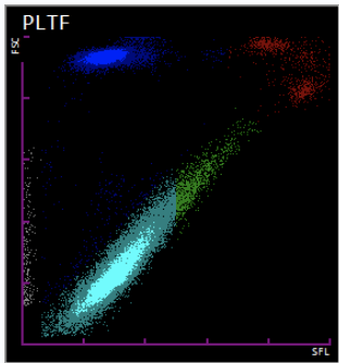
PLT-F is a true fluroscent count, and the secondary method on the XN-series analyzers. Immature platelet fraction: IPF represents those immature platelets that have been recently released by the bone marrow.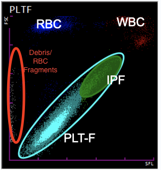
36
Q: What are the reportable parameters of PLT-F channel and what is the x & y-axis?
A: PLT (PLT-F), IPF (Platelets, P License)
X-axis = Side fluorescence, separating based on size of nucleus
Y-axis = Forward scatter, seprated based on cell size
X-axis = Side fluorescence, separating based on size of nucleus
Y-axis = Forward scatter, seprated based on cell size
37
RET Channel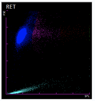
Immature Retic Fraction: The IRF is obtained as a ratio of reticulocytes count in the area with strong fluorescent light intesity in the RET scattergram (IRF zone), to the total reticulocyte count.
Reticulocyte Hemoglobin equivalent (RET-He): Measurement of the amount of hemoglobin in the immature red cells. It is unique to sysmex and it is derived using the reticulocyte scattered light signals and a proprietary sysmex calculation.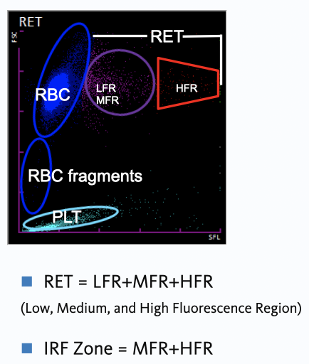
Reticulocyte Hemoglobin equivalent (RET-He): Measurement of the amount of hemoglobin in the immature red cells. It is unique to sysmex and it is derived using the reticulocyte scattered light signals and a proprietary sysmex calculation.
38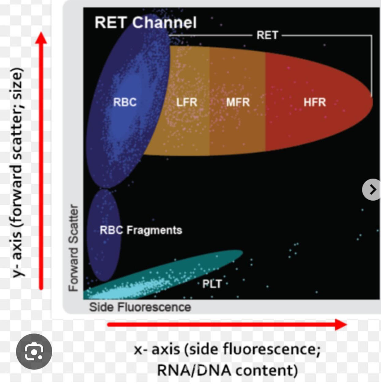
39
Q: What are the reportable parameters and the x & y-axis of the RET channel?
A: RET%, RET#, IRF, RET-He (Reticulocytes, R License)
X-axis = side fluorescence, separating based on size of nucleus
Y-axis = forward, separting based on cell size
X-axis = side fluorescence, separating based on size of nucleus
Y-axis = forward, separting based on cell size
40
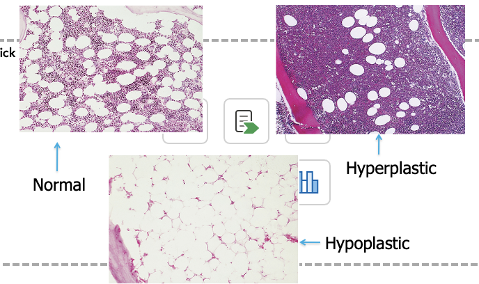
WPC channel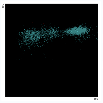
Provides improved differentiation of blast cell and abnormal lymphocyte flagging. Abnormal lymphocytes: Lysercell WPC reagent effeciently perforates the cell membrane of abnormal lymphocytes, allowing Fluorocell WPC reagent to effectively stain the cell nuclei, resulting in higher side fluorescence in the flow cell. Blasts are not effectively perforated by the Lysercell reagent which leads to a lower side fluorescence. 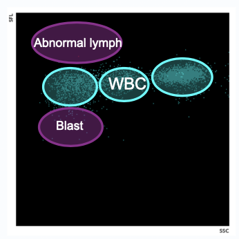
41
Q: What are the reportable parameters and the x & y-axis of the WPC channel?
42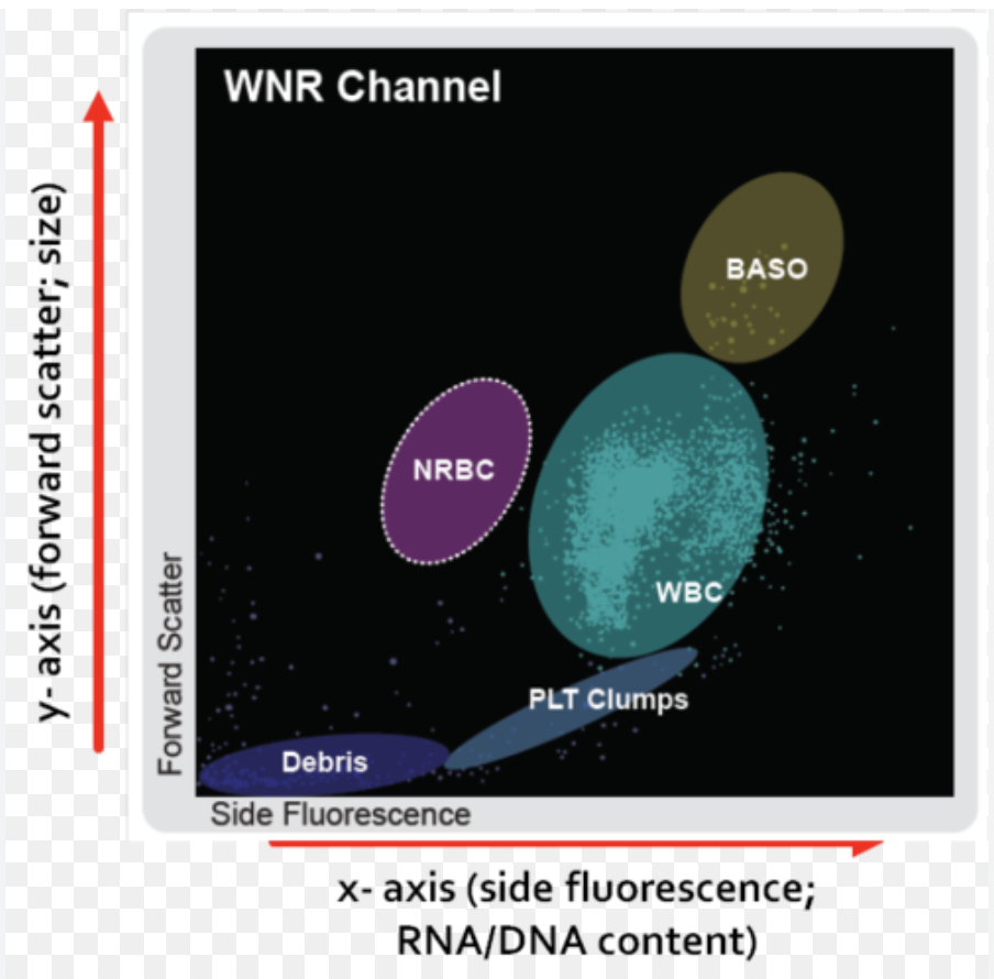
WNR Channel; STRD with every XN-CBC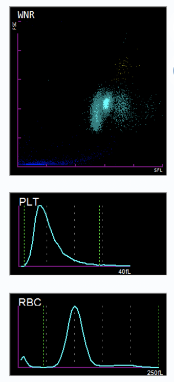
43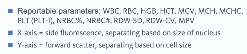
Q: What are the reportable parameters and the x & y-axis of the WNR channel?
44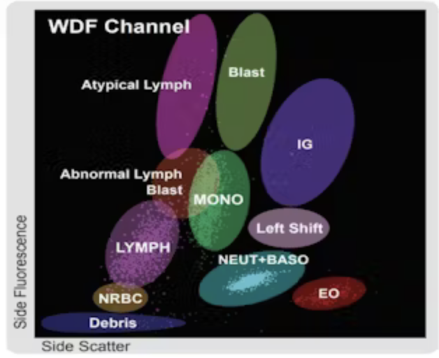
WDF Channel; STRD with every XN-DIFF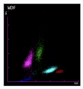
45
Q: What are the reportable parameters and the x & y-axis of the WDF channel?
46
Q: Condition that cause interference: Cold agglutinins
leads to falsely low RBC, Hgb and falsely high MCV, MCH, MCHC
47
Q: Conditions that cause interference: Lipemia and Icterus
falsely elevated Hgb, MCH, and MCHC
48
Q: Condition that cause interference: Hemolysis
49
Q: Condition that cause interference: Lysis-resistant RBCs with abnormal hemoglobins
50
Q: Conditions that cause interference: Microcytes or schistocytes
51
Q: Conditions that cause interference: nRBCs, megakaryocyte, fragments, or micromegakaryoblasts
52
Q: Condition that cause interference: PLT clumps
53
Q: Condition that cause interference: WBC> 100,000/microL
54
Q: Condition that cause interference: Leukemia, especially with chemotherapy
55
Q: Condition that cause interference: Old specimen
56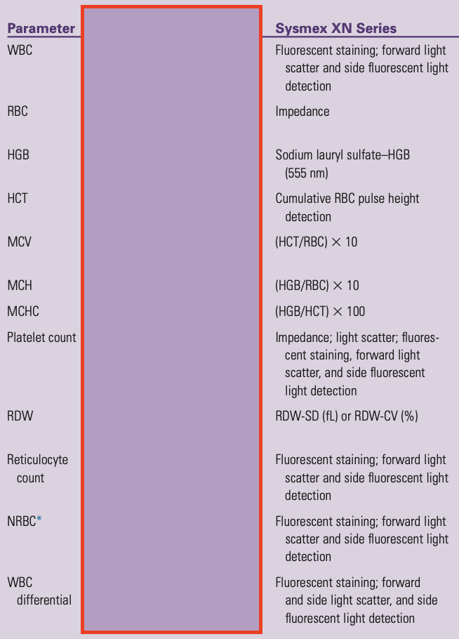
Q: Method for Hemogram, Reticulocyte, nRBC, and WBC Diff counts
57
Q: What is measured in the principle of the erythrocyte sedimentation rate (ESR)?
A: The rate of RBC settle is measured as the number of millimeters of clear plasma present at the top of the column after one hour (mm/hr).
58
Q: Why do RBCs settle in the ESR test?
A: RBCs settle because their density is greater than that of plasma.
59
Q: What alteration on RBC surfaces leads to rouleaux formation?
A: An alteration in the distribution of charges on the surface of RBCs results in the formation of aggregates known as rouleaux.
60
Q: What determines rouleaux formation in ESR?
A: Increased levels of plasma fibrinogen and globulins.
61
Q: What does ESR reflect?
A: ESR reflects changes in plasma proteins that accompany acute and chronic infections, some tumors, and degenerative diseases.
62
Q: What type of test is ESR considered?
A: ESR is a non-specific screening test.
63
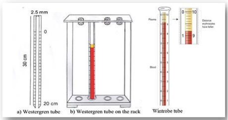
Q: What are the 4 stages of ESR?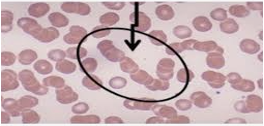
1. Negative charge on the RBC by zeta potential, if this charge is neutralized, rouleaux formation occurs
2. RBC rate fall through plasma
3. Packing of RBC
4. The distance is measured in mm/hr, after 1 hour
2. RBC rate fall through plasma
3. Packing of RBC
4. The distance is measured in mm/hr, after 1 hour
64
Q: What effect does inflammation have on RBCs?
A: Inflammation causes the cells to clump together.
65
Q: Why do clumps of RBCs settle faster than individual cells?
A: Clumps have a higher density than individual cells and therefore tend to settle at the bottom more rapidly.
66
Q: What does the ESR test measure?
A: ESR measures the rate at which RBCs separate from the plasma and fall to the bottom of the tube.
67
Q: What do higher ESR values indicate?
A: Higher values mean greater inflammation.
68
Q: What factors, besides inflammation, can affect ESR results?
A: Certain medications and drugs can affect a person’s ESR test result.
69
Q: Based on the Traditional Westergren Method, why can’t ESR procedures be calibrated?
A. Because they are susceptible to a variety of factors such as, temperature, hematocrit, erythrocyte mean corpuscular volume (MCV), and plasma viscosity.
70
Q. What are some interferences that may increase ESR values?
A.
- Fibrinogen
- Globulins (alpha 1 and alpha 2)
- Technical factors such as mechanical vibrations
- Elevated room temperature.
- Fibrinogen
- Globulins (alpha 1 and alpha 2)
- Technical factors such as mechanical vibrations
- Elevated room temperature.
71
Q. What are some interferences that may decrease ESR values?
A.
- Abnormally shaped RBC (sickle, spherocytosis)
- Hemolysis
- Clotted sample
- Presence of air bubble
- Low room temperature
- Delay in test performance
- Excess anticoagulant
- Specimen quality such as blood to anticoagulant ratio, age of specimen and fill volume
- Abnormally shaped RBC (sickle, spherocytosis)
- Hemolysis
- Clotted sample
- Presence of air bubble
- Low room temperature
- Delay in test performance
- Excess anticoagulant
- Specimen quality such as blood to anticoagulant ratio, age of specimen and fill volume
72
Q: What are the two traditional ESR methods and what do they require?
A: Westergren and Wintrobe. Both methods require manual pipetting, special materials, and long analysis times.
73
Q: What is the current ESR method used at UCLA Health?
A: The iSED/iSED ELITE Automated Erythrocyte Sedimentation Rate Analyzer.
74
Q: What technology does the iSed/iSed Elite use to measure rouleaux formation?
A: Uses advanced photometric rheology technology to measure the rouleaux formation, the earliest form of erythrocyte sedimentation
75
Q: What ultimately determines the sedimentation length in a Westergren tube?
A. The aggregation of RBCs during the rouleaux formation ultimately determines the length at which the red cells sediment in the Westergren tube.
76
Q: How does the iSED analyzer measure RBC aggregation compared to traditional ESR methods?
A. The iSeds analyzer consists of “directly” measuring the aggregation of the RBCs, while traditional ESR methods “indirectly” measure aggregation of the RBCs by recording the length at which the red cells setting in a Westergren tube.
77
Q: What does the micro-flow cell in the iSed/iSed ELITE analyzer capture?
A: It captures the critical kinetics of RBC aggregation in the testing environment, helping reduce the impact of factors that contribute to result variability.
78
Q: How is the sample prepared in the iSed/iSed ELITE analyzer?
A: The sample is automatically mixed in position.
79
Q: How does the iSed/iSed ELITE analyzer follow the progress of RBC aggregation?
A: A sensitive optical detector monitors light transmission through the sample over time.This produces a signal that is direct representation of the aggregation.
80
Q: What type of blood collection tube does the iSed/iSed ELITE analyzer directly use and how are results reported?
A. The analyzer directly uses primary EDTA blood collection tube and produces ESR results within 20 seconds. Results are reported in mm/hr and iSed/iSed ELITE performance is highly correlated with the Westergren method.
81
Q. What are the CBC sample requirements and what tests are included?
A. An unclotted sample in a EDTA tube. A CBC includes RBC, WBC, HGB, HCT, PLT, MCV, MCH and MCHC.
82
Q. What are the reference ranges for RBC count?
A. Ranges: Male 4.5-6.0 x 10^12/L, Female 4.5-5.1 x 10^12/L
83
A. What is the reference range for WBC count?
A. Ranges: 4.5-11 x 10^3/microL
84
Q. What kind of information can a WBC count provide?
A.
- Number of Total WBC can indicate infection
- High sub population can indicate type of infection
- Abnormal proliferation can indicate cancer
- Number of Total WBC can indicate infection
- High sub population can indicate type of infection
- Abnormal proliferation can indicate cancer
85
Q. What is the reference range for PLT count and what are PLTs involve in?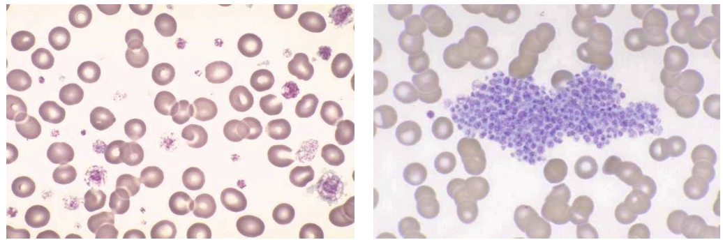
A. Range: 150-450 x 10^9 /L
Platelets are involved in clotting
Platelets are involved in clotting
86
Q. What is the reference range for Hemoglobin?
A. Ranges:
Male 14-17.5 g/dL
Female 12.3-15.3 g/dL
Male 14-17.5 g/dL
Female 12.3-15.3 g/dL
87
Q. What is hemoglobin responsible for and what happens when there is a mutation?
A. Responsible for oxygen carrying capacity. If there is a mutation, it affects oxygen carrying capacity and can increase number of RBCs
88
Q. What is the reference range for Hematocrit? 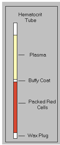
A. Ranges:
Male 41.5-50%
Female 35.9-44.6%
Male 41.5-50%
Female 35.9-44.6%
89
Q. What is the reference range for Mean Corpuscular volume (MCV) and how is it measured?
A.
- MCV is Average “size” of rbc
- HCT x 10/RBC
- Range 80-96 fL
- Indicates: Microcytes, Normocytes, Macrocytes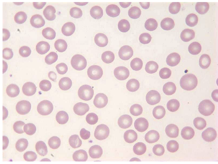
- MCV is Average “size” of rbc
- HCT x 10/RBC
- Range 80-96 fL
- Indicates: Microcytes, Normocytes, Macrocytes
90
Q. What is the reference range for Mean Corpuscular Hemoglobin (MCH) and how is it measured?
A.
- Average amount of hgb per rbc
- HGB X 10/ RBC
- Range: 27-33 pg
- Indicates: Hypochromic, Hyperchromic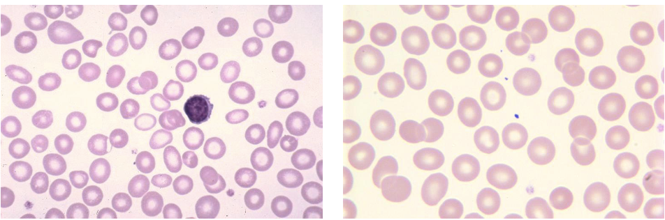
- Average amount of hgb per rbc
- HGB X 10/ RBC
- Range: 27-33 pg
- Indicates: Hypochromic, Hyperchromic
91
Q. What is the reference range for Mean Corpuscular Hemoglobin Concentration (MCHC) and how is it measured?
A.
- Average hgb in a given volume of rbc volume
- MCH/MCV x 100
- Range: 33-36 g/dL
- If high, suspect agglutination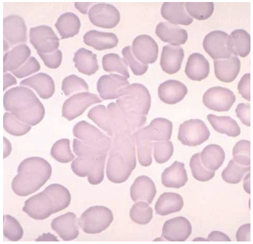
- Average hgb in a given volume of rbc volume
- MCH/MCV x 100
- Range: 33-36 g/dL
- If high, suspect agglutination
92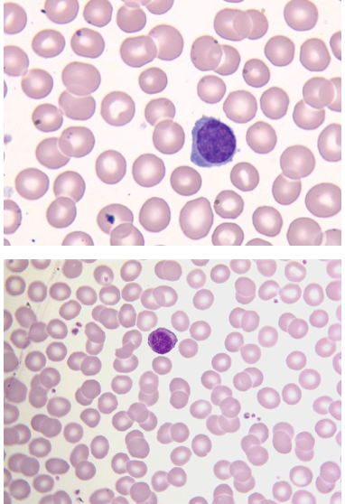
Normocytic, Normochromic
93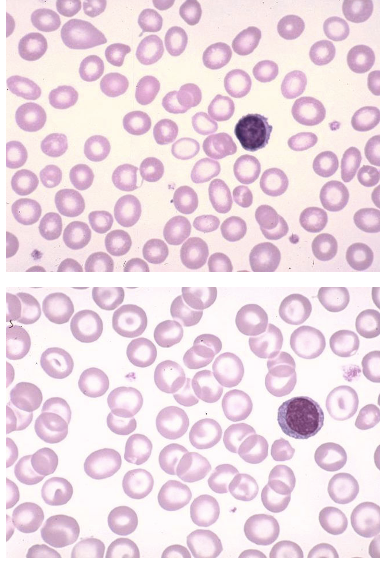
Microcytic, Hypochromic
94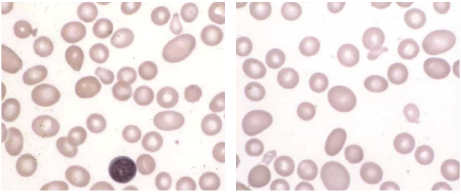
Macroovalocytes
95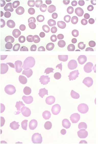
Spherocytes, Hyperchromic
96
Q: What is the reference range for RDW and what does it indicate?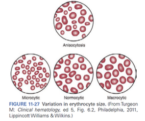
A:
- Indicates variation of RBC size
- Range 11.5%-14.5%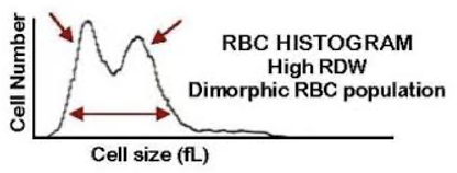
- Indicates variation of RBC size
- Range 11.5%-14.5%
97
Q: What is a PBS used for?
A.
- Check for to verify indices
- Look for clots
- Estimate platelets
- Look for “weird” rbcs and wbcs
- 100 WBC differential
- Check for to verify indices
- Look for clots
- Estimate platelets
- Look for “weird” rbcs and wbcs
- 100 WBC differential
98
Q: What can a CBC tell you?
A: If a patient has Anemia, Infection, Leukemia & Clotting
99
Q: What is the rule of 3 of normal RBCs?
A: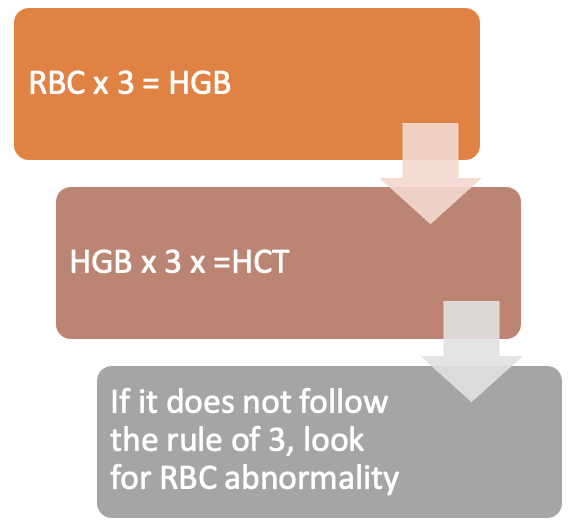
100
Q: What does elevated WBCs suggest?
A: WBCs help fight infection, therefore, elevated WBC count suggests infection.
101
Q: What is the difference between Relative and Absolute WBCs?
A:
- WBCC = WBC Count
- Instrument sorts into 5 types of cells
- By percentage = relative
- Percentage x WBCC = Absolute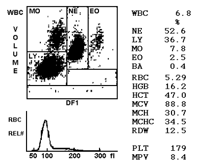
- WBCC = WBC Count
- Instrument sorts into 5 types of cells
- By percentage = relative
- Percentage x WBCC = Absolute
102
Q: What are the key characteristics of neutrophils?
A: Neutrophils are the most common white blood cell, making up an average of 56% of WBCs (range 33–71%), and they are elevated in bacterial infections.
103
Q: What are the key characteristics of Lymphocyte?
A: Lymphocytes get bigger as it "reacts", are activated against viruses and make up an average of 34% of WBCs.
104
Q: What are the key characteristics of Monocytes?
A: Monocytes are the largest leukocytes, they are phagocytic cells and make up an average of 2 to 10% of WBCs.
105
Q: What are the key characteristics of Eosinophils?
A: Eosinophils have a characteristic pink granules, are elevated in allergies and worm infections and make up 3% of WBCs.
106
Q: What are the key characteristics of Basophils?
A: Basophils are the smallest % of WBC (0.6%) and an increase of basos may indicate CML.
107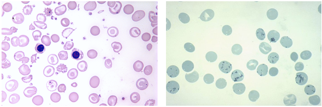
Reticulocyte/nRBC - Immature RBCs
108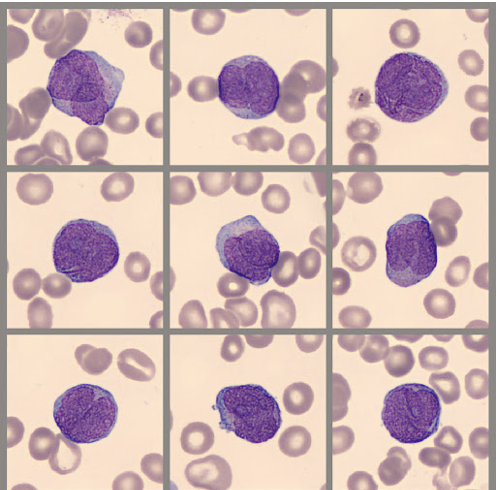
Leukemia (AML)
109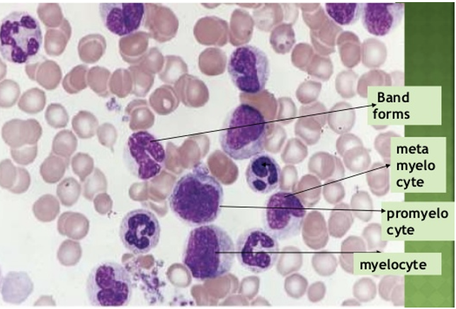
Leukemia (CML)
110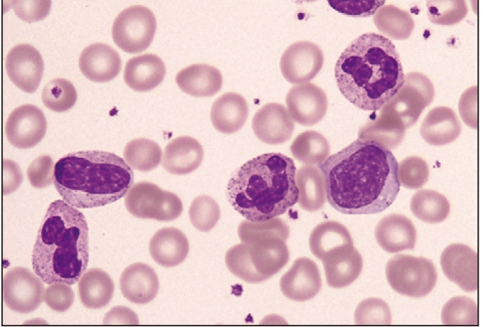
Left shift
111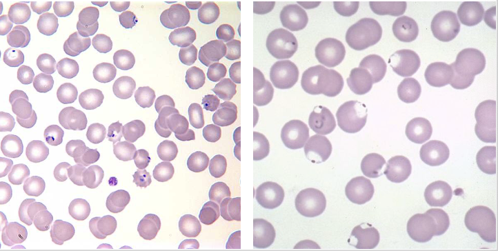
Malaria (RBC parasites)
112
113
114
Q: Which one has an error?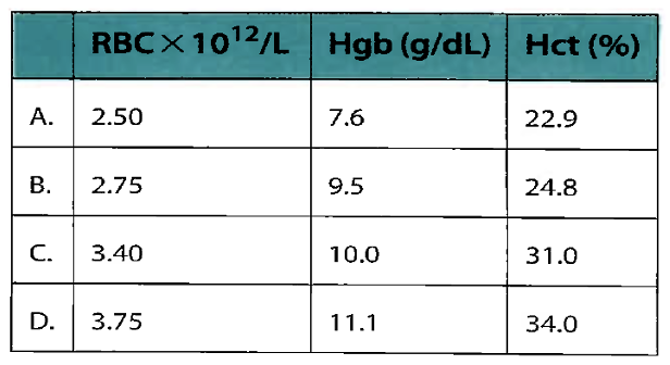
Answer: B (rule of 3)
115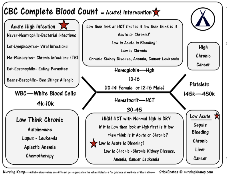
116
Q: What are the functions of Blood?
A: 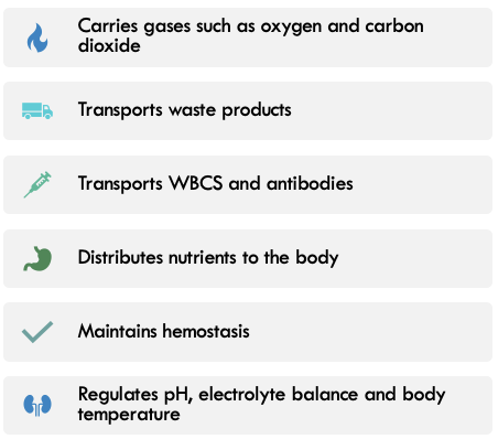
117
Q: What is blood comprised of?
A: Most of blood (~55%) is plasma or serum. Then, there is a layer of white blood cells or leukocytes. (~1%). Red blood cells or erythrocytes make up ~44% of blood. 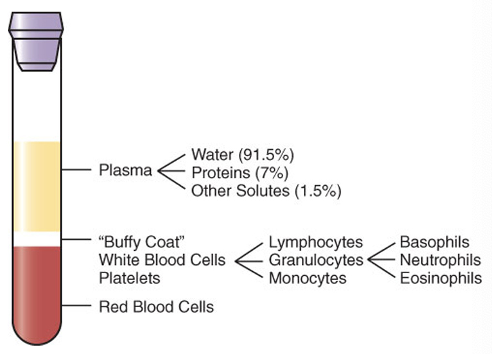
118
Q: What are some key characteristics of Erythrocytes?
A:
- Small, measuring around 7 um in diameter and 2 um thick
- Biconcave disc shape makes them flexible and can squeeze through small spaces such as capillaries
- Lose their nucleus when they mature, before it leaves the bone marrow
- Typically lives ~120 days
- A typical human has 5-6 liters of blood
- Small, measuring around 7 um in diameter and 2 um thick
- Biconcave disc shape makes them flexible and can squeeze through small spaces such as capillaries
- Lose their nucleus when they mature, before it leaves the bone marrow
- Typically lives ~120 days
- A typical human has 5-6 liters of blood
119
Q: What is Hemostasis?
A: The property of the circulation that maintains blood as a fluid within the blood vessels, and the systems ability to form a barrier(blood clot) to prevent excessive blood loss when the vessel is damaged.
120
Q: What is Hematopoiesis?
A: a term describing the formation and development of blood cells.
121
Q: What two processes are involved in maintaining hematopoiesis and what system is involved?
A: Loss, destruction and Self Renewal. This system involves: Proliferation, Differentiation and Maturation.
122
Q: When does hematopoiesis begin in embryonic development and what type of cells are produced first in the yolk sac?
A: Hematopoiesis begins as early as the nineteenth day after fertilization in the yolk sac of the embryo. Only erythrocytes are made which contain unique fetal hemoglobin.
123
Q: What happens to erythrocyte production around 6 weeks of gestation?
A: Yolk sac production of erythrocytes decreases and production of RBCs in the human embryo itself begins.
124
Q: Where is the blood made?
A:
- The fetal liver then becomes the chief site of blood cell production.
- Erythrocytes and the beginnings of WBC and Platelets occurs
- The spleen, kidney, thymus, and lymph nodes serve as minor sites of blood cell production.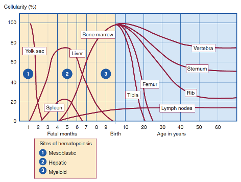
- The fetal liver then becomes the chief site of blood cell production.
- Erythrocytes and the beginnings of WBC and Platelets occurs
- The spleen, kidney, thymus, and lymph nodes serve as minor sites of blood cell production.
125
Q: When does the bone marrow become the primary site of hematopoiesis and how does it affect lymphocyte and thrombocyte production?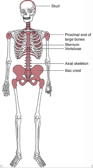
A: BM is primary site after ~ 6 months and leukocytes and thrombocyte production increases when BM switch.
126
Q: What is hematopoiesis called in the BM and what is it called in other areas?
A: Hematopoiesis in the bone marrow is called medullary hematopoiesis. Hematopoiesis in areas other then the bone marrow is called extramedullary hematopoiesis.
127
Q: Where is bone marrow located, and how is it divided in a normal adult?
A: Bone marrow is located inside spongy bone. In a normal adult, half of the BM is hematopoietically active (red marrow) and half is inactive, fatty marrow (yellow marrow).
128
Q: What does red marrow contain, and how does it change with age?
A: Red marrow contains erythroid, leukocyte, and platelet precursors. Early in life most marrow is red marrow, but it gradually decreases with age to 50% in adults.
129
Q: How can bone marrow respond in certain pathologic states?
A: It can increase its activity to 5–10 times its normal rate.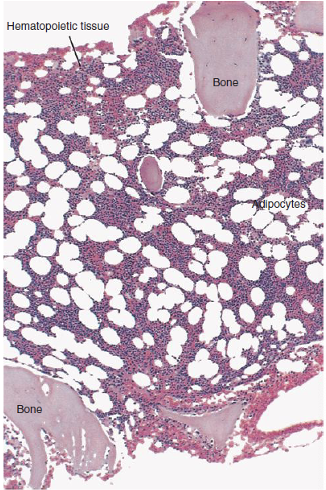
130
Q: In what conditions does abnormal hematopoiesis occur?
A: It occurs in conditions with increased or ineffective hematopoiesis.
131
Q: What pathologic states can cause abnormal hematopoiesis?
A:
- Acute blood loss: temporary replacement of yellow marrow.
- Severe chronic anemia: marrow may erode the bone itself.
- Malignant disease: both normal red marrow and fatty marrow may be replaced by proliferating abnormal cells.
- Acute blood loss: temporary replacement of yellow marrow.
- Severe chronic anemia: marrow may erode the bone itself.
- Malignant disease: both normal red marrow and fatty marrow may be replaced by proliferating abnormal cells.
132
Q: What can happen to hematopoietic tissue in certain conditions?
A: The hematopoietic tissue may become inactive or hypoplastic.
133
Q: What are possible causes of hypoplastic bone marrow?
A:
- Chemicals
- Genetics
- Myeloproliferative disease that replaces hematopoietic tissue with fibrous tissue
- Chemicals
- Genetics
- Myeloproliferative disease that replaces hematopoietic tissue with fibrous tissue
134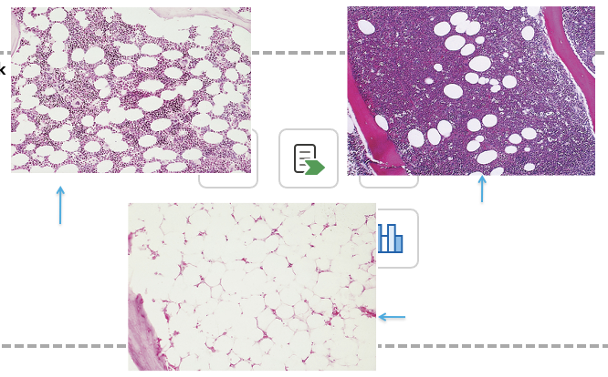
135
Q: How can hematopoietic cells be divided based on maturity?
A: Hematopoietic cells can be divided into three cellular compartments: pluripotential stem cells, committed progenitor stem cells, and mature cells.
136
Q: What is the function of pluripotential stem cells?
A: They are capable of self-renewal and differentiation into all blood cell lines.
137
Q: What are committed progenitor stem cells?
A: Stem cells destined to develop into distinct cell lines.
138
Q: What are mature hematopoietic cells?
A: They are mature cells with specialized functions.
139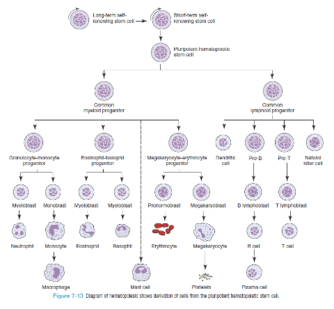
140
141
142
143
Q: What is the role of the mononuclear phagocytic system?
A: The mononuclear phagocytic system is involved in cellular destruction
144
Q: What components are included in the mononuclear phagocytic system?
A:
- Circulating blood monocytes
- Fixed macrophages in the bone marrow, liver, spleen, and lymph nodes
- Free macrophages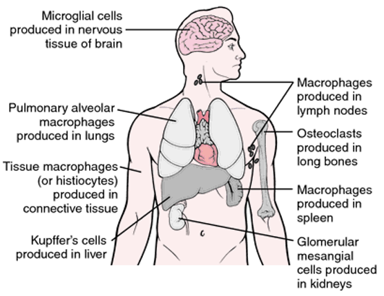
- Circulating blood monocytes
- Fixed macrophages in the bone marrow, liver, spleen, and lymph nodes
- Free macrophages
145
Q: What does the spleen contain the largest collection of in the body?
A: The spleen contains the largest collection of lymphocytes and mononuclear phagocytes in the body.
146
Q: What are the main functions of the spleen?
A:
- Filtering and destruction
- Removal of particles (pitting)
- Enforcing close contact of blood-borne antigens with lymphocytes and phagocytic cells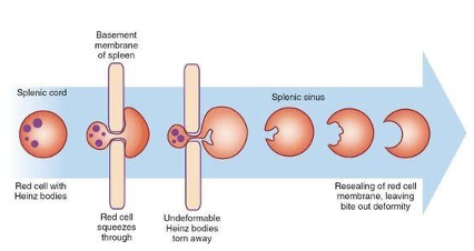
- Filtering and destruction
- Removal of particles (pitting)
- Enforcing close contact of blood-borne antigens with lymphocytes and phagocytic cells
147
Q: Cellular destruction: What can happen when the spleen sequesters cells?
A: It may lead to hypersplenism, an overactive spleen that removes blood cells too early and too quickly.
148
Q: What is the term for an enlarged spleen?
A: Splenomegaly
149
Q: What is the difference between primary and secondary splenomegaly?
A: Primary splenomegaly has no identified underlying disease, while secondary splenomegaly is caused by an underlying disorder.
150
Q: What are some causes of secondary splenomegaly?
A: Inflammatory diseases, infectious diseases, compensatory or workload hypertrophy (abnormal RBCs (spherocytes), and antibody-coated RBCs (ITP)).
151
Q: How can the effects of splenomegaly be relieved?
A: By splenectomy.
152
Q: What are the main components of the lymphatic system?
A: Lymph nodes, lymphatic vessels, and lymph.
153
Q: What are lymph nodes composed of, and what happens when antigens pass through them?
A: Lymph nodes are composed of lymphocytes, macrophages, and a reticular network. As antigens pass through, they encounter and stimulate immunocompetent lymphocytes to proliferate and differentiate into effector cells.
154
Q: What is lymph, and how is it formed?
A: Lymph is a thin watery fluid formed from plasma when it diffuses into tissue spaces.
155
Q: What is the function of the thymus in the lymphatic system?
A: It serves as a compartment for the maturation of T lymphocytes into immunocompetent T cells.
156
Q: Which hormone plays a role in T lymphocyte maturation in the thymus?
A: Thymosin
157
Q: What phagocytic cells are found in the liver, and what is their function?
A: Phagocytes known as Kupffer cells, which act as a filter for damaged or aged cells, though less efficiently than the phagocytic cells in the spleen.
158
Q: Besides filtering, what additional role can the liver play?
A: It is capable of extramedullary hematopoiesis.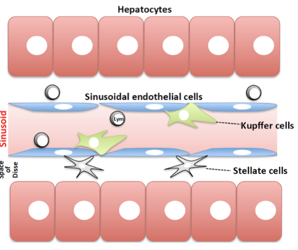
159
Q: Do mature blood cells have the ability to self-renew?
A: Mature blood cells have a limited lifespan and, except for lymphocytes. Mature lymphocytes are incapable of self-renewal.
160
Q: What is responsible for replacing peripheral hematopoietic cells?
A: pluripotential stem cells in the bone marrow.