Bacti - Orientation
Study these flashcardsFree · spaced repetition · iPhone, Android, Mac & web
All cards
1
What role does Gram Stain play in identifying bacteria?
Gram Stain is one of the most useful tools in microbiology. It aids in the identification of bacterial organisms by visualizing organisms and cells from a direct specimen.
2
What information does a Gram Stain provide?
The presence or absence of organisms and cells aid in empiric therapy of infectious processes and aid in the presumptive diagnosis of pneumococcal, meningococcal or gonococcal infections
3
What is the difference between methanol fixing and heat fixing in terms of cellular morphology?
Methanol fixing preserves cell morphology (visualizes WBC, RBC, epithelial cells, etc.). Heat fixing lyses the cellular elements.
4
What principle defines how a Gram Stain differentiates bacteria?
Differential stain based on dye retention and microscopic arrangement
5
What components are required to perform a Gram Stain and what does each one do?
Components:
Crystal Violet: primary stain, purple.
Gram’s Iodine: mordant.
Acetone-Alcohol: decolorizer.
Safranin: counterstain, pink
Crystal Violet: primary stain, purple.
Gram’s Iodine: mordant.
Acetone-Alcohol: decolorizer.
Safranin: counterstain, pink
6
What structural feature and staining appearance characterize Gram Positive organisms?
Thick peptidoglycan layer allows retention of crystal violet-iodine complex. Appears blue/purple.
7
What structural feature and staining appearance characterize Gram Negative organisms?
Thin peptidoglycan layer surrounded by outer lipopolysaccharide membrane. Appears pink / red.
8
How do you prepare a smear for a Gram Stain?
1. Place a small loopful of distilled water on a clean slide.
2. Touch a sterile inoculating loop to a colony, rub it into the droplet and create even suspension.
3. Let the smear dry at room temperature.
4. Heat-fix.
5. Cool to room temperature before staining
2. Touch a sterile inoculating loop to a colony, rub it into the droplet and create even suspension.
3. Let the smear dry at room temperature.
4. Heat-fix.
5. Cool to room temperature before staining
9
What is the step-by-step procedure for performing a Gram stain?
- Flood fixed smears with CV for 1 minute, then decant and rinse with water.
- Flood with gram’s iodine for 1 minute, then decant and rinse with water.
- Decolorize with acetone-alcohol solution, stopping when runoff becomes clear, remove excess decolorizer with water. (Adjust decolorizing time to thickness of smear and type of decolorizer used)
- Counterstain with safranin for 30 seconds to 1 minute, then decant and rinse with water. (Faint organisms may require longer counterstain time. Alternative stains such as carbol fuchsin may be used.)
- Let air dry or blot dry (if blot drying, be careful as to not rub and remove the smear from the slide).
*Stain time can be adjusted. there is no need to actually spend a lot of time or watch the clock as you perform gram stain.
- Flood with gram’s iodine for 1 minute, then decant and rinse with water.
- Decolorize with acetone-alcohol solution, stopping when runoff becomes clear, remove excess decolorizer with water. (Adjust decolorizing time to thickness of smear and type of decolorizer used)
- Counterstain with safranin for 30 seconds to 1 minute, then decant and rinse with water. (Faint organisms may require longer counterstain time. Alternative stains such as carbol fuchsin may be used.)
- Let air dry or blot dry (if blot drying, be careful as to not rub and remove the smear from the slide).
*Stain time can be adjusted. there is no need to actually spend a lot of time or watch the clock as you perform gram stain.
10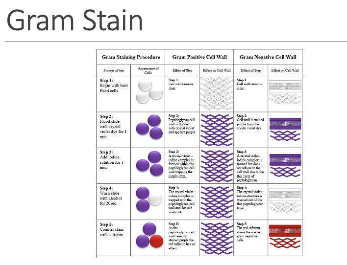
11
What are the characteristics of Staphylococci?
Gram Positive Cocci:
Spheres in irregular clusters, singly, pairs or short chains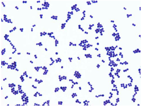
Spheres in irregular clusters, singly, pairs or short chains
12
What are the characteristics of Streptococci?
Gram Positive Cocci:
spheres in pairs, short or long chains. Typically more elongated than staph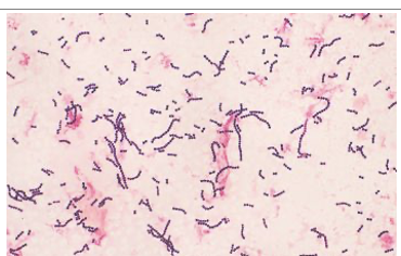
spheres in pairs, short or long chains. Typically more elongated than staph
13
What are the characteristics of Pneumococci?
Gram Positive Cocci:
resemble strep, but with lancet shape, capsule may be observed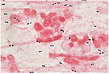
resemble strep, but with lancet shape, capsule may be observed
14
What morphological features characterize Corynebacterium?
straight or slightly curved. V, L, Y formations. Pleomorphic, diphtheroid-like (club shaped)
15
What morphological features characterize Listeria?
small rounds with rounded ends, but possibly short coccal forms
16
How do Lactobacilli vary in appearance?
vary from short plump palisading to long slender rods
17
What morphological features characterize Clostridium?
large fat in single, pairs or chains. Frequently gram variable. Spores not always visible
18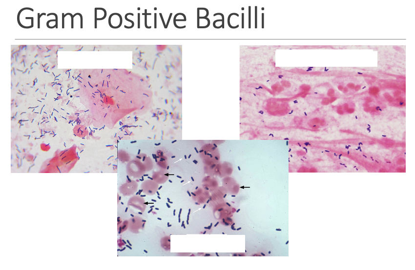
19
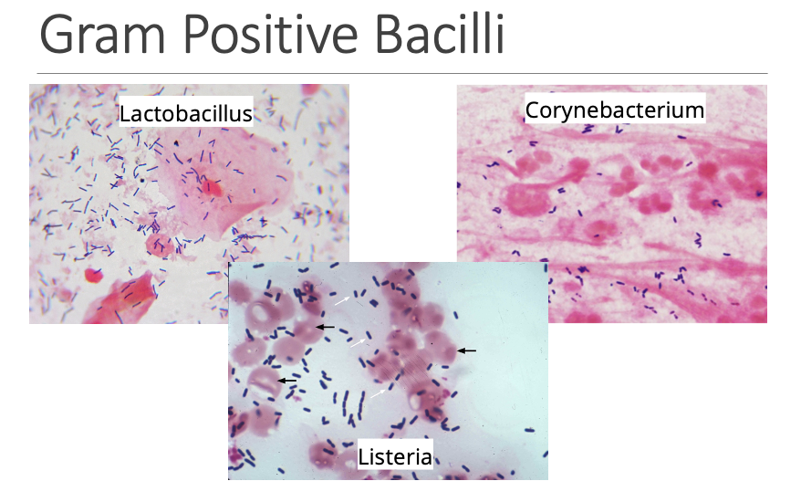
What is the overall challenge when identifying Gram Negative organisms?
Usually impossible to identify based strictly on gram stain
20
How are the shapes and sizes of Enteric bacilli and Pseudomonas characterized?
larger and more elongated
21
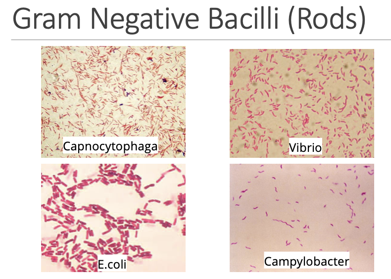
How is the morphology of Vibrio characterized?
may be curved
22
How is the appearance of Campylobacter characterized on a gram stain?
small curved, faint staining
23
Which forms are associated with Haemophilus?
small pleomorphic or coccobacillary
24
How are Pasteurella, Brucella, and Bordetella characterized in terms of size and form?
small, short, almost coccoid
25
How is the structure of Capnocytophaga defined?
thin long with pointed ends
26
Which morphology is assigned to Moraxella and Acinetobacter?
coccobacillary
27
How is Moraxella catarrhalis specifically classified?
Moraxella catarrhalis is true cocci
28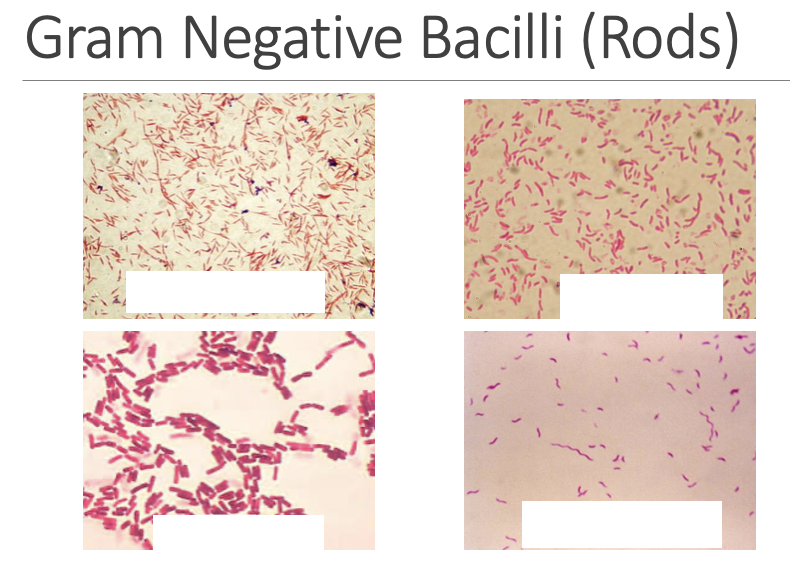
29
What are the characteristics of Neisseria?
Gram Negative Cocci:
spheres in single or pairs. Pairs often have “kidney bean” or “coffee bean” shape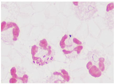
spheres in single or pairs. Pairs often have “kidney bean” or “coffee bean” shape
30
What factors can cause Gram stain characteristics of an organism to vary?
Organisms may look atypical in very young, old, dead or degenerating cultures and in specimens collected post-antibiotic therapy.
Must evaluate gram stain quality (thickness, stain time, stain precipitate)
Must evaluate gram stain quality (thickness, stain time, stain precipitate)
31
How is Sheep Blood Agar (BA/BAP/SBA) classified in terms of selectivity?
Primary, non-selective
32
What components make up Sheep Blood Agar?
5% sheep blood and trypticase soy agar
33
What hemolytic reactions can be identified using BAP?
Alpha: green around colony.
Beta: complete clearing around colony.
Gamma/Non-hemolytic: no hemolysis around colony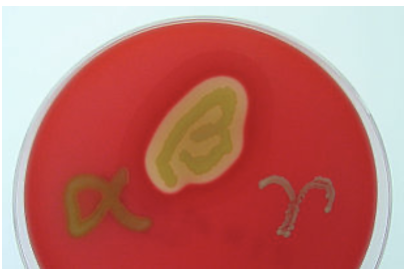
Beta: complete clearing around colony.
Gamma/Non-hemolytic: no hemolysis around colony
34
Which fastidious organisms are recovered on Chocolate Agar (CA/CAP/CHOC)?
Recovers fastidious organism such as Neisseria gonorrhoeae and Haemophilus species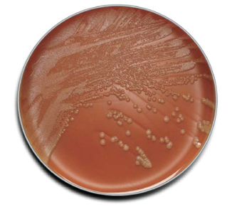
35
How is CHOC prepared, and what factors are released during preparation?
Same preparation as BAP, but RBCs are lysed releasing hemin (X factor) and NAD (V factor)
36
How do some manufacturers further enrich Chocolate Agar?
with IsoVitalex
37
Why will certain lipophilic organisms not grow on CHOC?
Lacks lipids. Lipophilic organisms such as certain species of Corynebacterium (i.e., Corynebacterium jeikeium) will not grow
38
What selective agents in MacConkey Agar (MAC) inhibit gram positive flora?
Bile salt and crystal violet inhibit gram positive flora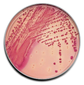
39
Which differential indicator is used in MacConkey Agar?
Neutral red pH indicator
40
How do lactose fermenters react on MacConkey Agar and what color change occurs?
Lactose fermenters produce organic acid which reduce pH, turning neutral red reddish pink. The neutral red is absorbed by the bacteria which appears bright pink on the agar
41
How are lactose-positive and lactose-negative organisms designated on MacConkey Agar?
Lactose positive (Lactose fermenter, LF). Lactose negative (Non-lactose fermenter, NLF)
42
What reaction occurs with strong lactose fermenters involving bile salts?
Strong lactose fermenters produce enough acid to cause precipitation of bile salts resulting in pink halo surround colonies
43
What defining microscopic and colony features characterize Staphylococcus?
The gram positive cocci appear in clusters. The colonies are typically flatter, more opaque and are catalase positive
44
What defining microscopic and colony features characterize Streptococcus?
The gram positive cocci appear in pairs/chains and the colonies are typically raised, more translucent. Catalase negative.
45
What types of infections can Staphylococcus aureus cause in humans?
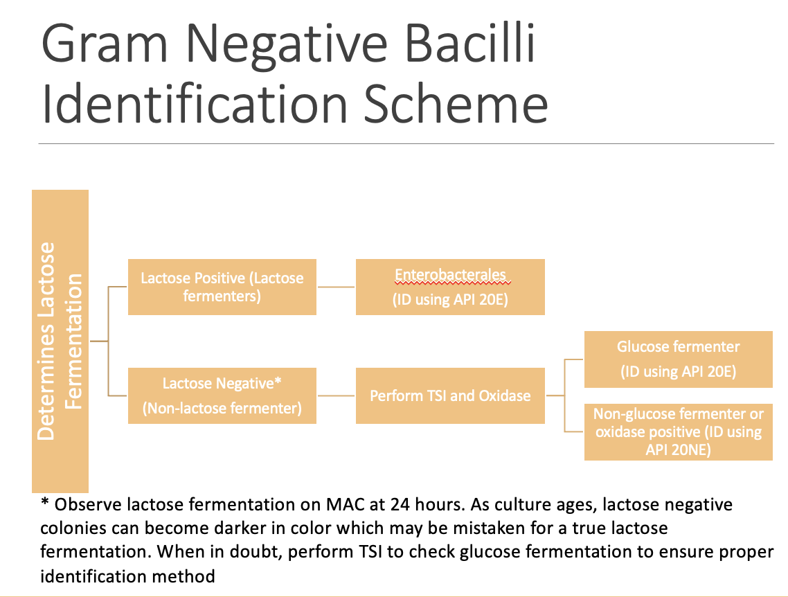
Important human pathogen that can cause a variety of infections ranging from skin infections (cellulitis, abscess, carbuncles) to systemic illnesses (bacteremia, endocarditis, meningitis, osteomyelitis)
46
What condition is associated with Staphylococcus aureus and who is affected?
Staphylococcal Scalded Skin Syndrome: exfoliation of the skin in neonates and young children
47
Which toxin-mediated syndrome is linked to Staphylococcus aureus?
Toxic Shock Syndrome via TSST-1 (assoc. tampons)
48
What immunologic effects are triggered by TSST-1 in Staphylococcus aureus?
Stimulate T-cells and induce TNF and cytokine interleukin-1
49
What food-related illness is linked to Staphylococcus aureus?
Food poisoning via enterotoxin
50
Which antimicrobial resistance patterns are associated with hospital-acquired Staphylococcus aureus?
Nosocomial pathogen of hospitalized patients. Methicillin-resistant SA (MRSA). Vancomycin-intermediate and –resistant (VISA, VRSA)
51
Where does SA commonly colonize, and in what situations is screening performed?
Colonizes the nares (screens in-patient, surgical patients for MRSA by culture or PCR)
52
What colonial morphology is characteristic of SA?
Gram Stain: GPCCl - Gram-Positive Cocci in Clusters
Dense, large, smooth, butyrous colonies. Typically, golden/yellow. Beta-hemolytic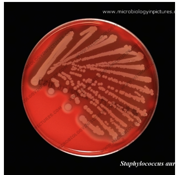
Dense, large, smooth, butyrous colonies. Typically, golden/yellow. Beta-hemolytic
53
Which biochemical reactions are positive in SA?
Catalase (+)
Slide coagulase (+)
Tube coagulase (+)
Slide coagulase (+)
Tube coagulase (+)
54
Which alternative test may laboratories use instead of the slide coagulase test for Staphylococcus aureus, and what does it detect?
Laboratories may opt for Staphaurex™ Latex Agglutination test (detects Protein A) as substitute for slide coagulase test
55
Which primary infection is associated with Staphylococcus saprophyticus and what specific conditions are included?
Primarily causes acute UTI in young, sexually active females.
Cystitis, urethritis, pyelonephritis
Cystitis, urethritis, pyelonephritis
56
How can Staphylococcus saprophyticus bacteremia occur?
S. saprophyticus bacteremia seen as complication of UTI
57
Which additional infections involving the urinary tract or prostate are associated with Staphylococcus saprophyticus?
Urethritis, catheter-associated UTI, prostatitis in elderly men
58
What possible etiologic role may Staphylococcus saprophyticus have in males?
Possible agent of non-gonococcal urethritis in males
59
Which colonial morphology features are characteristic of Staphylococcus saprophyticus?
Gram Stain: GPCCl
Large, smooth colonies, usually white. Non-hemolytic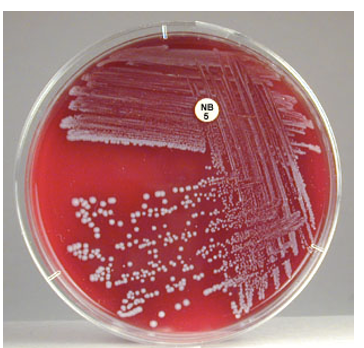
Large, smooth colonies, usually white. Non-hemolytic
60
Which biochemical reactions are associated with Staphylococcus saprophyticus?
Catalase (+).
Slide and tube coagulase (-)
Novobiocin Resistant (zone of inhibition <=16 mm)
Slide and tube coagulase (-)
Novobiocin Resistant (zone of inhibition <=16 mm)
61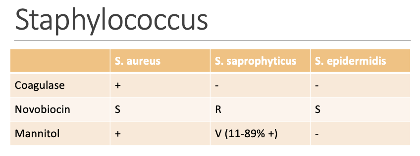
62
What selective property is associated with Mannitol Salt Agar (MS)?
high salt concentration inhibit most gram negative and some gram positive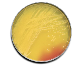
63
What differential property is associated with Mannitol Salt Agar (MS)?
organism that ferments mannitol produce yellow colonies
64
How does the Catalase Test differentiate Staphylococci from Streptococci and Enterococci?
Staphylococci (+) vs Streptococci/Enterococci (-)
65
Which groups of organisms can also be differentiated using the Catalase Test?
Can be used to differentiate among gram positive rods and some fastidious organisms
66
What is the procedure for the Catalase Test?
Streak colony onto slide and place a drop of hydrogen peroxide on it
- Positive: Rapid production of bubbles(effervescence)
- Negative: No or weak bubbling
- Catalase present in RBC, avoid carryover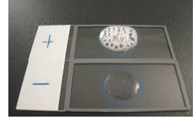
- Positive: Rapid production of bubbles(effervescence)
- Negative: No or weak bubbling
- Catalase present in RBC, avoid carryover
67
What chemical reaction represents the Catalase Test?
2H₂O₂ -- CATALASE --> 2H₂O + O₂
68
What does the Coagulase Test detect and which materials are used for the reaction?
Clotting of coagulase plasma (rabbit plasma and EDTA) by enzyme coagulase
S. aureus (+)
Other staph (-)*
S. aureus (+)
Other staph (-)*
69
What type of coagulase is detected by the slide coagulase method?
Slide coagulase detects bound coagulase (clumping factor)
70
What type of coagulase is detected by the tube coagulase method?
Tube coagulase detects extracellular free coagulase
71
How is a slide Coagulase test performed using a colony and rabbit plasma?
- Emulsify a portion of colony (using loop or wooden stick) into rabbit plasma
- Macroscopic clumping in 10 seconds is positive
- Macroscopic clumping in 10 seconds is positive
72
What must be done when a slide Coagulase test is negative?
Negative slide coagulase must be confirmed by tube coagulase method
73
How is a Tube Coagulase test performed?
- Emulsify several colonies (using loop or wooden stick) into 0.5 mL rabbit plasma
- Incubate at 35-37oC
- Observe at 1-4 hours for positive result, if negative incubate at room temperature overnight
- Incubate at 35-37oC
- Observe at 1-4 hours for positive result, if negative incubate at room temperature overnight
74
Which factor produced by Staphylococcus aureus can alter tube Coagulase results?
Staphylokinase producing SA can revert result to negative
75
How is the initial bacterial suspension prepared for the Novobiocin Test?
Using a swab, prepare 0.5 McF suspension of bacteria to be tested in 2mL 0.85% saline.
76
How is the swab prepared before streaking the Mueller Hinton agar plate in the Novobiocin Test?
Using a new swab, soak it in the suspension and wring out excess solution
77
How is the Mueller Hinton agar plate inoculated during the Novobiocin Test?
Streak the swab soaked with 0.5 McF suspension onto Mueller Hinton agar plate, creating a lawn (streaks should be tight with no gaps in between)
78
How is the novobiocin disk applied during the Novobiocin Test?
Drop a novobiocin disk on to the surface of the streaked agar
79
What incubation conditions are required for the Novobiocin Test?
Incubate in ambient air at 35–37°C overnight
80
How is resistance or susceptibility determined in the Novobiocin Test?
Measure the diameter zone size:
Resistant: <=16 mm.
Susceptible: >16 mm
Resistant: <=16 mm.
Susceptible: >16 mm
81
What are some differences between Staphylococcus v. Micrococcus?
Both Staphylococci and Micrococci are gram positive cocci in clusters. Micrococci tend to be larger in size, form tetrads, may be yellow pigmented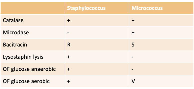
82
How are Streptococci characterized?
Gram positive cocci in pairs/chains. May appear rod-like from agar plate media
83
Which medium is essential for identifying hemolysis patterns in Streptococci?
Hemolysis on Sheep Blood Agar is key to identifying Streptococci
84
How is alpha hemolysis defined for Streptococci and which species are associated with it?
Alpha: Partial/incomplete hemolysis, greening around the colony.
- S. pneumoniae and S. viridans
- S. pneumoniae and S. viridans
85
How is beta hemolysis defined for Streptococci and which species produce it?
Beta: Complete lysis of RBC, clearing zone around colony.
- S. pyogenes (GAS/BSA) and S. agalactiae (GBS/BSB)
- S. pyogenes (GAS/BSA) and S. agalactiae (GBS/BSB)
86
How is gamma hemolysis defined and which species demonstrates this pattern?
Gamma: Non-hemolytic, no hemolysis around colony.
- Enterococcus faecalis
- Enterococcus faecalis
87
What characteristics define Streptolysin O?
Oxygen labile
Antigenic
Stab agar to enhance hemolysis
Antigenic
Stab agar to enhance hemolysis
88
What characteristics define Streptolysin S?
Oxygen stable
Non-antigenic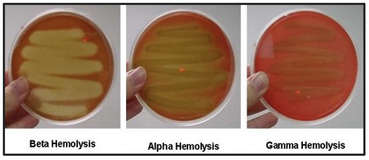
Non-antigenic
89
Where is Streptococcus pneumoniae normally found in the human body?
Normal upper respiratory flora
90
Which infections are associated with Streptococcus pneumoniae?
Otitis media and bacteremia in infants and children
91
Which major respiratory infection is commonly caused by Streptococcus pneumoniae?
Community-acquired bacterial pneumonia
92
Which serious central nervous system infection is frequently caused by Streptococcus pneumoniae and has a vaccine available?
Common cause of meningitis (vaccine available)
93
Which additional infections can be caused by Streptococcus pneumoniae?
Sinusitis, conjunctivitis, arthritis, endocarditis
94
What gram stain appearance is associated with Streptococcus pneumoniae?
Gram positive diplococci, lancet-shaped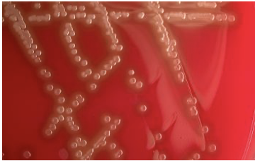
95
What colony morphology features are characteristic of Streptococcus pneumoniae?
alpha, glistening, gray; mucoid in strains has capsule
umbilicated (depressed center) due to autolytic enzyme
umbilicated (depressed center) due to autolytic enzyme
96
Which biochemical reactions identify Streptococcus pneumoniae?
Catalase (-).
Optochin (P Disk) - S.
Bile (10% sodium deoxycholate) soluble
Optochin (P Disk) - S.
Bile (10% sodium deoxycholate) soluble
97
Which body sites normally contain Streptococcus viridans as part of their flora?
respiratory, gastrointestinal, urogenital, skin
98
Which infections are commonly caused by Streptococcus viridans?
Common cause of subacute bacterial endocarditis (heart valves), abscesses, bacteremia (subsequent to dental procedures)
99
What gram stain characteristics are associated with Streptococcus viridans?
Gram Stain: GPCPrch, sometimes elongated and rod-like
100
Which colony morphology features are described for Streptococcus viridans?
minute to small, gray, domed, smooth or matte, alpha or gamma hemolytic
101
Which biochemical reactions characterize Streptococcus viridans?
Catalase (-).
Optochin R.
Bile insoluble.
Bile-esculin (+/-).
6.5% NaCl (-).
PYR (-).
LAP (+)
Optochin R.
Bile insoluble.
Bile-esculin (+/-).
6.5% NaCl (-).
PYR (-).
LAP (+)
102
How do optochin and bile solubility assist in identifying Streptococcus viridans?
Optochin and bile solubility helps differentiates from Streptococcus pneumoniae
103
How do bile esculin and 6.5% NaCl reactions help differentiate Streptococcus viridans from other alpha-hemolytic Streptococci?
Bile esculin and 6.5% NaCl helps different from other alpha-hemolyticus Streptococci (including Enterococci)
104
What does the Optochin (P Disk, Taxo P) test differentiate?
Differentiates Streptococcus pneumoniae from other alpha hemolytic Streptococci
105
What is the chemical nature and mechanism of action of Optochin?
Optochin = ethylhydrocupreine hydrochloride (5.0 ug) interferes with ATPase and production of ATP
106
What result indicates a positive Optochin test?
zone of inhibition ≥ 14mm in diameter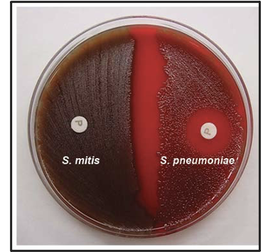
107
What requirement applies to equivocal Optochin results?
Equivocal: any ZOI <14mm should be confirmed by bile solubility test
108
What mechanism does the Bile Solubility Test rely on in Streptococcus pneumoniae?
Activates autolysin produced by S. pneumoniae which enhances lytic reaction.
109
How do Streptococcus pneumoniae and other alpha-hemolytic streptococci differ in bile solubility?
S. pneumoniae (+/soluble).
Other alpha-hemolytic streptococci (-/insoluble)
Other alpha-hemolytic streptococci (-/insoluble)
110
How is the Bile Solubility Test performed?
Add drop of 2% sodium deoxycholate into well-isolated colonies on SBA
Incubate with lid up at 35°C for 30 min
Incubate with lid up at 35°C for 30 min
111
What indicates a positive or negative Bile Solubility Test?
Positive: colony disintegrates, imprint may remain
Negative: intact colonies
Negative: intact colonies
112
Which illness caused by Streptococcus pyogenes accounts for most cases in school-age children?
Streptococcal pharyngitis
113
Which additional upper-respiratory infections are associated with Streptococcus pyogenes?
Tonsillitis, sinusitis
114
What treatment guidance applies when Streptococcus pyogenes infections require therapy?
Usually self-limiting, but if treatment is necessary penicillin is still the treatment of choice
115
Which superficial cutaneous infections are linked to Streptococcus pyogenes?
Implicated in other superficial cutaneous infections: impetigo (red skin lesions), erysipelas (soft tissues), cellulitis (spreading inflammation)
116
Which severe toxin-mediated condition is associated with Streptococcus pyogenes Group A?
Group A streptococcus toxic shock-like syndrome
117
Why is isolation of Streptococcus pyogenes(Group A) from a non-respiratory site significant, and what condition may it be implicated in?
Isolation of S. pyogenes from non-respiratory site is a significant finding and is reported to the clinician (implicated in necrotizing fasciitis)
118
Which clinical manifestations define acute rheumatic fever as a complication of Streptococcus pyogenes(Group A) infections?
carditis, polyarthritis, subcutaneous nodules, chorea (neurologic – muscular spasms, weakness)
119
What pathologic changes define acute glomerulonephritis as a complication of Streptococcus pyogenes (Group A) infections?
inflammation of the glomeruli leads to hypertension, hematuria, proteinuria
120
What mechanism is thought to underlie complications following Streptococcus pyogenes infections?
Complications mostly thought to be through formation of antistreptococcal antibodies that cross-react with antigens on various organs
121
What gram stain characteristics are associated with Streptococcus pyogenes (Group A) and what is the colony morphological features?
Gram Stain: GPCPrCh
Colony morphology: Grayish white, transparent to translucent, matte or glossy small to pinpoint colonies with large zone of beta hemolysis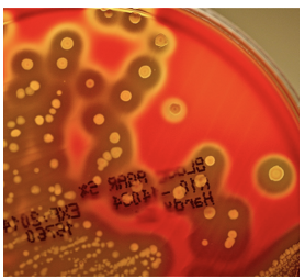
Colony morphology: Grayish white, transparent to translucent, matte or glossy small to pinpoint colonies with large zone of beta hemolysis
122
Which biochemical reactions identify Streptococcus pyogenes (Group A)?
Catalase (-)
Lancefield Group A
PYR (+)
Bacitracin (A Disk, Taxo A) - susceptible
SXT (Trimethoprim/sulfamethoxazole) - resistant
Bile-esculin (-)
6.5% Nacl (-)
LAP (+)
Lancefield Group A
PYR (+)
Bacitracin (A Disk, Taxo A) - susceptible
SXT (Trimethoprim/sulfamethoxazole) - resistant
Bile-esculin (-)
6.5% Nacl (-)
LAP (+)
123
Which hemolysins are produced by Streptococcus pyogenes(Group A)?
Produces Streptolysin S (oxygen-stable) and Streptolysin O (oxygen-labile)
124
Where is Streptococcus agalactiae(Group B) normally found as part of the human flora?
Normal flora of gastrointestinal tract
125
How can Streptococcus agalactiae(Group B) be carried in females, and what risk does this pose to newborns?
Female may carry GBS as vaginal flora. Can cause sepsis and meningitis in neonates
126
Which infections are associated with Streptococcus agalactiae(Group B) in postpartum or adult patients?
Postpartum fever, osteomyelitis, wound infections, endocarditis
127
What gram stain characteristics and colony morphology are associated with Streptococcus agalactiae (Group B)?
Gram Stain: GPCPrCh
Colony morphology: larger colonies than Grp A, translucent to opaque; flat, glossy, narrow zone of BH, some strains are gamma.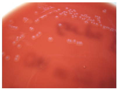
Colony morphology: larger colonies than Grp A, translucent to opaque; flat, glossy, narrow zone of BH, some strains are gamma.
128
Which biochemical reactions characterize Streptococcus agalactiae (Group B)?
Biochemical:
Catalase (-)
Lancefield Group B
PYR (-)
Bacitracin (R)
SXT (R)
Bile-esculin (-)
6.5% NaCl (+)
LAP (+)
CAMP (+)
Hippurate (+)
Catalase (-)
Lancefield Group B
PYR (-)
Bacitracin (R)
SXT (R)
Bile-esculin (-)
6.5% NaCl (+)
LAP (+)
CAMP (+)
Hippurate (+)
129
What purpose does the Lancefield Antigen system serve in streptococcal identification?
Distinguish beta-hemolytic streptococcus
130
How is antigen extracted for Lancefield latex agglutination testing?
antigen is extracted from organism using nitrous acid extraction procedure or physical grinding and react with group-specific antisera
131
What antigens are associated?
Lancefield antigen: A, B, C, F, G
132
Which organism is now associated with Lancefield Group F?
Group F is now associated with Streptococcus anginosus group (small colony beta streptococcus)
133
How does the PathoDX Strep Grouping Kit function in beta-hemolytic streptococci grouping?
Antibody from latex particles reacts and agglutinates with streptococcal antigens extracted from bacterial cell wall.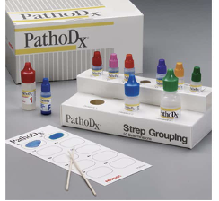
134
Which cross-reactions may occur due to shared antigenicity in beta-hemolytic streptococci grouping?
Limitations: organisms that share similar antigenicity may cross-react, e.g.:
pneumoniae with Strep C latex
Listeria monocytogenes with Strep B/G
Streptococcus porcinus types as group B
Viridans group Streptococcus maybe have more than 1 demonstrable group antigen
A, C, G Streptococci must be differentiated from minute A, C, F, G Streptococcus anginosus
pneumoniae with Strep C latex
Listeria monocytogenes with Strep B/G
Streptococcus porcinus types as group B
Viridans group Streptococcus maybe have more than 1 demonstrable group antigen
A, C, G Streptococci must be differentiated from minute A, C, F, G Streptococcus anginosus
135
What steps are required for the extraction phase of the PathoDX Strep Grouping Kit procedure?
Extraction Method Procedure
Extraction:
Add 2 drops each of Reagent 1 and 2 to test tube
Pick 1-4 beta colonies (more maybe used for younger/smaller colonies) with wooden stick and mix into the reagent
Add 4 drops of Reagent 3 and mix by tapping the tube
Extraction:
Add 2 drops each of Reagent 1 and 2 to test tube
Pick 1-4 beta colonies (more maybe used for younger/smaller colonies) with wooden stick and mix into the reagent
Add 4 drops of Reagent 3 and mix by tapping the tube
136
How is the testing phase performed when using the PathoDX Strep Grouping Kit?
Testing:
Add 1 drop of your extracted bacterial antigen to 1 drop of selected strep grouping latex on a PathoDX reaction card
Mix with applicator stick
Observe for agglutination
Add 1 drop of your extracted bacterial antigen to 1 drop of selected strep grouping latex on a PathoDX reaction card
Mix with applicator stick
Observe for agglutination
137
How is the Direct Method Procedure performed using the PathoDX Strep Grouping Kit?
Direct Method Procedure:
Rub colonies onto PathoDx reaction card (mechanically releases the antigen on the cell wall)
Add selected strep grouping latex and mix as per previous procedure
Non-specific clumping may be observed due to increased amount of organism used
Rub colonies onto PathoDx reaction card (mechanically releases the antigen on the cell wall)
Add selected strep grouping latex and mix as per previous procedure
Non-specific clumping may be observed due to increased amount of organism used
138
What biochemical reaction defines the PYR Test?
Pyrrolidonyl aminopeptidase enzyme hydrolyzes L-pyrrolidonyl-β-naphthylamide to β-naphthylamide
139
How does the color developer function in the PYR Test?
Addition of color developer para-dimethylaminocinnamaldehyde complex with free naphthylamide to produce red color
140
What is the step-by-step procedure for performing the PYR Test?
Moisten PYR disk with small amount of water
Rub small amount of colony to be tested onto the disk
Wait 2 minutes
Add a drop of color developer on to the disk
Rub small amount of colony to be tested onto the disk
Wait 2 minutes
Add a drop of color developer on to the disk
141
How are PYR Test results interpreted?
Positive – pink color. Negative – no color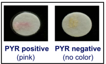
142
What organisms can be differentiated using the Bacitracin (Taxo A) disk test?
Differentiates
1. β-hemolytic Grp A streptococci (S) from other β-hemolytic streptococci (R). 2. Staphylococcus (R) from Micrococcus (S)
1. β-hemolytic Grp A streptococci (S) from other β-hemolytic streptococci (R). 2. Staphylococcus (R) from Micrococcus (S)
143
What is the antibiotic and mechanism involved in the Bacitracin (Taxo A) disk test?
Antibiotic: bacitracin (0.04 units). Inhibits synthesis of bacterial cell walls
144
What preparatory steps are required before placing the Bacitracin (Taxo A) disk on the agar?
Streak lawn on BAP with 2–3 colonies of isolate. Place bacitracin disk on surface of agar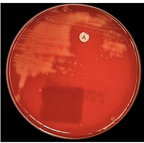
145
What incubation conditions are required for the Bacitracin (Taxo A) disk test?
Incubate plate for 18–24h at 35–37°C in:
- streptococci: 5–10% CO₂.
- staphylococci: ambient air
- streptococci: 5–10% CO₂.
- staphylococci: ambient air
146
How is a positive (susceptible) result defined in the Bacitracin (Taxo A) disk test?
Positive (Susceptible): zone of inhibition >7–10mm
147
What infections can Enterococcus species cause?
Enterococcus species can cause UTI, bacteremia, endocarditis, intrabdominal and pelvic infections, wound and soft-tissue infections, neonatal sepsis.
148
What gram stain characteristics and colony morphology are associated with Enterococcus species?
Gram Stain: GPCPrCh
Colony morphology:
- Small, cream, or white, round, convex, smooth, entire
- Typically gamma but can be beta or alpha.
Colony morphology:
- Small, cream, or white, round, convex, smooth, entire
- Typically gamma but can be beta or alpha.
149
Which biochemical reactions characterize Enterococcus species?
Biochemical:
Catalase (-) some have peroxidase that slowly catalyzes H2O2 – not truly positive
Bile-esculin (+)
6.5% NaCl (+)
PYR (+)
Lancefield group D (not typed)
LAP (+)
Catalase (-) some have peroxidase that slowly catalyzes H2O2 – not truly positive
Bile-esculin (+)
6.5% NaCl (+)
PYR (+)
Lancefield group D (not typed)
LAP (+)
150
What does the 6.5% NaCl (Salt) Tolerance Test differentiate?
Differentiates enterococci (+) from non-enterococci (–).
151
What does the 6.5% NaCl broth contain?
Heart infusion broth containing 6.5% NaCl, small amount of glucose, and pH indicator bromocresol purple.
152
How do you inoculate the 6.5% NaCl test?
Inoculate from 18–24h culture into 6.5% NaCl broth at 35–37°C in ambient air.
153
How do you interpret a 6.5% NaCl tolerance result?
Positive: visible turbidity in broth, with or without color
change from purple to yellow
Negative: no turbidity and no color change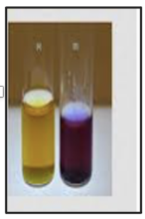
Negative: no turbidity and no color change
154
What is the purpose of the Bile-Esculin Test?
Presumptive ID of enterococci and organisms in the Streptococcus bovis group.
155
What does the Bile-Esculin Test detect?
Ability to grow in the presence of 4% bile and hydrolyze esculin to esculetin (Esculetin reacts with Fe3+ to form dark brown / black precipitate).
156
How do you inoculate and incubate the Bile-Esculin Test?
Inoculate onto the surface of a slant and incubate at 35–37°C in ambient air.
157
How do you interpret a Bile-Esculin result?
Positive: black slant.
Negative: no color change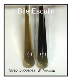
Negative: no color change
158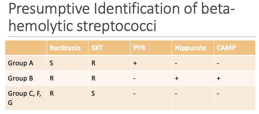
159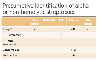
160
What types of infections are Corynebacterium species(Gram Positive Bacilli) normally associated with?
Most coryneform bacteria are considered part of normal flora (skin, urogenital, respiratory)
161
What are the Gram stain characteristics of Corynebacterium species?
Pleomorphic, diphtheroid, non-spore-forming gram positive rod (PLGPR)
Palisading/picket fence/chinese letters/forming V, Y, L arrangement
Palisading/picket fence/chinese letters/forming V, Y, L arrangement
162
What is the colony morphology of Corynebacterium species and its biochemical reaction?
Varies depending on the species. Catalase strongly positive.
163
How are pleomorphic gram-positive rods that are catalase positive typically reported?
Can be reported as Corynebacterium species, unless further speciation is needed.
164
What infections are caused by Corynebacterium jeikeium?
Bacteremia and sepsis, valve endocarditis, meningitis, pneumonia, and soft-tissue infections.
165
What types of healthcare-associated infections are linked to Corynebacterium jeikeium?
Nosocomial infections and infections associated with prosthetic device implants.
166
When coryneform bacteria are isolated from normally sterile body sites, what organism should be ruled out?
Corynebacterium jeikeium.
167
What does Corynebacterium jeikeium look like on Gram stain?
May appear coccobacillary instead of a pleomorphic gram-positive rod.
168
What is the colony morphology of Corynebacterium jeikeium?
Pinpoint colonies; lipophilic and grows poorly on chocolate agar due to absence of lipids.
169
What are the key biochemical reactions of Corynebacterium jeikeium?
CAT (+)
MOT (-)
URE (-), NIT (-), Glucose (+)
MOT (-)
URE (-), NIT (-), Glucose (+)
170
What is notable about Corynebacterium jeikeium’s antimicrobial profile?
Multidrug resistant but remains susceptible to vancomycin.
171
What are the key shared characteristics of all Enterobacterales (formerly Family Enterobacteriaceae)?
All members have the following characteristics:
Ferments glucose
Oxidase negative (exception of Plesiomonas)
Nitrate (+)
Growth on Mac
Ferments glucose
Oxidase negative (exception of Plesiomonas)
Nitrate (+)
Growth on Mac
172
What is the most common Enterobacterales isolate?
Escherichia coli.
173
Which Enterobacterales species are not part of normal GI flora?
Most species are normal GI tract flora except Salmonella, Shigella and Yersinia
174
What infections can Enterobacterales cause?
May cause UTIs, gastroenteritis, septicemia, food poisoning, wound infections, pneumonia, and meningitis.
175
Which organisms are H₂S positive?
Salmonella, Citrobacter freundii, Proteus (except P. penneri, which is variable).
176
Which Enterobacterales are non-motile?
Motility (-): Shigella, Klebsiella, Yersinia (35oC)
177
Which organisms are VP positive?
Klebsiella, Enterobacter, Serratia.
178
Which organisms are gelatin positive / DNase positive?
Serratia and Proteus.
179
Which organisms are ADH positive?
Salmonella (70%), Citrobacter (65%), Enterobacter cloacae.
180
Which organisms are anaerogenic (do not produce gas from glucose fermentation)?
Shigella, Salmonella typhi, Yersinia, Providencia (except P. alcalifaciens).
181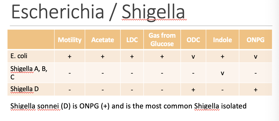
182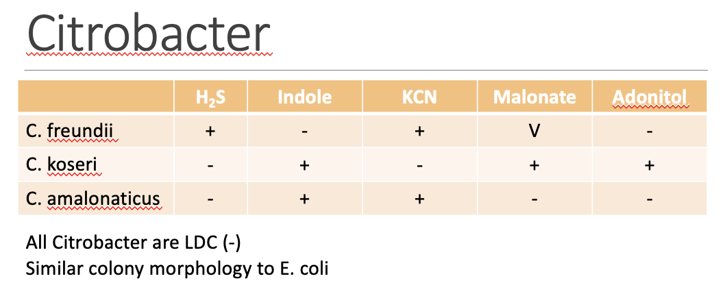
183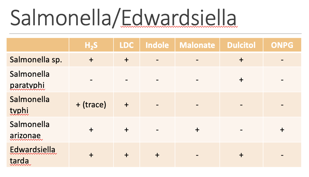
184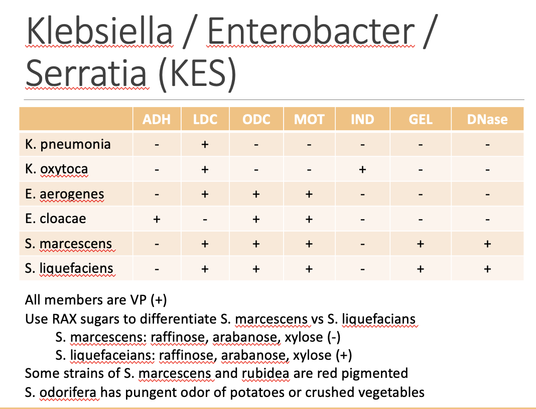
185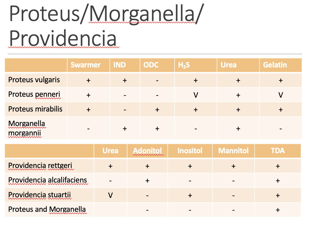
186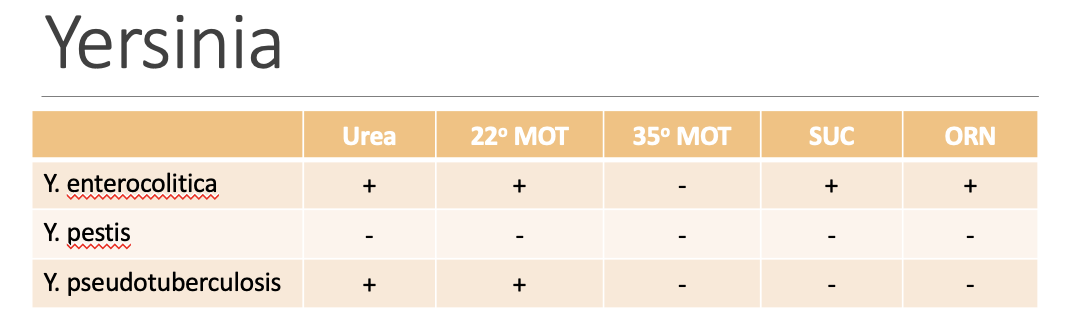
187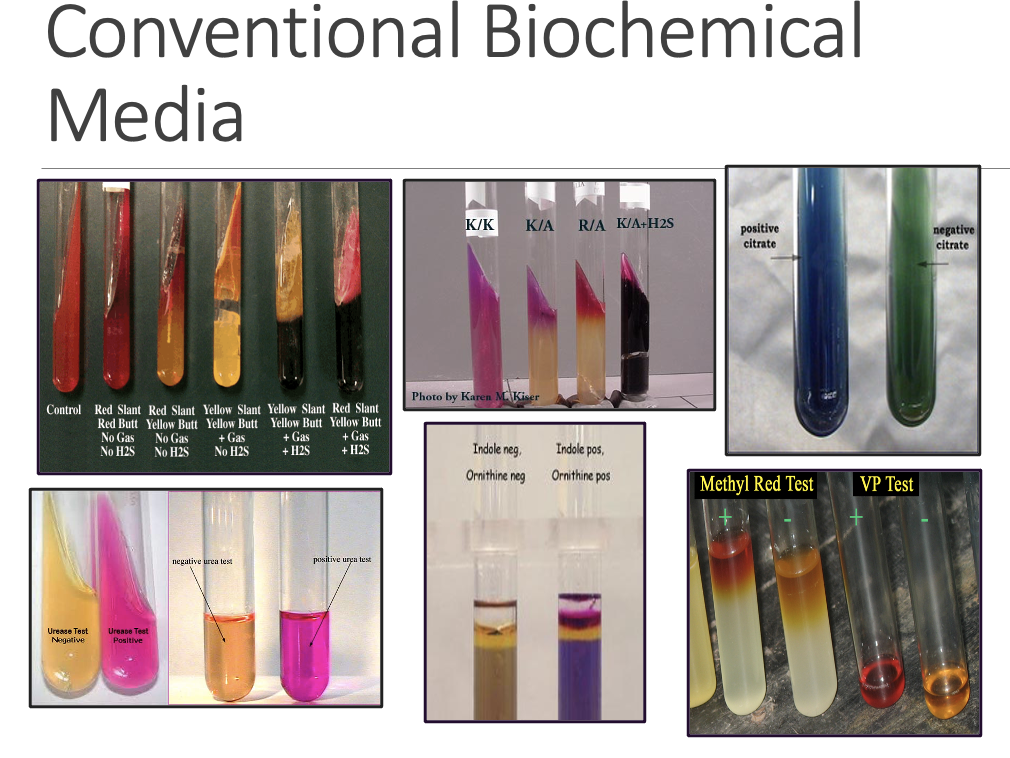
188
What does the TSI (Triple Sugar Iron) Agar test detect?
-Carbohydrate utilization (glucose, lactose, sucrose)
-Gas production (CO2)
-H2S production
-KIA (Kligler Iron Agar) is essentially same as TSI, except does not contain sucrose
-Gas production (CO2)
-H2S production
-KIA (Kligler Iron Agar) is essentially same as TSI, except does not contain sucrose
189
What are the key components of the TSI medium?
Sugar: Lactose, Sucrose, Glucose in 10:10:1
pH indicator: Phenol Red
H2S indicator: Ferrous sulfate (sodium thiosulfate as source of sulfur) complex with H2S to form black precipitate
pH indicator: Phenol Red
H2S indicator: Ferrous sulfate (sodium thiosulfate as source of sulfur) complex with H2S to form black precipitate
190
What color is an uninoculated TSI tube?
Uninoculated tube is red
191
How do you inoculate a TSI tube?
Stab the butt once and then streak the slant. incubate tube at 35-37°C in ambient air for 18-24 h with loosen cap
192
How are TSI reactions recorded and what do the primary color changes indicate (A, K, H₂S, gas)?
Record result as slant / butt
Yellow color = Acid (A)
Pink / red color = Alkaline (K)
Formation of black precipitate = H2S positive (*H2S forms under acidic condition only)
Bubbles in the media = gas produced (Denotes with “G”)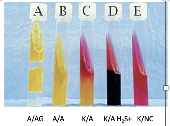
Yellow color = Acid (A)
Pink / red color = Alkaline (K)
Formation of black precipitate = H2S positive (*H2S forms under acidic condition only)
Bubbles in the media = gas produced (Denotes with “G”)
193
What does lactose or sucrose fermentation look like on TSI?
Yellow slant.
194
What does glucose fermentation look like on TSI?
Yellow butt.
195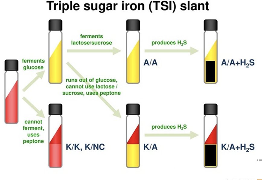
196
What does LIA (Lysine Iron Agar) detect?
Lysine decarboxylation, lysine deamination, and H₂S production.
197
What are the main components of LIA?
Lysine, dextrose, peptone, ferric ammonium citrate, and sodium thiosulfate.
198
What is the indicator used in LIA?
Bromocresol purple.
199
How are LIA results recorded?
Record result as slant / butt
- Yellow color = Acid (A)
- Purple color = Alkaline (K)
- Red color slant = Red (R)
- Formation of black precipitate = H2S positive
*H2S cannot be detected in LIA if lysine decarboxylase is negative (an acid production in the butt may suppress H2S formation)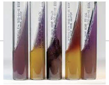
- Yellow color = Acid (A)
- Purple color = Alkaline (K)
- Red color slant = Red (R)
- Formation of black precipitate = H2S positive
*H2S cannot be detected in LIA if lysine decarboxylase is negative (an acid production in the butt may suppress H2S formation)
200
How do you interpret LIA results?
Lysine decarboxylation produces cadaverine turns the butt alkaline again (returns to purple) - Alkaline Butt (Purple) = LDC +
Lysine deamination produce α-ketocarboxylic acid, which reacts with iron to form a red slant - Red Slant = LDA (+)
Lysine deamination produce α-ketocarboxylic acid, which reacts with iron to form a red slant - Red Slant = LDA (+)
201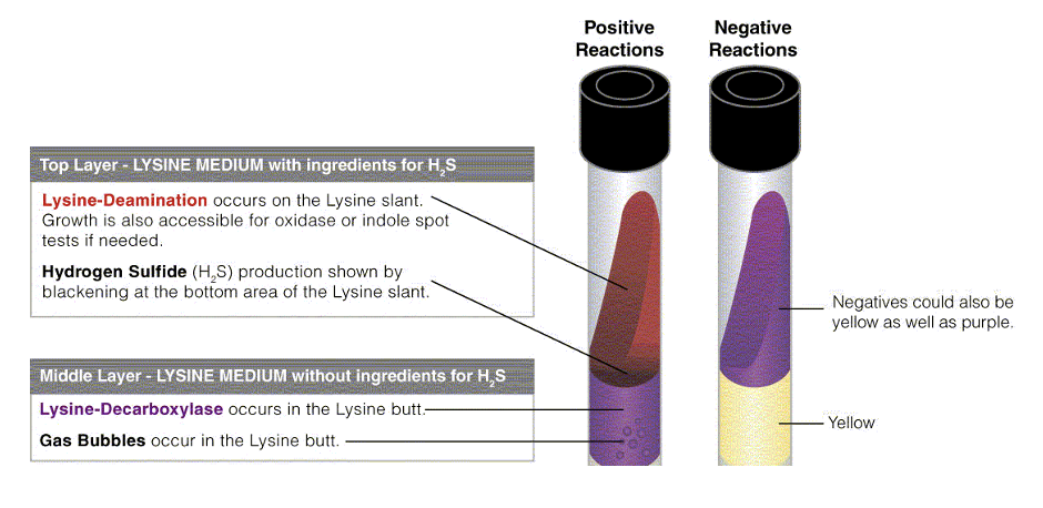
202
What does urease do in the urea slant test?
Enzyme urease hydrolyze urea to ammonia producing pink color change
203
What is the indicator in the urea slant?
Phenol Red.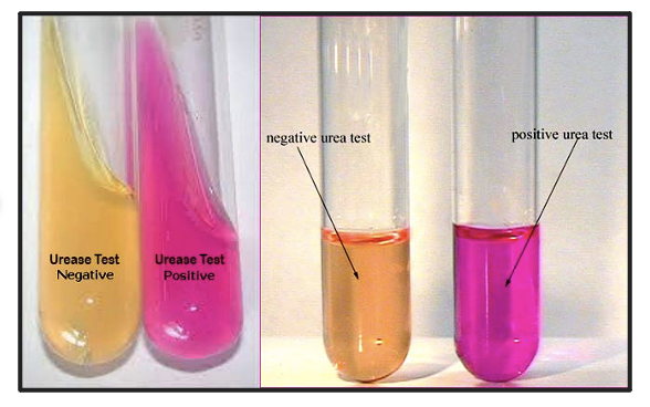
204
What is Christensen’s urea agar used for?
less buffered, more suitable for organism that produce less urease such as Klebsiella, Enterobacter, Brucella
205
What is Stuart’s urea broth used for?
Detecting high urease–producing organisms, such as Proteus (heavily buffered and virtually selective).
206
What does the OMI / Motility Indole Ornithine (MIO) medium demonstrates?
Ornithine decarboxylase
Motility - Turbidity from the stab line
Indole production - Red color after addition of Kovac’s Reagent
Motility - Turbidity from the stab line
Indole production - Red color after addition of Kovac’s Reagent
207
What is the indicator used in MIO medium?
Bromocresol purple.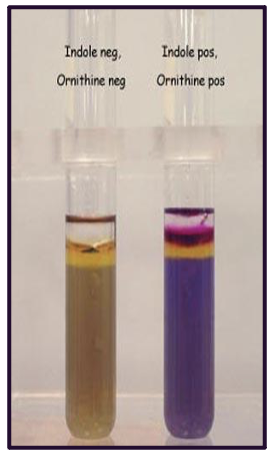
208
What happens when an organism ferments glucose in OMI medium?
It turns the medium yellow (acidic).
209
What does ornithine decarboxylase produce, and what color does it change into?
It produces putrescine, turning the medium purple (alkaline).
210
How is motility detected in OMI medium?
By growth extending outward from the line of inoculation.
211
What enzyme produces indole, and how is it detected?
If the organism has enzyme tryptophanase, it will produce indole from tryptophan. Addition of Kovacs Reagent (p-dimethylaminobenzaldehyde) to the surface (3-4 drops) produces red color band on the media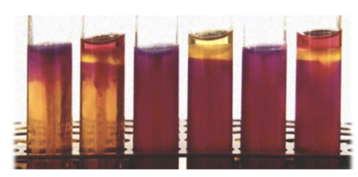
212
What does the Simmons Citrate test determine?
Whether an organism can utilize ammonium dihydrogen phosphate and sodium citrate as the sole nitrogen and carbon source.
213
What happens when citrate is utilized?
Alkaline products are produced, turning the slant blue.
214
What is the indicator in the citrate test?
Bromothymol blue.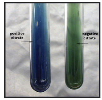
215
What does the MR-VP broth determine?
Distinguish which metabolic pathway is used to break down dextrose / glucose
216
What does Methyl Red (MR) in a MR-VP Broth indicate?
Methyl Red: mixed acid pathway producing acidic end products (lactic, acetic, formic acid)
217
What does Voges-Proskauer (VP) in a MR-VP Broth indicate?
Voges-Proskauer: butylene glycol pathway producing neutral end product (acetoin)
218
Can an organismuse use both pathways in MR-VP Broth?
Organism that utilizes one pathway will not use the other
219
How is the MR-VP test performed?
Inoculate MR-VP broth and incubate at 35–37°C in ambient air, then split broth into aliquots for separate MR and VP tests.
220
What does the Methyl-Red (MR) test detect?
Mixed acid fermentation.
221
What indicator is used in the MR test?
Methyl red indicator (1-2 drops per 1 mL broth)
at pH 4.4: MR = red (+)
at pH6.2: MR = yellow (-)
at pH 4.4: MR = red (+)
at pH6.2: MR = yellow (-)
222
What do shades of orange results indicate in the MR test?
Negative or inconclusive.
223
Why does the MR test stay red in positive reactions?
Methyl red will stay red in the
Presence of acid end products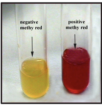
224
What does the VP test detect?
Detects neutral end-products of glucose digestion via butylene-glycol pathway
225
What reagents are used in the VP test?
Alpha-naphthol and potassium hydroxide (KOH).
226
What is the procedure for a VP test?
1.cAdd 0.6mL (6 drops) of alpha-naphthol and 0.3mL (3 drops) of KOH to 0.5 mL of MRVP broth
2.cShake well after addition of each reagent
3. Observe for 5 min
2.cShake well after addition of each reagent
3. Observe for 5 min
227
What is a positive or negative VP test result?
Interpretation
Positive: red (acetoin production)
Negative: yellow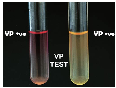
Positive: red (acetoin production)
Negative: yellow
228
What enzyme is detected in the indole test?
Tryptophanase, which hydrolyzes tryptophan to form indole.
229
What is the biochemical pathway of indole formation?
Tryptophan → indolepyruvic acid (intermediate) → indole + pyruvate + ammonium.
230
What is in Kovac’s reagent?
p-dimethylaminobenzaldehyde (p-DMAB), isoamyl alcohol, and hydrochloric acid.
231
What indicates a positive indole reaction using Kovac’s reagent?
A red-colored compound forms at the surface.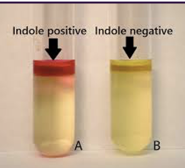
232
Which indole test method is more sensitive—tube or spot?
Tube method using Kovac’s reagent is more sensitive than the spot indole test.
233
What is the most sensitive indole test method?
Tube method using Ehrlich’s reagent (requires extraction with xylene).
234
What reagent is used in the Spot Indole Test?
p-dimethylaminocinnamaldehyde (DMACA).
235
What color is produced when indole reacts with DMACA?
Indole combines with DMACA to form blue-green compound
236
How is the Spot Indole Test performed?
Saturate filter paper with spot indole reagent
- use wooden applicator stick to rub portion of org on paper
- observe for a color change; most turn blue w/in 30 sec
** Cannot be performed on mixed culture (diffusion cause false positive)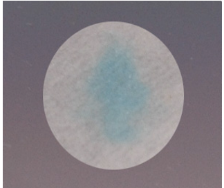
- use wooden applicator stick to rub portion of org on paper
- observe for a color change; most turn blue w/in 30 sec
** Cannot be performed on mixed culture (diffusion cause false positive)
237
What is IMViC used for?
Older biochemical grouping/mnemonic used to differentiate Enterobacterales.
238
What does IMViC stand for?
Indole, Methyl Red, Voges-Proskauer, Citrate.
Example:
E. coli: ++--
(Indole positive, MR positive, VP negative, citrate negative)
Klebsiella pneumonia: --++
Proteus mirabilis: -+-+
Citrobacter freundii: -+-+
Enterobacter aergoenes: --++
Example:
E. coli: ++--
(Indole positive, MR positive, VP negative, citrate negative)
Klebsiella pneumonia: --++
Proteus mirabilis: -+-+
Citrobacter freundii: -+-+
Enterobacter aergoenes: --++
239
Why is IMViC not sufficient alone for species identification?
Other biochemical tests are necessary to fully differentiate bacteria species.
240
What does the API 20E system identify?
Identifies Enterobacterales and other non-fastidious gram-negative rods (can detect some non-glucose fermenters).
241
How is the API 20E system inoculated and incubated?
Inoculate 1 colony into 5 mL of 0.85% saline and incubate at 35°C in O₂ for 18–24 hours.
242
What reagents are used in the API 20E system for TDA, IND, and VP tests?
TDA(Tryptophan Deaminase): 10% ferric chloride (forms a red complex with indolepyruvic acid).
IND: Kovac’s reagent (1 drop).
VP: Alpha-naphthol + 40% KOH (1 drop each).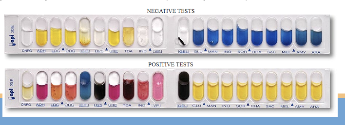
IND: Kovac’s reagent (1 drop).
VP: Alpha-naphthol + 40% KOH (1 drop each).
243
What does the ONPG test differentiate?
Differentiates late lactose fermenters from non–lactose fermenters.
244
What two functions must lactose fermenters normally have?
must be able to transport carbohydrate (via β-galactoside permease) and hydrolyze lactose to glucose and galactose (via β-galactosidase)
245
How does ONPG enter the bacterial cell?
ONPG can enter the cell without permease.
246
What reaction occurs during the ONPG test?
ONPG → galactose + o-nitrophenol (yellow).
247
What does a positive ONPG test indicate?
Presence of β-galactosidase.
248
Which Shigella species is ONPG positive?
Shigella sonnei.
249
Is ONPG available on API 20E?
Yes, ONPG is included in the API 20E panel.
250
What are the four serological antigens of Enterobacterales and what are their heat stability characteristics?
O (somatic) antigen – cell wall, heat stable;
K (envelop) antigen – capsular, heat labile;
H (flagellar) antigen – heat labile;
Vi antigen – capsular antigen of Salmonella typhi, heat labile.
K (envelop) antigen – capsular, heat labile;
H (flagellar) antigen – heat labile;
Vi antigen – capsular antigen of Salmonella typhi, heat labile.
251
Which genera require additional serological identification?
Salmonella and Shigella species require additional serological identification after full identification by biochemical methods
252
What does the Wellcolex Salmonella test use for detection?
Wellcolex Salmonella reacted with two test reagents consisting of a mixture of suspensions of red, blue and green latex particles, each of which is coated with antibody specific for different Salmonella serogroups (A, B, C, D, E ,G) based on O antigen as well as Vi antigen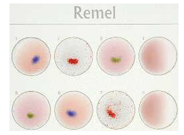
253
What percentage of human Salmonella isolates are detected by the Wellcolex test?
98% of human Salmonella isolates.
254
What additional testing can Public Health labs perform?
They can further test the H antigen for epidemiological purposes.
255
Which Shigella species correspond to serogroups A, B, C, and D?
Each serological group of Shigella corresponds to a specific specie
Shigella dysenteriae (A)
Shigella flexneri (B)
Shigella boydii (C)
Shigella sonnei (D)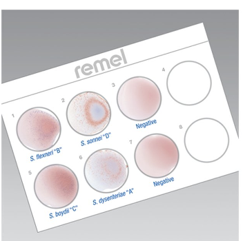
Shigella dysenteriae (A)
Shigella flexneri (B)
Shigella boydii (C)
Shigella sonnei (D)
256
257
258
What TSI reaction do non-glucose fermenters produce?
Non-glucose fermenting organism produces K/NC (no change) reaction on TSI
259
What are the three most commonly isolated non-glucose fermenting organisms?
Pseudomonas aeruginosa (1st), Acinetobacter baumannii (2nd), and Stenotrophomonas maltophilia (3rd).
260
How are non-glucose fermenters identified after confirmation by TSI?
Once confirmed as a non-glucose fermenter by TSI, identification by API20NE
261
What is the most commonly isolated non-fermenter?
Pseudomonas aeruginosa
262
What is the colony morphology of Pseudomonas aeruginosa?
Metallic sheen and grape / corn tortilla odor
263
What are the major biochemical characteristics of Pseudomonas aeruginosa?
Oxidase positive, acetamide positive, room temperature motility positive
264
When is a positive oxidase test sufficient to identify Pseudomonas aeruginosa?
If metallic sheen present, a positive oxidase test is sufficient
265
How should Pseudomonas aeruginosa be confirmed if no metallic sheen is present?
If no metallic sheen present, confirm ID with a positive oxidase and acetamide test
266
What is the substrate used in the oxidase test?
Tetramethyl-p-phenylenediamine dihydrochloride
267
What enzyme does the oxidase test detect?
Cytochrome oxidase, which oxidizes the substrate to indophenol (dark purple end product)
268
What is the procedure of the oxidase test?
- Moisten filter paper with 1% tetramethyl-p-phenylenediamine dihydrochloride
- Use platinum wire or wooden stick to rub small portion of colony (< 24h) into filter paper
- Nickel-based alloy wires containing chromium and iron (nichrome) – may cause false positive results due to oxidation products from flaming
- Observe for color change to deep blue/purple in 10 sec
- Don’t perform the test on selective / glucose containing media (fermentation products inhibit oxidase = false negative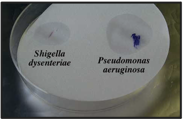
- Use platinum wire or wooden stick to rub small portion of colony (< 24h) into filter paper
- Nickel-based alloy wires containing chromium and iron (nichrome) – may cause false positive results due to oxidation products from flaming
- Observe for color change to deep blue/purple in 10 sec
- Don’t perform the test on selective / glucose containing media (fermentation products inhibit oxidase = false negative
269
What does the Acetamide Slant detect?
Enzymatic hydrolysis of acetamide with production of NH₃.
270
What indicators are used in the Acetamide Slant?
Phenol Red or Bromothymol Blue.
271
What is a positive Acetamide Slant reaction?
Green to blue with Bromothymol Blue, or dark pink to red on the slant with Phenol Red.
272
What environments can Acinetobacter baumannii be found in?
Moist natural and hospital environments; multidrug-resistant strains may be recovered in hospitals.
273
What can be seen in a Gram stain for Acinetobacter baumannii?
Gram-negative coccobacilli (almost cocci in some cases).
274
What is the colony morphology of Acinetobacter baumannii?
Smooth, opaque, raised, creamy, and smaller than Enterobacterales; lactose-negative but colonies may show a purplish hue that can be mistaken as lactose positive.
275
What are the key biochemical characteristics of Acinetobacter baumannii?
Oxidase negative, room-temperature motility negative, full ID using API 20NE.
276
Where can Stenotrophomonas maltophilia be found?
It can be found in moist natural and hospital environments.
277
What sites does Stenotrophomonas maltophilia commonly colonize?
Common colonizer of non-sterile sites (e.g., respiratory tract).
278
What are the biochemical characteristics of Stenotrophomonas maltophilia?
Oxidizes maltose faster than glucose; oxidase negative; room temperature motility positive; full ID using API 20NE.
279
Why is susceptibility testing for Stenotrophomonas maltophilia not routinely performed?
Intrinsically resistant to many antibiotics; Trimethoprim/sulfmethoxazole is treatment of choice
280
What does API 20NE identify?
Identification of non-fastidious, non-enteric gram negative rods (e.g., Pseudomonas, Acinetobacter, Aeromonas, Vibrios, etc.).
281
How many tests are included in API 20NE?
8 conventional tests and 12 assimilation tests (bacteria grow if they can utilize substrate).
282
What inoculum is required for API 20NE?
0.5 McFarland suspension in 2 mL 0.85% NaCl (18–24 hours old growth).
283
What manufacturer’s instructions must be followed for inoculating the API 20NE test strip?
Requires API AUX Medium for assimilation tests; perform separate oxidase test; see next page for NO₃ test; for TRP test add 1 drop of James reagent.
284
What does the nitrate reduction test determine?
Determines if bacteria reduce nitrate to nitrite.
285
What is the nitrate reduction reaction?
NO₃ (nitrate) -- nitrate reductase → NO₂ (nitrite).
286
Which group of organisms are all nitrate positive?
All Enterobacteriaceae are nitrate positive.
287
What reagents are used in the nitrate reduction test and how do they produce the red azo dye?
The reagents are sulfanilic acid and N,N-dimethyl-alpha-naphthylamine; sulfanilic acid reacts with nitrite to form a diazonium salt, which then combines with alpha-naphthylamine to produce a red azo dye.
288
What does a red color change mean in the nitrate test?
Red color change indicates nitrate positive.
289
What does no corlor change indicate in a nitrate reaction test?
If no color change: either no reduction or reduction of nitrate further to N2
Add zinc: If NO3 still present, Zn will reduce it and form red color
Red after Zn = Nitrate negative
No color change after Zn = Nitrate positive
Add zinc: If NO3 still present, Zn will reduce it and form red color
Red after Zn = Nitrate negative
No color change after Zn = Nitrate positive
290