WBC Lecture 3: Myeloproliferative disorders, FCM
Study these flashcardsFree · spaced repetition · iPhone, Android, Mac & web
All cards
1
A 34-year-old woman came to the physician with a 2-month history of increasing weakness, persistent nonproductive cough, fever and chills accompanied by night sweats, and a 13-pound weight loss over a 6-month period. Results of chest radiographs and purified protein derivative test (for tuberculosis) were negative. The patient was treated with ciprofloxacin and her cough improved, but she continued to grow weaker and was able to consume only small quantities of food. The patient appeared pale and cachectic. Tenderness and fullness were present in the left upper quadrant, and the spleen was palpable below the umbilicus. No hepatomegaly or peripheral adenopathy was noted. Her laboratory results were as follows:
What do you expect to see in the BM investigation?
What are next steps and the expected results?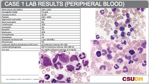
What do you expect to see in the BM investigation?
What are next steps and the expected results?
1. What do you expect to see in the BM (bone marrow) investigation?
Findings expected:
- Hypercellular marrow: The bone marrow is packed with cells, particularly granulocytic precursors at all stages of maturation.
- Marked myeloid hyperplasia: Striking increase in myeloid-to-erythroid (M:E) ratio, often 10–50:1 (normal is ~2–4:1).
- Left shift: Abundant myeloblasts, promyelocytes, myelocytes, and metamyelocytes.
- Erythroid hypoplasia: Suppressed red cell precursors.
- Megakaryocytic hyperplasia: Often small, abnormal megakaryocytes.
- Basophilia and eosinophilia: Both seen in the bone marrow and peripheral blood.
- Chromosomal / genetic abnormality: Philadelphia chromosome t(9;22)(q34;q11), resulting in BCR-ABL fusion gene.
2. What are next steps and the expected results?
Next diagnostic steps:
1. Cytogenetic testing (karyotyping or FISH):
- Look for the Philadelphia chromosome t(9;22).
- Expected result: Positive in >95% of CML cases.
2. Molecular testing (RT-PCR or quantitative PCR):
- Detect and quantify the BCR-ABL fusion gene transcript.
- Expected result: Strong positive confirming diagnosis.
3. Additional supportive tests:
- Leukocyte alkaline phosphatase (LAP) score is very low in CML (as seen in this patient: 20, with reference 40–130).
- This helps distinguish CML from a leukemoid reaction (where LAP is high).
Expected interpretation:
- This 34-year-old woman’s presentation (fever, night sweats, weight loss, splenomegaly, marked leukocytosis with left shift, basophilia, eosinophilia, low LAP, and hyperuricemia) is highly consistent with Chronic Myeloid Leukemia (CML).
- Confirmatory cytogenetic and molecular tests would establish the diagnosis.
- Next steps in management would likely include targeted therapy (e.g., tyrosine kinase inhibitor such as imatinib).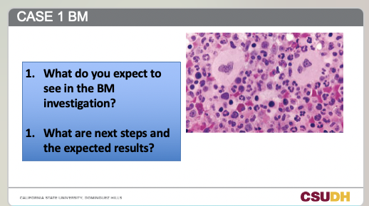
Findings expected:
- Hypercellular marrow: The bone marrow is packed with cells, particularly granulocytic precursors at all stages of maturation.
- Marked myeloid hyperplasia: Striking increase in myeloid-to-erythroid (M:E) ratio, often 10–50:1 (normal is ~2–4:1).
- Left shift: Abundant myeloblasts, promyelocytes, myelocytes, and metamyelocytes.
- Erythroid hypoplasia: Suppressed red cell precursors.
- Megakaryocytic hyperplasia: Often small, abnormal megakaryocytes.
- Basophilia and eosinophilia: Both seen in the bone marrow and peripheral blood.
- Chromosomal / genetic abnormality: Philadelphia chromosome t(9;22)(q34;q11), resulting in BCR-ABL fusion gene.
2. What are next steps and the expected results?
Next diagnostic steps:
1. Cytogenetic testing (karyotyping or FISH):
- Look for the Philadelphia chromosome t(9;22).
- Expected result: Positive in >95% of CML cases.
2. Molecular testing (RT-PCR or quantitative PCR):
- Detect and quantify the BCR-ABL fusion gene transcript.
- Expected result: Strong positive confirming diagnosis.
3. Additional supportive tests:
- Leukocyte alkaline phosphatase (LAP) score is very low in CML (as seen in this patient: 20, with reference 40–130).
- This helps distinguish CML from a leukemoid reaction (where LAP is high).
Expected interpretation:
- This 34-year-old woman’s presentation (fever, night sweats, weight loss, splenomegaly, marked leukocytosis with left shift, basophilia, eosinophilia, low LAP, and hyperuricemia) is highly consistent with Chronic Myeloid Leukemia (CML).
- Confirmatory cytogenetic and molecular tests would establish the diagnosis.
- Next steps in management would likely include targeted therapy (e.g., tyrosine kinase inhibitor such as imatinib).
2
What are myeloproliferative neoplasms (MPN)?
Clonal hematopoietic disorders caused by gene mutations in HSCs, leading to increased proliferation of and accumulation of mature erythrocytes, granulocytes, and platelets.
3
What is the WHO classification of MPN?
Chronic myeloid leukemia (CML), Polycythemia vera (PV), Essential (primary) thrombocythemia (ET), Primary myelofibrosis (PMF).
4
Which MPNs commonly have the JAK2 mutation?
PV, ET, and PMF (more frequent in PV).
5
Which chromosome abnormality is characteristic of CML?
Philadelphia (Ph) chromosome (t(9;22)) only in CML.
6
In myeloproliferative neoplasms, what predominates?
In all these neoplasms one line predominates.
7
What is the M:E ratio in CML?
M:E ratio 10:1
8
What percentage of patients have hyperleukocytosis in CML?
Approximately 15% of patients exhibit hyperleukocytosis
9
What Total WBC count defines hyperleukocytosis and what risk is it associated with?
(total WBC counts greater than 300 × 109/L), risk of leukostasis
10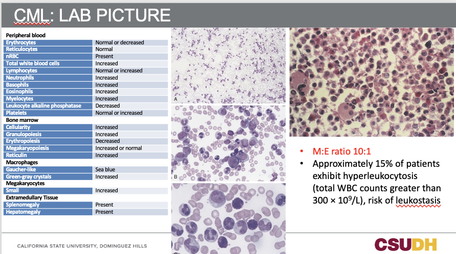
11
What are other laboratory findings in CML?
Hyperuricemia, uricosuria, risk of secondary gout, uric acid stones
12
What cytogenetic analysis is performed in CML?
Cytogenetic analysis for detection of t(9;22)
13
How is the BCR-ABL1 transcript detected?
Detection of the BCR-ABL1 transcript using qRT-PCR
14
How is the BCR-ABL1 fusion gene detected?
Detection of the BCR-ABL1 fusion gene using fluorescence in situ hybridization (FISH)
15
How is the LAP score in CML?
LAP score decreased in CML
16
How is the LAP test performed?
PBS incubated with the substrate of ALP (naphthol-phosphate substrate and diazo dye at an alkaline pH)
17
What forms during the LAP test?
A colored precipitate formed on the granules of neuts and bands
18
How many cells are counted in LAP scoring and how are they scored?
100 neutrophils and bands counted, and a score (from 0–4+) given depending on color intensity, score multiplied by the number of cells and added up
19
What is an example of an LAP score calculation?
For example, 5 cells with 4+ staining, 5 cells with 3+, 25 cells with 2+, 45 cells with 1+, and 20 cells with 0 staining, will provide a LAP score of 130
20
What is the normal range of the LAP score?
Normal range: 15–170
21
What is the therapy response and genetic change in CML blast crisis?
Poor response to therapy and appearance of more chromosomal changes
22
What hematologic change occurs in CML blast crisis?
More prominent anemia
23
What happens to neutrophils and basophils in CML blast crisis?
Decrease in number of mature neuts and more basophils
24
What platelet abnormalities are seen in CML blast crisis?
Fewer platelets, more abnormal platelets, micromegakaryocytes, and megakaryocytic fragments
25
What happens to blasts and disease transformation in CML blast crisis?
Increase of circulating blast count from 10% to 19% and transformation into either acute lymphoid or acute myeloid leukemia
26
What are the key features of Polycythemia Vera?
Panmyelosis and JAK2 mutation
27
How is JAK2 related to cell signaling?
JAK2 is related to STAT (signal transducers and activators of transcription) protein which are activated following binding of cytokines (EPO here) leading to cell proliferation and differentiation
28
What happens with mutated JAK2?
Mutated JAK2 causes constitutive activation of the JAK/STAT signaling pathway, (independent of EPO)
29
What are the WHO criteria for PV diagnosis?
Three major: I–Elevated Hb and HCT, II–BM hypercellularity with trilineage growth (panmyelosis), III–Identification of JAK2 mutation
30
What is the minor WHO criterion for PV diagnosis?
Low serum EPO levels
31
What is the frequency of Pseudo-Gaucher cells in PV?
Pseudo-Gaucher cells are rare
32
What percentage of patients manifest BM panmyelosis in PV?
Approximately 80% of patients manifest BM panmyelosis
33
What can 100% of BM volume exhibit in PV?
100% of BM volume may exhibit hematopoietic cellularity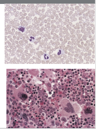
34
What organ changes and complications occur in PV?
Splenomegaly, hepatomegaly, generalized vascular engorgement, and circulatory disturbances increase the risk of hemorrhage, tissue infarction, and thrombosis
35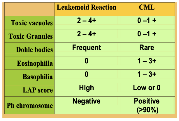
36
What happens to red cell mass in Polycythemia Vera?
Red cell mass always increased
37
What happens to EPO and arterial O₂ saturation in Polycythemia Vera?
EPO is decreased and arterial O₂ saturation is normal
38
What additional blood findings are seen in Polycythemia Vera?
Also has leukocytosis (>12) and increased PLTS (>400)
39
What bone marrow and organ findings are seen in Polycythemia Vera?
BM myelopoiesis and splenomegaly (>75%)
40
What happens to red cell mass in Hypoxia?
Red cell mass can be increased
41
What bone marrow finding is seen in Hypoxia?
In BM, only erythroid hyperplasia
42
What happens to EPO and arterial O₂ saturation in Hypoxia?
EPO is increased and arterial O₂ saturation is decreased
43
Polycythemia Vera vs. Hypoxia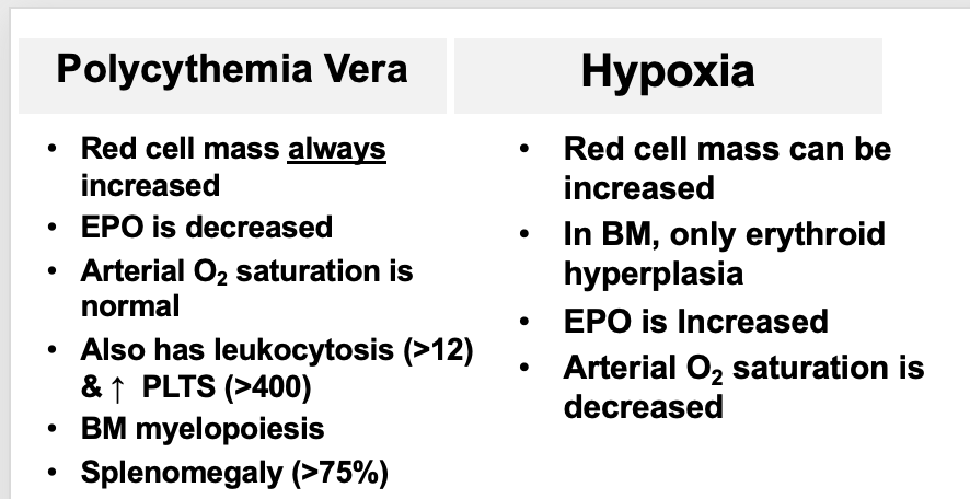
44
What characterizes ET in terms of thrombocytosis?
Marked and persistent thrombocytosis; platelets range from 600–2000 × 10⁹/L (>450); large masses of platelet aggregates; abnormal platelet morphology including giant and bizarre forms and megakaryocyte fragments
45
What are the WBC and RBC findings in ET?
WBCs ± elevated (can see neutrophilia with slight left shift); RBCs normal or slightly decreased (rare NRBC)
46
How is ET often discovered?
Often discovered in routine CBC
47
What are possible patient presentations and symptoms in ET?
Patients can present with vascular occlusion or bleeding; symptoms include headache, dizziness, throbbing / burning in hands and feet, mucosal bleeding, seizures, and myocardial infarction
48
What mutations occur in ET and how does it compare to PV?
Most of the mutations described in PV also occur in ET but usually at a lower frequency
49
What are the WBC and RBC findings in ET lab picture?
WBCs ± elevated – neutrophilia with slight left shift; RBCs are normal or slightly decreased, rare NRBC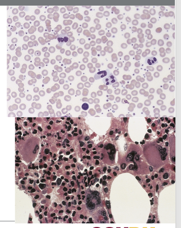
50
What are the platelet findings in ET lab picture?
Platelets greatly increased; generally ‘normal’ looking but can vary in size, shape, granulation; some giant platelets; and sometimes in clusters
51
What are the results of platelet function studies and platelet aggregation in ET?
Platelet function studies are abnormal and platelet aggregation is abnormal
52
What happens when platelet count is reduced to normal in ET?
When the PLT count is reduced to normal, the platelet function returns to normal
53
What complications can occur in ET?
Complications include hemorrhage (bleeding tendencies – abnormal PLTs) or thrombosis (usually with PLTs > 2000 × 10⁹/L)
54
What abnormal cellular process initiates PMF?
Abnormal clone of megakaryocytes stimulate the bone marrow fibroblasts to produce collagen
55
What factors and changes occur in bone marrow during PMF?
Fibroblastic growth factors released (PDGF-β); normal bone marrow tissue is gradually replaced with a fibrous scar-like material (fibrosis); dry tap BM during biopsy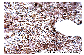
56
What is the long-term effect of fibrosis in PMF?
Over time, this leads to progressive bone marrow failure
57
What does progressive bone marrow failure result in for PMF?
Results in extramedullary hematopoiesis
58
What mutation is involved in PMF and how common is it?
As with PV and ET, the JAK2 V617F mutation is involved and is found in 60% of PMF patients
59
What does the peripheral blood film show in PMF?
Immature granulocytes and NRBCs, teardrops, giant platelets, and other bizarre RBC shapes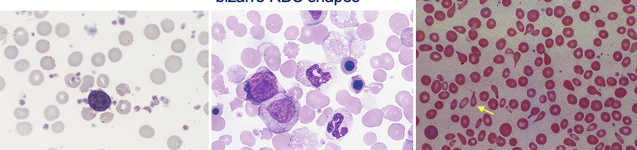
60
What platelet and megakaryocyte findings are seen in PMF?
Increased platelets and a micromegakaryocyte
61
What is tear drop poik and what causes it?
Tear drop poik is the result of narrowing and rigidity of splenic sinusoids
62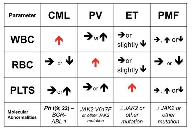
63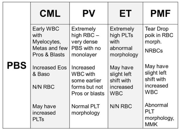
64
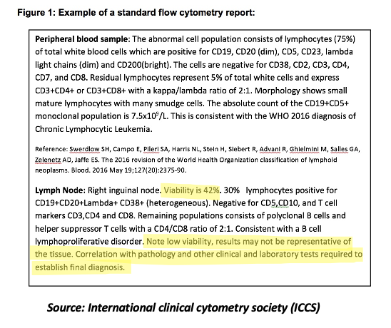
What specimens are most commonly analyzed for FC in hematology?
- Bone marrow (aspirate), in heparin
- Bone marrow FNB
- Peripheral blood, in EDTA
- Lymphoid tissues
- Body cavity fluids and solid tissues when they are suspected to harbor a hematologic malignancy
- Bone marrow FNB
- Peripheral blood, in EDTA
- Lymphoid tissues
- Body cavity fluids and solid tissues when they are suspected to harbor a hematologic malignancy
65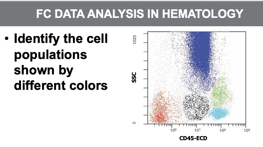
Blue: Neutrophils
Red: Red cells and PLTs
Black: Blasts
Aqua: Mature lymphocytes
Green: Monocytes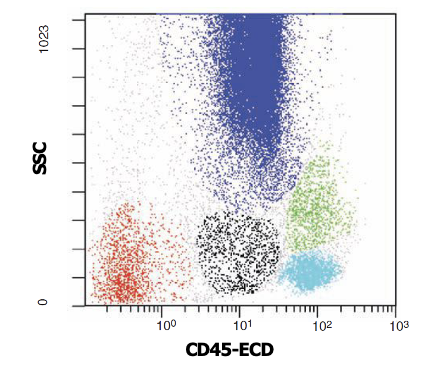
Red: Red cells and PLTs
Black: Blasts
Aqua: Mature lymphocytes
Green: Monocytes
66
67
68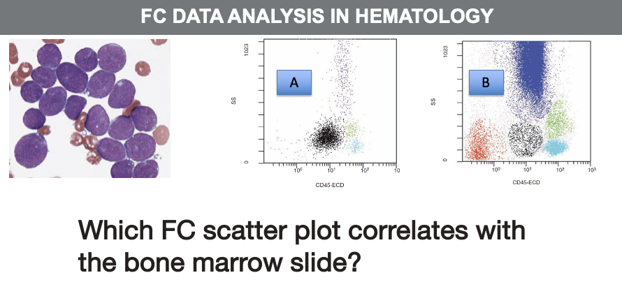
Figure A
69
70
71
72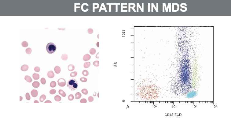
73
74
75
76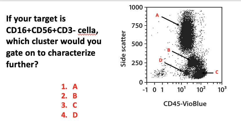
3. C
77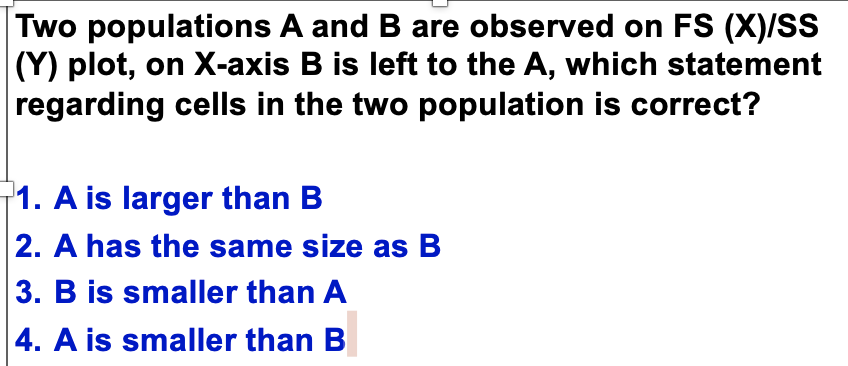
1. A is larger than B and 3. B is smaller than A is correct
78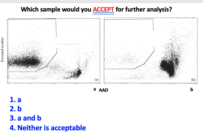
1. a
79
What immune-related conditions can be analyzed by FC in hematology?
Immune deficiency (SCID, HIV, ……)
80
What stem cell marker is enumerated using FC in hematology?
Stem cell enumeration (CD34)
81
What markers are analyzed for PNH using FC in hematology?
PNH (CD55, CD59)
82
What cell types and dyes are used for FC in hematology?
Reticulocytes (RNA binding dyes, e.g. TO)
83
What antibodies are used for FMH testing in FC?
FMH (Ab against HbF, RhD, or both, or CA)
84
What can FC analyze in the cell cycle?
Cell cycle analysis (DNA content)
85
What transplantation application uses FC in hematology?
Transplantation (HLA, flow cross match: donor lymphocytes + patient’s serum, then incubate with antihuman IgG F(ab)/FITC)
86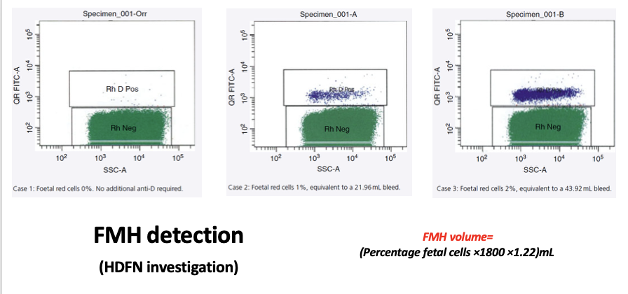
87
What makes specimens unacceptable for FC due to storage issues?
WB or BM samples stored for a long time with the anti-coagulants; WB or BM aspirates are NOT delivered to the lab while fresh or are collected in inappropriate anticoagulants
88
What makes tissue samples unacceptable for FC?
Tissue samples (BM FNB, LNB) are not transferred to the lab in appropriate media
89
What processing steps can compromise cell viability?
Using harsh vortex on cell pellets and using aggressive mechanical disruption of tissues
90
What handling of cell pellets can reduce viability?
Cell pellets are kept as dry for a long time or highly concentrated cell suspension is used
91
How can cell viability be improved during sample QC?
Keeping cells on ice can improve the viability as this slows down necrosis and the cell metabolism
92
Can FCM be run on body fluids?
Yes, FCM can be run on body fluids like CSF
93
Can frozen specimens be used for FCM?
Yes, frozen specimens can be used
94
What factors influence the quality of a specimen?
The quality of the specimen will be influenced by transferring to the lab (time, condition), storage in the lab (time, condition), type of anti-coagulant, and staining procedure
95