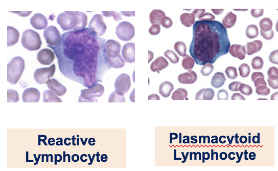
WBC Lecture 2: BM, NON-Malignant WBC Disorders
Study these flashcardsFree · spaced repetition · iPhone, Android, Mac & web
All cards
1
What is evaluated in bone marrow examination?
Cellularity, maturation of myeloid & erythroid series (M:E ratio), cell distribution (Differential), eosinophils / basophils, megakaryocytes
2
Which other cells and findings are assessed in bone marrow evaluation?
Presence of other cells: Lymphocytes, plasma cells, histiocytes, osteoclasts, fibroblasts, iron stores, abnormal infiltrates, and fibrosis
3
What parameters are included in aspirate evaluation?
Relative cellularity, differential and M:E, iron stores, megakaryocyte evaluation, metastatic tumor cells, and lymphoid aggregates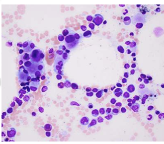
4
How is a bone marrow aspirate specimen examined?
Examined under low power (x10) to detect presence of spicules
5
What are spicules and how do they stain?
Spicule is relatively intact marrow consisting of hematopoietic and stromal cells; stain dark blue/purple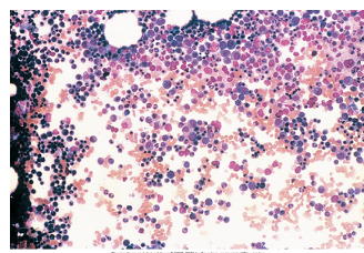
6
What features are assessed in aspirate specimens?
General cellularity, abnormal cell clusters, and quality of aspirate using megakaryocytes
7
How is aspirate differential performed and how many cells are counted?
Performed under x100 magnification; 300–500 nucleated cells are counted
8
Which cells are included in aspirate differential count?
All nucleated cells: myeloid series – blasts – segs (neut, eos, baso), erythroid series, lymphs, monos, plasma cells, and early cells as well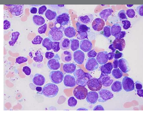
9
Why is aspirate differential important?
It is a more reliable differential and determines the M:E ratio
10
What do most cells in normal BM consist of?
Most cells in the normal BM consist of myeloid and erythroid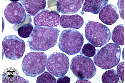
11
What should be present in BM differential count?
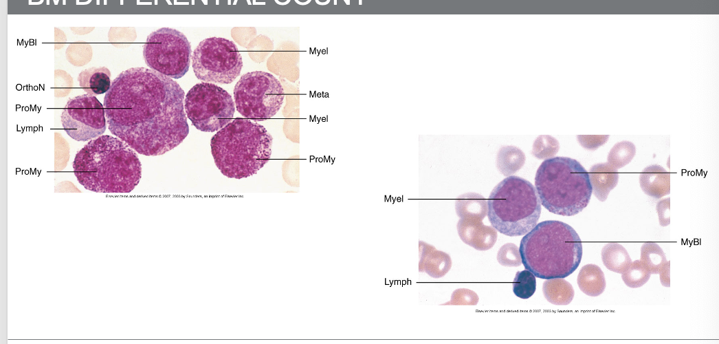
All stages of development should be present but within their anticipated distribution
12
What is required in BM differential count?
Count and differentiate all cell lines and maturation stages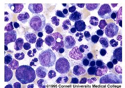
13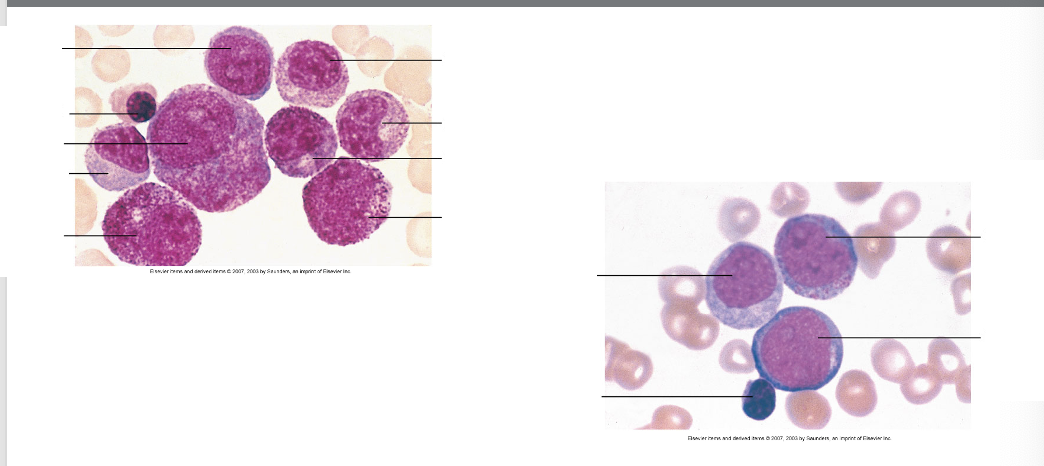
14
How is the M:E ratio derived and what is normal?
Derived from the differential; normal M:E ratio is 1.5:1 – 3.3:1
15
Why does granulopoietic tissue occupy more space than erythroid precursors?
The granulopoietic tissue occupies 1.5 to 3.3 times more space than the erythroid precursors as a result of shorter survival of granulocytes in the circulation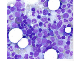
16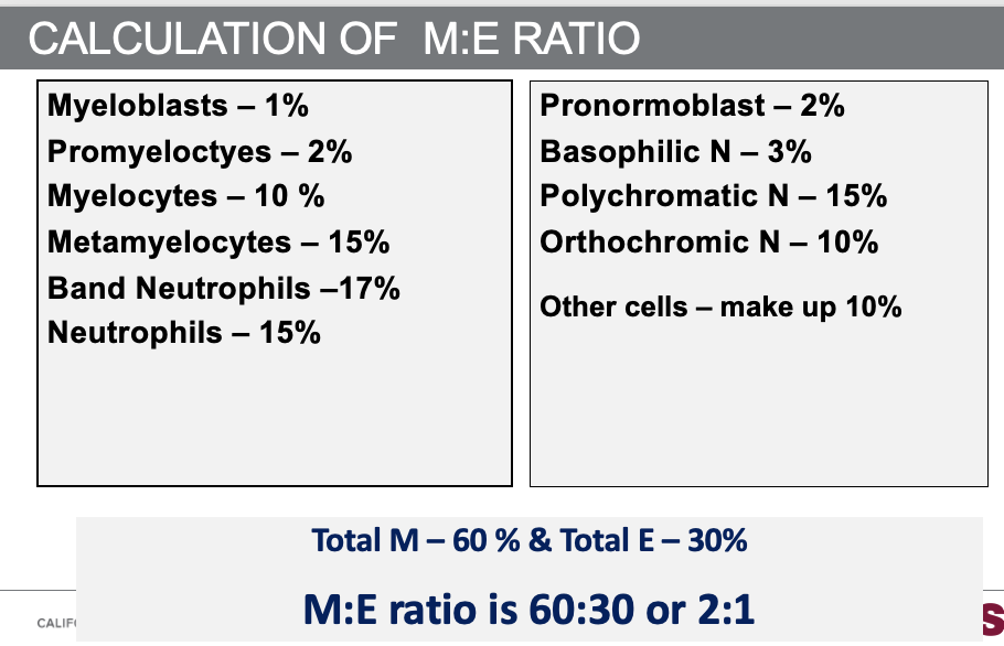
Calculation of M:E Ratio
17
How is iron reported in bone marrow?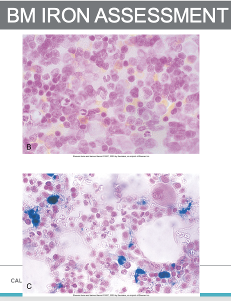
Iron reported as absent, decreased, adequate, moderately increased, or markedly increased
18
How are megakaryocytes evaluated in bone marrow?
By the number seen per low power field (10x); 2–10/lpf is normal
19
Why are megakaryocytes not counted in differential?
They are too unevenly distributed to count in differential
20
What do clusters or low counts indicate?
Clusters may indicate hyperplasia; fewer than 2 megakaryocytes/LPF may indicate hypoplasia or poor quality aspirate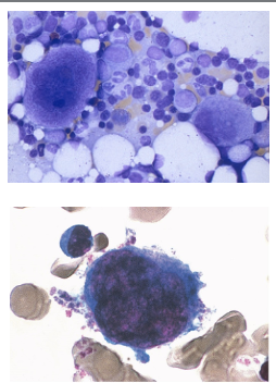
21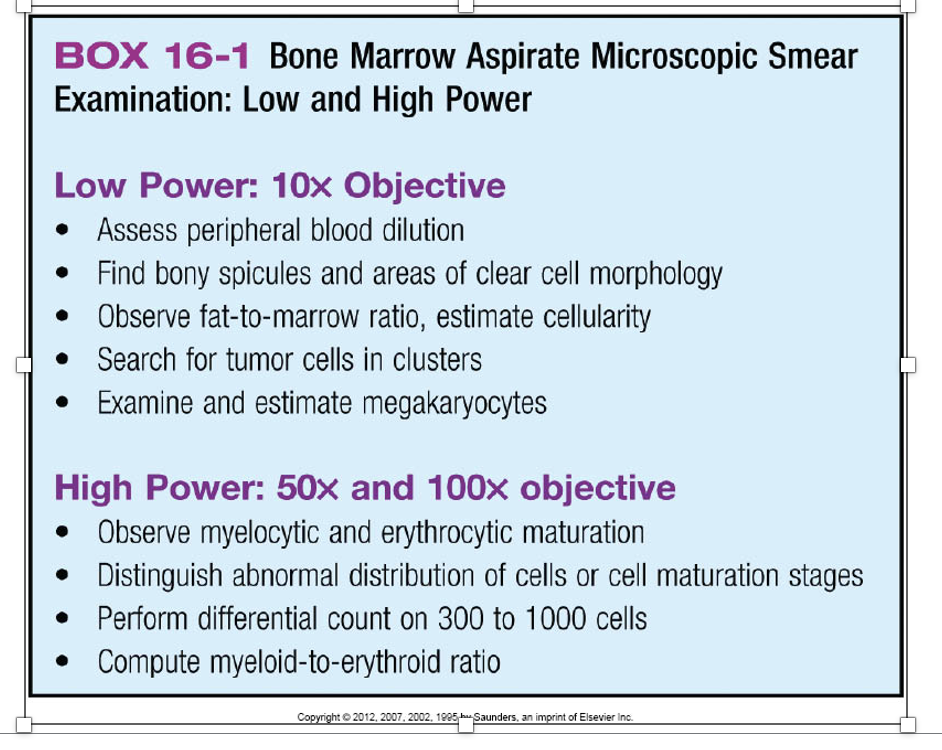
22
What is cellularity in core biopsy and how is it measured?
Cellularity is the ratio of all nucleated hematopoietic cells (ANC) to fat cells
23
What is normal cellularity in adults?
Cellularity in adults is approximately 50% (1:1)
24
What rule is used to estimate normal cellularity by age?
100 – age ±10
25
How is cellularity reported?
Reported as increased, normal, or decreased
26
What BM cellularity is this?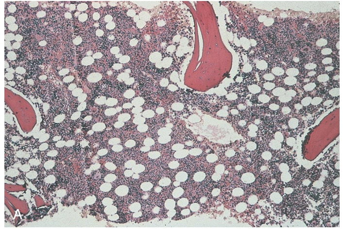
Normal 1:1
27
What is hypercellular marrow?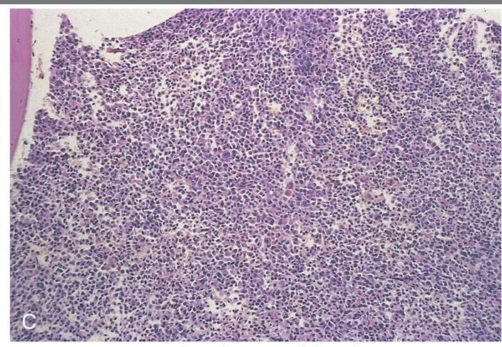
Hypercellular marrow 10:1
28
What is hypocellular marrow?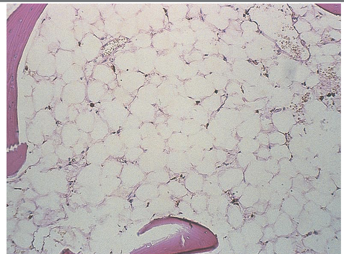
Hypocellular marrow 1:10
29
What patient and sample details are included in bone marrow report?
Patient history, age, sex, description of samples received
30
What peripheral blood data are included in bone marrow report?
CBC, ESR, reticulocyte count, manual differential, PBS
31
What bone marrow details are reported?
Differential count, description of the cellularity, M:E ratio, status of the iron stores, description of histological sections of biopsy and diagnostic conclusion.
32
What is included in the diagnostic conclusion and sample report?
sample report includes normal values for HGB, HCT, MCV, MCH, WBC, platelet; abbreviations PB (peripheral blood), BMA (bone marrow aspirate), BMBX (bone marrow biopsy), DX (diagnosis)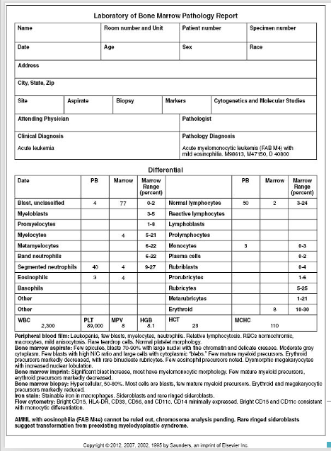
33
What is this called in a Bone Marrow smear?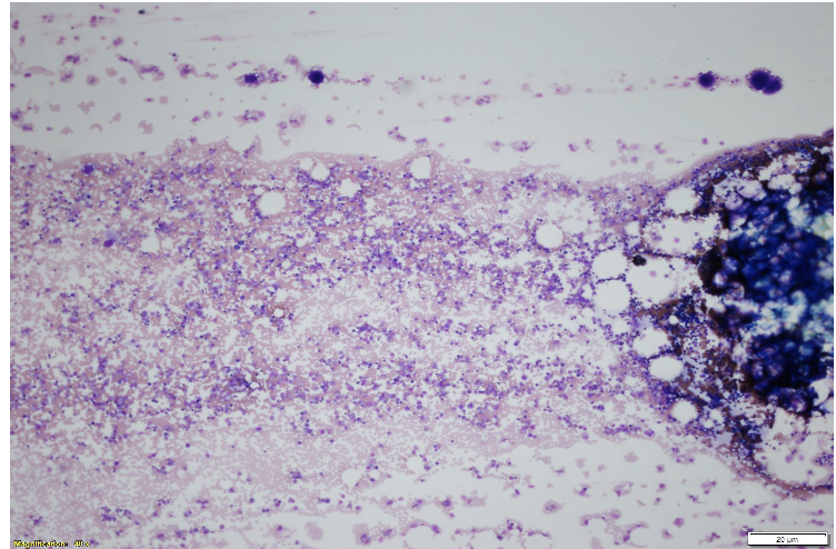
Spicule
34
What are these? What do you see here?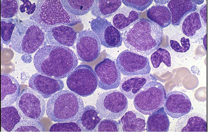
Myeloid Precursors
35
What do you see here?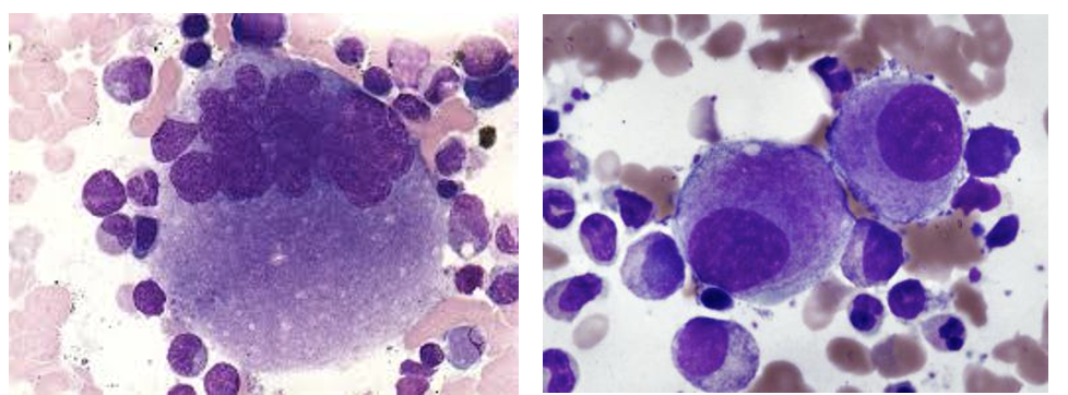
Early and late megakaryocytes
36
What do you see here?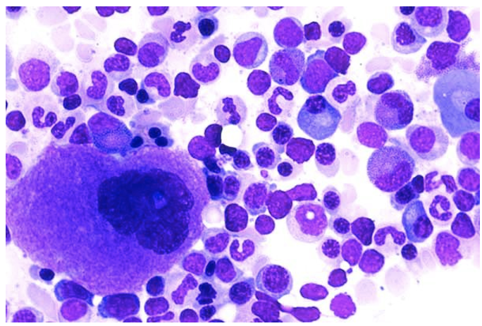
Megakaryocyte, plasma cells, neutrophils, band and metas, NRBC & slightly earlier RBC
37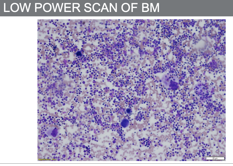
38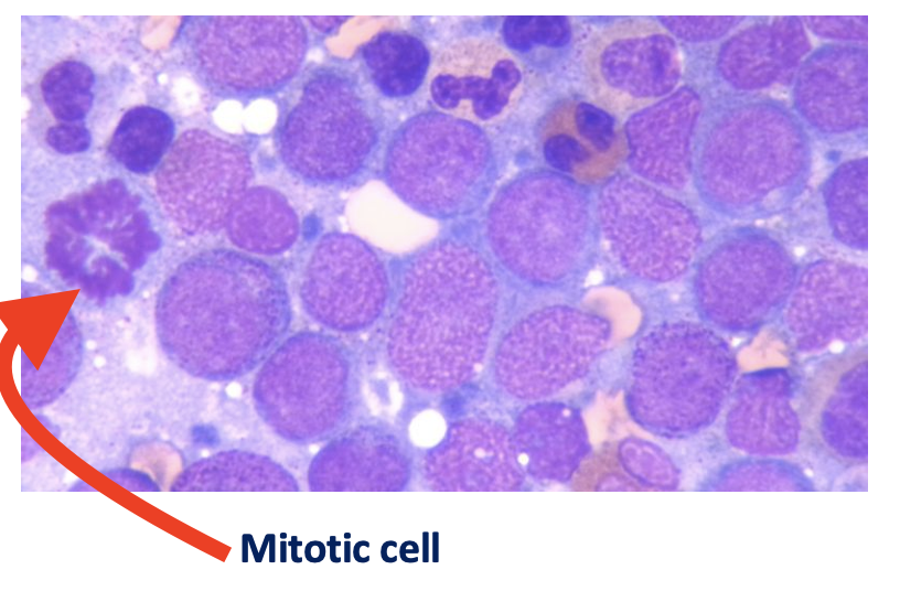
What do you see here?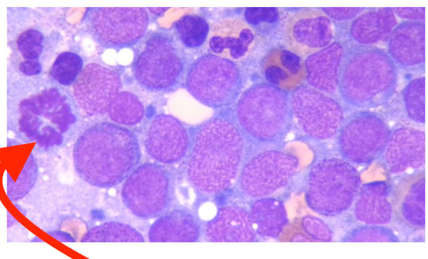
39
What do you see here?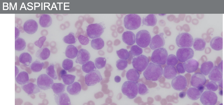
Lack of mature neuts.
Very hypercellular – monotonous cell population.
Very hypercellular – monotonous cell population.
40
What do you see here?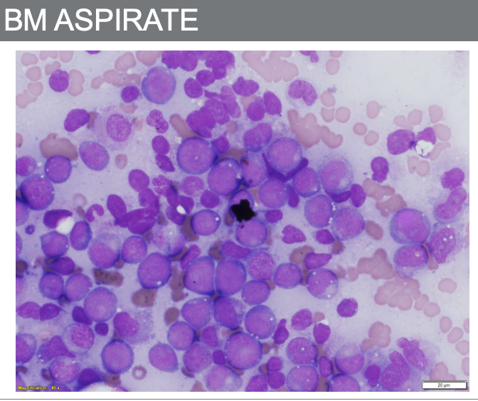
Mostly blasts
41
What do you see here?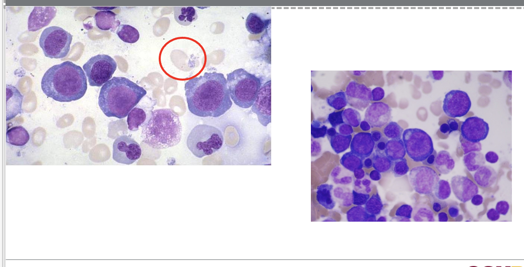
Megaloblastic anemia RBC precursors
42
What do you see here?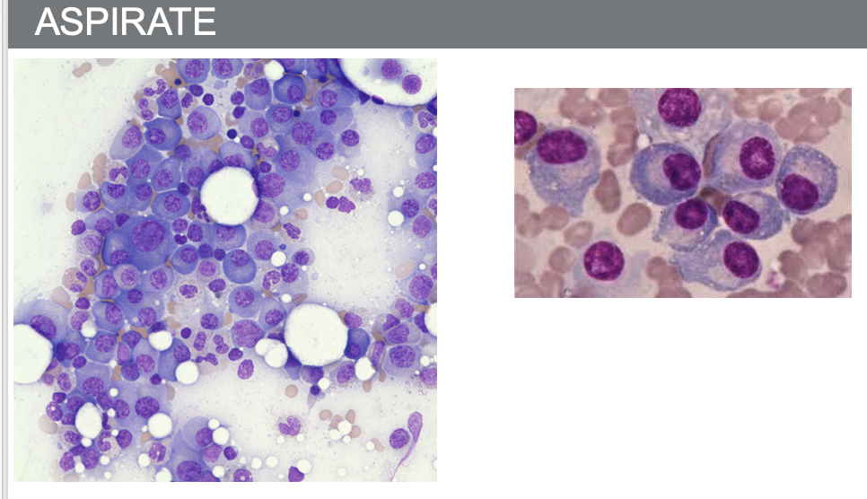
Plasma cells
43
44
What is a benign shift in neutrophils?
A benign shift in neuts from marginal into circulatory pool
45
What causes benign neutrophil shift?
Stress, trauma, labor, strenuous exercise, shock, burns, or increase in epinephrine
46
What are pathological conditions causing neutrophil shift?
Pathological conditions shift in neuts from marginal into circulatory pool, increased BM production, release of neutrophils from storage pool to PB, infection
47
What is Left Shift and what does it indicate?
Left shift describes an increased number of immature cells as an indicator of infection
48
Which immature cells increase in Left Shift?
Bands, metamyelocytes, and possibly myelocytes
49
What causes Left Shift and what is it seen with?
Increased release from storage pool; usually seen with neutrophilia and toxic changes
50
What is a leukemoid reaction?
A reactive leukocytosis above 50 × 10³/microL with neutrophilia and a marked left shift; bands and often metas/myelos, could have rare pro or blast
51
What are leukemoid reactions mostly a result of?
Severe and/or chronic infection (TB, pneumonia), metabolic disease, inflammation, response to a malignancy
52
What picture may appear with leukemoid reactions?
Leukoerythroblastic picture
53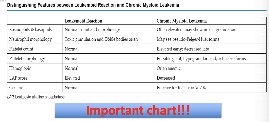
54
What defines a leukoerythroblastic picture?
Presence of immature neutrophils, nRBCs, and teardrop RBCs in the same sample
55
Is leukoerythroblastic picture always with neutrophilia?
Often accompanied by neutrophilia, but not always
56
What does a leukoerythroblastic reaction point to?
Possibility of a space-occupying lesion in the bone marrow
57
Which lesions are associated with leukoerythroblastic reactions?
Metastatic tumor, fibrosis, lymphoma, leukemia
58
What is a leukoerythroblastic reaction strongly associated with?
Primary myelofibrosis
59
What do qualitative granulocyte disorders involve?
Nucleus and/or cytoplasm
60
What is the origin of qualitative granulocyte disorders?
They may be acquired or inherited
61
What nuclear alterations can be seen in acquired granulocyte anomalies?
Hyposegmentation, hypersegmentation, pyknotic forms, and necrobiotic forms
62
What cytoplasmic alterations can be seen in acquired granulocyte anomalies?
Toxic granulation, degranulation, vacuolization (with or without engulfed matter) and Döhle bodies
63
What causes hypersegmented neutrophils?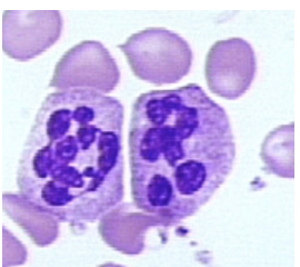
Chronic infections, megaloblastic anemia, and drugs
64
What causes hyposegmented neutrophils and what is seen?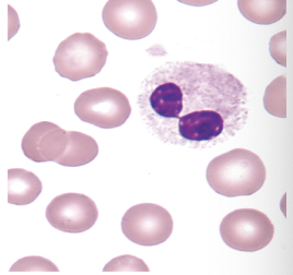
Myelodysplastic syndromes. Asynchrony of nuclear maturation (Very clumped chromatin and No segmentation)
65
What are pyknotic nuclear abnormalities?
‘Dying’ cell with nucleus very dark/dense, filaments still visible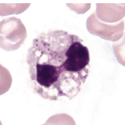
66
What are necrotic nuclear abnormalities?
Dead cell with no filaments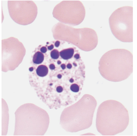
67
What abnormalities occur in neutrophilic cytoplasm?
Neutrophilic cytoplasmic abnormalities with absence of granules (hypo- or agranular, “degranulation,”) and abnormal granules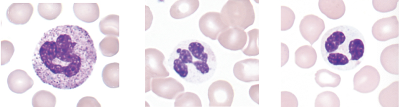
68
What reactive morphologic changes occur in neutrophils?
Toxic granulation, Döhle bodies, and cytoplasmic vacuoles
69
Why are toxic changes in neutrophils not always pathologic?
While these changes may be considered “abnormal,” they usually reflect a normal, reactive response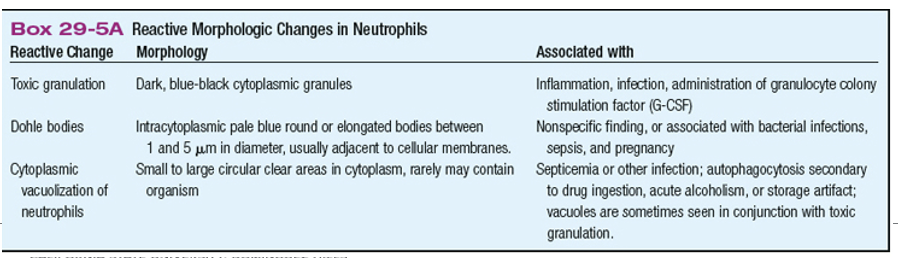
70
What is toxic vacuolization?
Often seen with toxic granulation, it can indicate phagocytosis and may contain bacteria or other material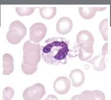
71
What are Döhle bodies and how do they appear?
Pale blue, oval inclusions at periphery; grey to light blue on Wright stain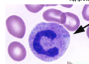
72
Which cells contain Döhle bodies?
Only neutrophils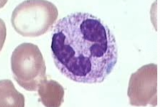
73
What are Döhle bodies composed of and in what conditions are they associated with?
Remnants of ribosomal RNA. Bacterial infection, sepsis, and pregnancy.
74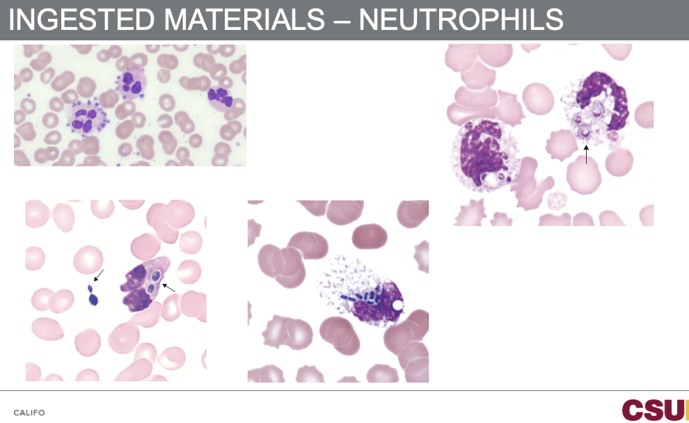
75
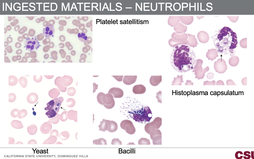
What materials can be ingested by neutrophils?
Histoplasma capsulatum, platelet satellitism, yeast, and bacilli
76
What are examples of inherited qualitative abnormalities?
Pelger-Huët anomaly, Alder-Reilly anomaly, May-Hegglin anomaly, and Chediak-Higashi syndrome
77
What nuclear abnormality defines Pelger-Huët anomaly and what is the inheritance pattern?
Hyposegmentation of the neutrophil nucleus. Autosomal dominant
78
Is Pelger-Huët anomaly symptomatic?
It is asymptomatic and fully functional
79
What does the heterozygous state of Pelger-Huët look like?
Bi-lobed nucleus, dense heterochromatin, incomplete nuclear segmentation, also called “dumbbell” or “pince-nez”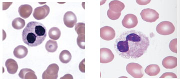
80
What does the homozygous state of Pelger-Huët look like?
Nucleus is round or oval with no segmentation (rare)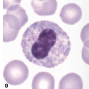
81
How does nuclear chromatin appear in Pelger-Huët anomaly?
Exceptionally coarse and condensed, showing it is a mature cell
82
What other cells may Pelger-Huët anomaly be confused with?
Band or myelocyte
83
What percentage of neutrophils are affected in Pelger-Huët anomaly?
70 to 90% of neutrophils are affected
84
What is pseudo-Pelger-Huët anomaly and which conditions is it seen?
A drug-induced form of Pelger-Huët. Seen in HIV, tuberculosis, Mycoplasma pneumonia, and severe bacterial infections
85
What disorders can pseudo-Pelger-Huët be secondary to?
Myelodysplastic syndromes, acute leukemias, and chronic myeloproliferative disorders
86
What is the inheritance pattern of Alder-Reilly anomaly?
Autosomal recessive
87
What cytoplasmic abnormality occurs in Alder-Reilly anomaly?
Abnormally large metachromatic granules in the cytoplasm of granulocytes, monocytes, and lymphocytes 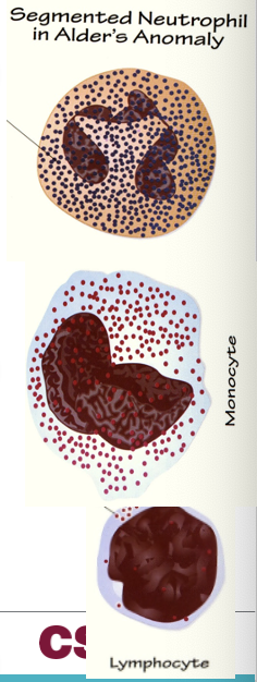
88
What causes the granules seen in Alder-Reilly Anomaly?
Incomplete degradation of mucopolysaccharides (Mucopolysaccharide deposits (granules) in most cells)
89
Does Alder-Reilly anomaly affect leukocyte function?
Leukocyte function is unaffected
90
How do Alder-Reilly granules appear in WBCs?
Deep-purple to lilac granules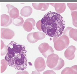
91
What may Alder-Reilly granules resemble?
Promyelocytes or heavy toxic granulation
92
How is Alder-Reilly distinguished from toxic granulation?
Neutrophilia, Döhle bodies, and left shift = toxic granulation; Alder-Reilly granules can also be found in lymphocytes and monocytes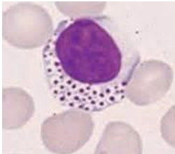
93
How common is Alder-Reilly anomaly?
Very rare
94
What abnormalities in platelets are seen in May-Hegglin anomaly?
Giant and bizarre platelets that may be hypogranular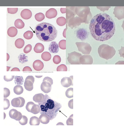
95
What is the inheritance pattern and rarity of May-Hegglin anomaly?
Autosomal dominant (rare)
96
What type of disorder is May-Hegglin anomaly?
Platelet disorder
97
What leukocyte abnormality may be present in May-Hegglin anomaly?
Leukopenia with large basophilic inclusions
98
What type of thrombocytopenia occurs in May-Hegglin anomaly?
Variable thrombocytopenia with giant platelet forms
99
What are the symptoms in May-Hegglin anomaly?
Patients are usually asymptomatic or have mild bleeding disorders
100
What is the inheritance pattern of Chediak-Higashi syndrome and how common and severe is it?
Autosomal recessive. Rare and fatal.
101
What abnormality defines Chediak-Higashi syndrome?
Characterized by abnormal fusion of granules in most cells that contain granules throughout the body
102
How do the fused granules appear?
Large and mostly dysfunctional
103
Which cells are affected in Chediak-Higashi syndrome?
Hematopoietic cells are affected
104
Where else are disease manifestations seen?
Hair, skin, adrenal and pituitary glands, and nerves
105
What phagocytic defect is seen in Chediak-Higashi syndrome and what immune defect results?
Dysfunctional phagocytosis and inefficient bacterial destruction. Increased susceptibility to infection (immunodeficient), albinism and peripheral neuropathy.
106
What platelet defect is present in Chediak-Higashi syndrome?
Platelet dense granule abnormality which has a tendency to bleed
107
What abnormality occurs in melanocytes?
Variable albinism
108
When do neurologic complications develop?
Neurologic complications develop through childhood
109
What granules are seen in WBCs in Chediak-Higashi syndrome?
Giant lysosomal granules in all WBCs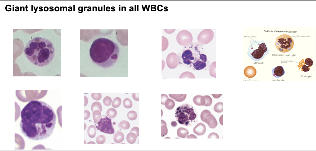
110
What is Chronic Granulomatous Disease (CGD)?
A rare X-linked or autosomal recessive condition
111
What is the neutrophil morphology in CGD?
Usually normal
112
What defect occurs in phagocytes in CGD?
Decreased ability to produce NADPH oxidase needed to kill microorganisms
113
What infections are common in CGD?
Recurring bacterial and fungal infections
114
What happens to bacteria ingested by phagocytes in CGD?
They are not killed on ingestion but remain encased in granulomas to localize them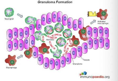
115
What can granulomas do in CGD?
Rupture and release live bacteria and pus
116
What is the WBC morphology in CGD?
Normal morphology or neutrophilia with toxic changes as seen in bacterial infection
117
What diagnostic tests are used for CGD?
Nitroblue tetrazolium (NBT) reduction test or flow cytometry
118
When do reactive changes occur in monocytes?
Reactive changes during infection, recovery from BM aplasia or post GM-CSF
119
What nuclear and cytoplasmic changes occur in reactive monocytes?
Nuclear contortion, increased cytoplasmic volume, and increased numbers of cytoplasmic granules
120
What evidence of phagocytic activity may be seen in reactive monocytes?
Cytoplasmic vacuolation, intracellular debris, and irregular cytoplasmic borders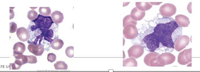
121
What diseases show functional abnormalities in macrophages?
Lysosomal storage diseases
122
What causes lysosomal storage diseases?
Enzyme deficiencies
123
What happens to phagocytized material in lysosomal storage diseases and where is it seen?
It accumulates in cells due to incomplete degradation. Seen in macrophages and monocytes found in BM & spleen.
124
What is Gaucher Disease and what does it do?
Most common lysosomal lipid storage disease. Unmetabolized lipid accumulates in the macrophages throughout the body.
125
Are Gaucher cells seen in the BM?
The BM contains Gaucher cells, macrophages that have abundant fibrillar blue gray cytoplasm with a wrinkled appearance.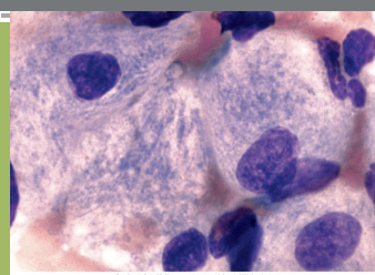
126
What does Niemann Pick Disease cause?
Lipid buildup in macrophages. The BM contains Niemann-Pick cells, macrophages that contain foamy cytoplasm packed with lipid-filled lysosomes that appear as vacuoles after staining 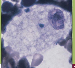
127
What causes infectious mononucleosis and what percentage of the world population does EBV infect?
Epstein-Barr Virus (EBV). EBV infects >90% of world population (usually young children).
128
Which cells does EBV preferentially infect?
B lymphocytes by attaching to CD21 receptors
129
What immune response occurs in infectious mononucleosis?
Lymphocyte proliferation including NK cells, T CD4, and T CD8
130
What antibodies are produced in infectious mononucleosis?
Production of heterophile antibodies in serum (react with RBCs from horse or sheep: principle of Mono test)
131
What is the hallmark of infectious mononucleosis?
Reactive lymphocytes
132
What is the WBC count in infectious mononucleosis?
Usually elevated (10–30 × 10⁹/L)
133
What type of lymphocytosis is seen in infectious mononucleosis?
Absolute lymphocytosis
134
How does lymphocyte morphology appear in infectious mononucleosis?
Wide variation in morphology