Stool Culture
Study these flashcardsFree · spaced repetition · iPhone, Android, Mac & web
All cards
1
Q: What transport medium is preferred for Shigella and inhibits Campylobacter?
A: Buffered glycerol saline
2
Q: How many stool specimens should be collected per day?
One specimen per day.
3
Q: How can the probability of pathogen recovery be increased in stool cultures?
A: By collecting 3 specimens on separate days.
4
Q: When should stool specimens ideally be collected during an infection?
A: During the acute stage of infection.
5
Q: What transport media can be used for stool specimens?
Clean, leak-proof container
Buffered glycerol saline (preferred for Shigella, inhibit Campylobacter)
Cary-Blair medium
Stool enrichment broth
Buffered glycerol saline (preferred for Shigella, inhibit Campylobacter)
Cary-Blair medium
Stool enrichment broth
6
Q: What do transport media such as Cary-Blair increase?
A: Survival of organisms without multiplying
7
Q: What does transport media prevent?
A: Overgrowth of normal flora and acidification of stool
8
Q: What can result in improper specimen preservation?
A: Overfilling
9
Q: How long is fresh stool not in transport medium acceptable at room temperature?
A: Within 1 hour at room temperature
10
Q: How long is fresh stool in transport medium acceptable at room temperature?
A: Within 24 hours at room temperature
11
Q: How long is fresh stool in transport medium acceptable at 4°C?
A: 96 hours at 4°C
12
Q: What alternative specimen uses rectal swabs?
A: Rectal swabs in Cary-Blair medium or buffered glycerol saline
13
Q: What alternative specimens use duodenal, colostomy, or ileostomy contents?
A: Duodenal, colostomy, ileostomy contents in leak-proof container or transport vial
14
Q: What specimen is acceptable for suspected shigellosis?
A: Sigmoidoscopy specimen – flexible scope into the sigmoid colon
15
What is the rejection criteria for stool collection?
Specimen too old (beyond acceptable time range)
More than 1 specimen per day
Adult and pediatric patient hospitalized for >3 days (unless HIV+ or cluster epidemic within institution)
Overfilled transport media
Transport media indicator turns yellow (acidic)
Hard, solid stools that cannot be sampled
Dry rectal swabs
Stools with urine or interfering substances (barium, castor oil, Metamucil)
More than 1 specimen per day
Adult and pediatric patient hospitalized for >3 days (unless HIV+ or cluster epidemic within institution)
Overfilled transport media
Transport media indicator turns yellow (acidic)
Hard, solid stools that cannot be sampled
Dry rectal swabs
Stools with urine or interfering substances (barium, castor oil, Metamucil)
16
Why can't toilet paper be used to obtain stool sample?
may contain barium salt
17
What media is used for stool analysis?
BAP, MAC and usually one or more of the following:
HE
XLD
CHROMagar Salmonella
SS
HE
XLD
CHROMagar Salmonella
SS
18
Which broth may be used to detect small numbers of Salmonella or Shigella?
Selenite-F or GN broth
19
Salmonella or Shigella is commonly useful ID for which occupations? and what is the result for PCR?
Day car worker, food service employees; PCR positive cultures
20
What does Hektoen Enteric (HE) differentiate? and what are the following color results mean:
Yellow to salmon color:
Blue-green color:
Black colonies or blue-green with black center: ?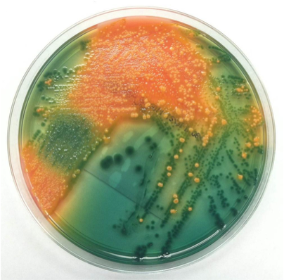
Yellow to salmon color:
Blue-green color:
Black colonies or blue-green with black center: ?
Salmonella and Shigella from other Enterics ;
Yellow to salmon color = ferment one or more carbohydrate
Blue-green color = does not ferment the carbohydrate available
Black colonies or blue-green with black center = production of H2S
Yellow to salmon color = ferment one or more carbohydrate
Blue-green color = does not ferment the carbohydrate available
Black colonies or blue-green with black center = production of H2S
21
What is Campy CVA selective for? and what are the inhibitors?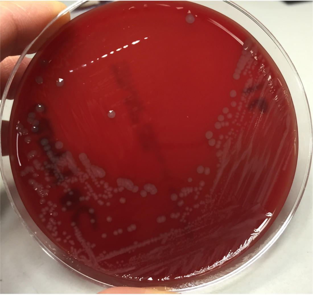
Campylobacter species;
Inhibitor: trimethoprim, vancomycin, polymyxin B, cefoperazone, Amphotericin B inhibit gram positive, enterics and yeast
Inhibitor: trimethoprim, vancomycin, polymyxin B, cefoperazone, Amphotericin B inhibit gram positive, enterics and yeast
22
What is TCBS (Thiosulfate Citrate Bile Salts Sucrose) Agar selective for?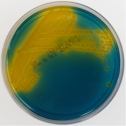
Vibrio species
23
TCBS (Thiosulfate Citrate Bile Salts Sucrose) Agar results
if YELLOW?
if BLUE-GREEN?
if YELLOW?
if BLUE-GREEN?
Yellow = Sucrose fermenters (i.e. Vibrio cholerae, Vibrio alginolyticus)
Blue-green = Sucrose non-fermenters (i.e. Vibrio vulnificus, Vibrio parahaemolyticus)
Blue-green = Sucrose non-fermenters (i.e. Vibrio vulnificus, Vibrio parahaemolyticus)
24
What is the CIN (Cefsulodin, Irgasan, Novobiocin) agar selective for?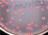
selective and differential isolation of Yersinia and Aeromonas species
25
What is MacConkey Agar with Sorbital (SMAC) selective for?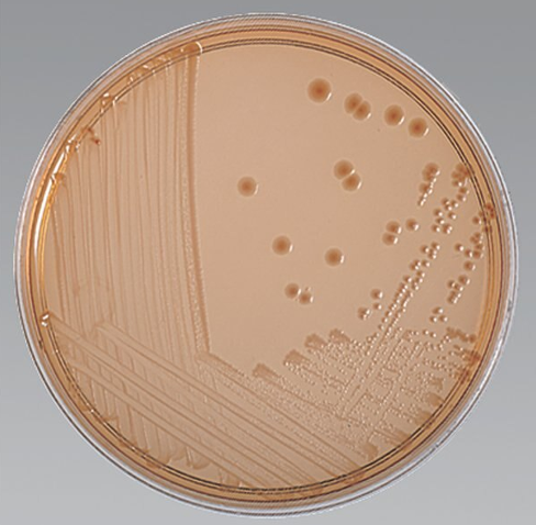
Selective and differential media for enterohemorrhagic E.coli O157:H7
26
Q: What is EMB (Eosin Methylene Blue) agar used for, and how does E. coli appear on it?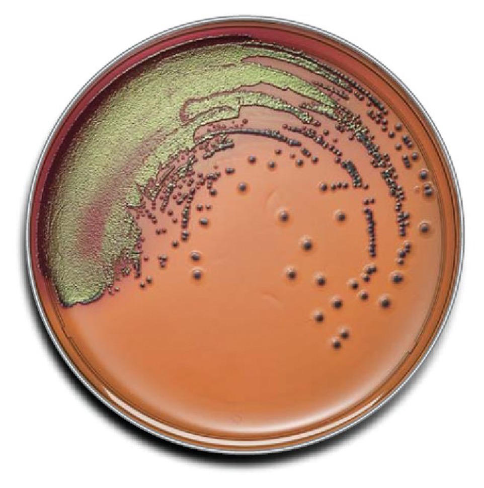
A: EMB agar is selective and differential for the isolation of Gram-negative bacilli;
it contains lactose and sucrose as carbohydrates and the dyes eosin Y and methylene blue.
Fermentation of carbohydrates causes bacteria to take up the eosin–methylene blue complex, producing brown or blue-black colonies.
E. coli forms blue-black colonies with a green metallic sheen.
it contains lactose and sucrose as carbohydrates and the dyes eosin Y and methylene blue.
Fermentation of carbohydrates causes bacteria to take up the eosin–methylene blue complex, producing brown or blue-black colonies.
E. coli forms blue-black colonies with a green metallic sheen.
27
What is SS (Salmonella Shigella) Agar used for?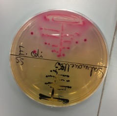
it's a differential and selective media for Salmonella and Shigella
28
Q: What are the characteristics and purpose of XLD (Xylose Lysine Deoxycholate) agar, and how do organisms appear on it?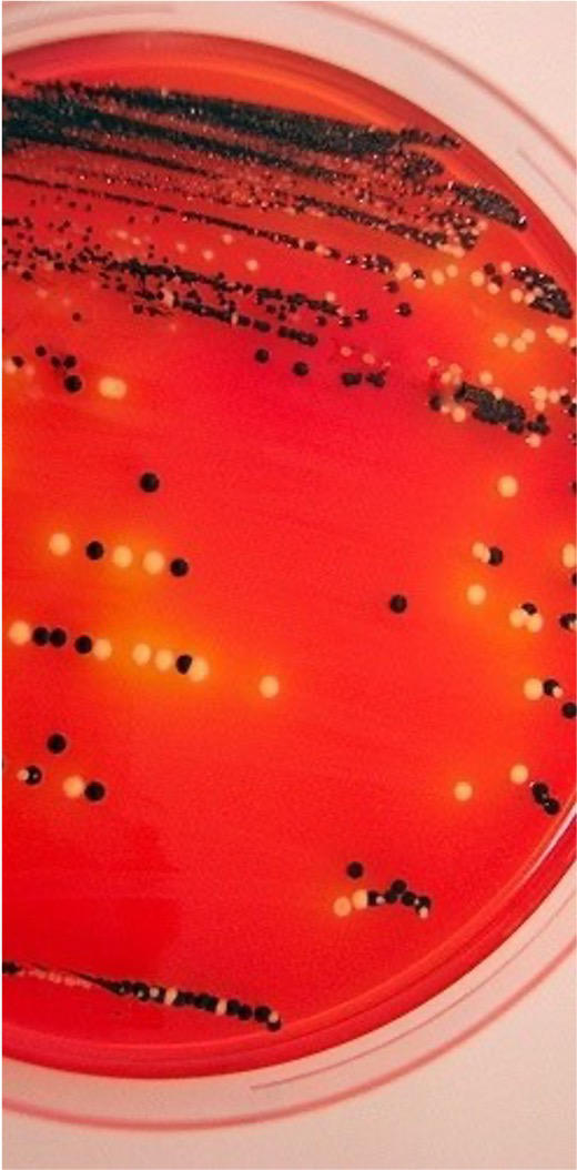
A: XLD agar is a selective and differential medium for Gram-negative rods (GNRs) that allows growth of Salmonella and Shigella.
It contains sodium deoxycholate to inhibit Gram-positive organisms;
xylose, lactose, and sucrose as carbohydrates (fermented by most organisms except Shigella);
Lysine to detect lysine decarboxylation (important for Salmonella);
and sodium thiosulfate with ferric ammonium citrate to detect H₂S production.
Xylose/Lactose/Sucrose fermenters and Lysine decarboxylase negative = Yellow
Salmonella/Shigella appears red (w/ or w/o black centers for Salmonella)
It contains sodium deoxycholate to inhibit Gram-positive organisms;
xylose, lactose, and sucrose as carbohydrates (fermented by most organisms except Shigella);
Lysine to detect lysine decarboxylation (important for Salmonella);
and sodium thiosulfate with ferric ammonium citrate to detect H₂S production.
Xylose/Lactose/Sucrose fermenters and Lysine decarboxylase negative = Yellow
Salmonella/Shigella appears red (w/ or w/o black centers for Salmonella)
29
Q: What are the characteristics and purpose of Bismuth Sulfite (Wilson–Blair) agar, and how do organisms appear on it?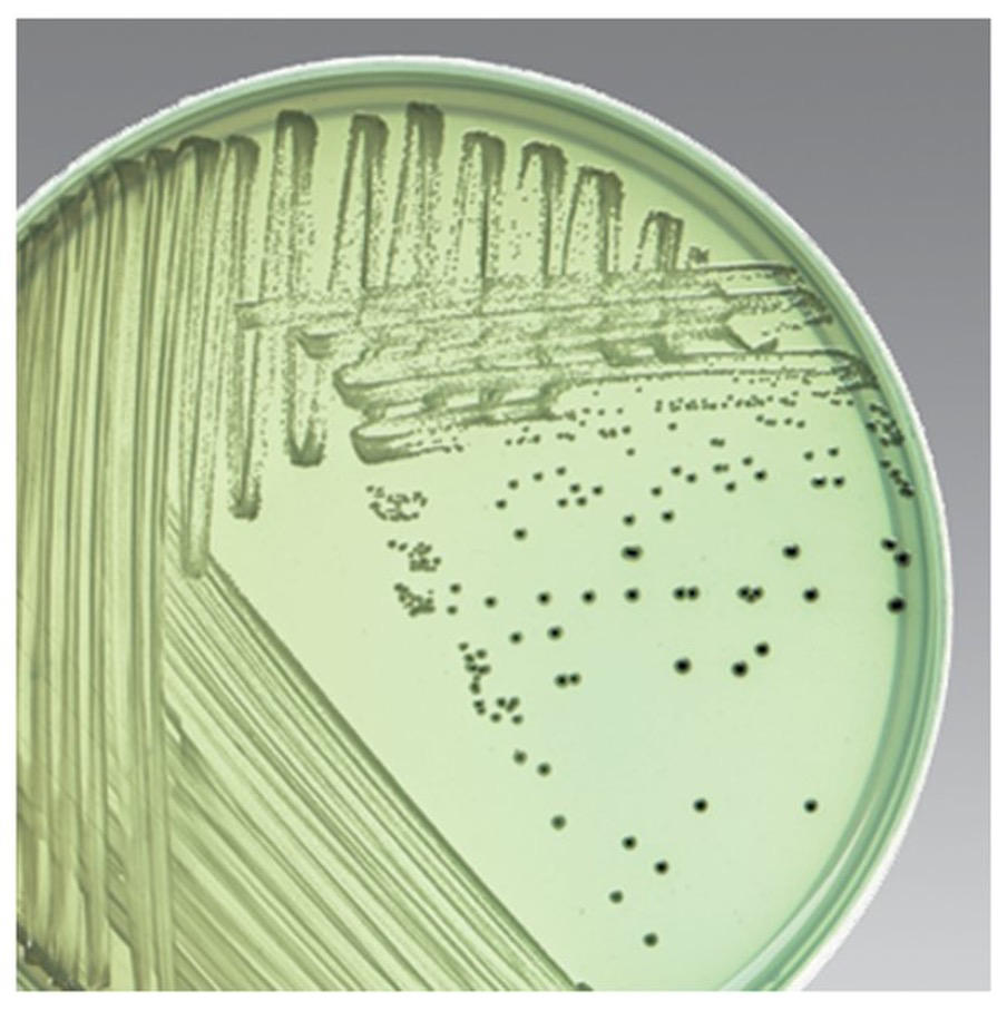
Isolation of Salmonella species, especially S. typhi
Inhibitory agent: Brilliant green and bismuth sulfite
Differential agent: ferrous sulfate
Black metallic: Salmonella typhi
Black: Other H2S positive organism
Green: H2S negative
Inhibitory agent: Brilliant green and bismuth sulfite
Differential agent: ferrous sulfate
Black metallic: Salmonella typhi
Black: Other H2S positive organism
Green: H2S negative
30
Q: What are the characteristics and purpose of Brilliant Green agar, and how do organisms appear on it?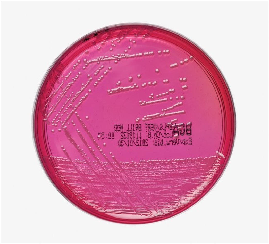
Not good for recovering S. typhi
Inhibitory agent: Brilliant green
pH indicatory: phenol red
Lactose/sucrose fermenter: yellow to yellow-green with yellow halo
Lactose/sucrose non-fermenter: red halo
Inhibitory agent: Brilliant green
pH indicatory: phenol red
Lactose/sucrose fermenter: yellow to yellow-green with yellow halo
Lactose/sucrose non-fermenter: red halo
31
Q: What is the purpose of stool cultures?
A: To screen for colony morphologies resembling potential pathogens (lactose negative, black colonies, etc.).
32
Q: What should be done if suspicious colonies are seen on stool culture?
A: Perform a 3-tube screen to rule out pathogens biochemically.
33
Q: What tests are included in the 3-tube screen for stool cultures?
A: TSI, LIA, and URE.
34
Q: When should organisms from stool cultures be fully identified?
A: When all biochemical tests match.
35
Q: What additional testing is performed if Salmonella and/or Shigella are identified?
A: Serological testing.
36
Q: How long and under what conditions are stool cultures held?
A: 48 hours at 35–37 °C in ambient air.
37
Q: Which organisms are considered pathogens in stool cultures?
A: Campylobacter;
Salmonella;
Shigella;
Yersinia enterocolitica / Yersinia pseudotuberculosis;
Vibrio spp.;
pathogenic E. coli (e.g., O157:H7).
Salmonella;
Shigella;
Yersinia enterocolitica / Yersinia pseudotuberculosis;
Vibrio spp.;
pathogenic E. coli (e.g., O157:H7).
38
Q: Which organisms are considered potential pathogens when predominant in stool cultures?
Aeromonas;
Plesiomonas shigelloides;
Staphylococcus aureus;
yeast;
Pseudomonas aeruginosa;
Edwardsiella tarda.
Plesiomonas shigelloides;
Staphylococcus aureus;
yeast;
Pseudomonas aeruginosa;
Edwardsiella tarda.
39
Q: What is the most common etiologic agent of gastroenteritis in the United States?
A: Campylobacter species.
40
Q: Which Campylobacter species are most commonly associated with infection?
A: Campylobacter jejuni (most common) and Campylobacter coli.
41
Q: From what types of specimens can Campylobacter be isolated?
A: Stool, and some species may also be isolated from blood.
42
Q: What are the common sources of Campylobacter infection?
A: Contaminated food, water, and milk.
43
Q: What is the most common source of Campylobacter infection in the U.S.?
A: Chicken.
44
Q: When is Campylobacter infection most common seasonally?
A: Late summer and early fall.
45
Q: What is the Gram stain appearance of Campylobacter species?
A: Small, curved Gram-negative rods (GNR) that may appear S-shaped (seagull-like) or as long spirals.
46
Q: How does Campylobacter stain with safranin during Gram staining?
A: It stains poorly with safranin.
47
Q: What adjustment can improve visualization of Campylobacter during Gram stain?
A: Counterstain with safranin for 2–3 minutes.
48
Q: What is an alternative counterstain that can be used for Campylobacter?
A: Carbol fuchsin.
49
Q: How is the colony morphology of Campylobacter described overall?
A: Variable.
Flat, gray, irregular shape colonies that are dry or moist
Flat, gray, round convex with entire edges
Spready growth similar to Proteus
Flat, gray, irregular shape colonies that are dry or moist
Flat, gray, round convex with entire edges
Spready growth similar to Proteus
50
Q: What primary media is used to culture Campylobacter species?
A: Campy CVA, examined after 48 hours of incubation.
51
Q: What other media can be used to culture Campylobacter species?
A: Skirrow, Butzler, and CCDA.
52
Q: What incubation conditions are required for Campylobacter species?
A: Microaerophilic conditions with 5% O₂ and 10% CO₂ at 42 °C.
53
Q: Why is incubation at 42 °C useful for isolating Campylobacter?
A: Normal fecal flora are suppressed at this temperature.
54
Q: What is the definitive biochemical test for Campylobacter identification?
Definitive: Hippurate (+)
OX (+), CAT (+)
Wet mount: darting motility
Cephalothin (R), Nalidixic acid (S)
OX (+), CAT (+)
Wet mount: darting motility
Cephalothin (R), Nalidixic acid (S)
55
Q: What test is classically used to differentiate Campylobacter jejuni from Campylobacter coli?
A: Rapid hippurate test.
56
What important limitation exists with using hippurate testing for C. jejuni?
A: Some strains of C. jejuni are hippurate negative.
57
Q: What is the basic morphology of Helicobacter pylori?
A: A spiral-shaped organism resembling Campylobacter.
58
Q: What diseases are associated with Helicobacter pylori?
A: Gastritis and peptic ulcer.
59
Q: What is the most common approach for detecting Helicobacter pylori?
Direct exam and detection is most common
1. Gastric biopsy performed
Place biopsy in urea-containing media (CLO test)
2. Urea breath Test
1. Gastric biopsy performed
Place biopsy in urea-containing media (CLO test)
2. Urea breath Test
60
Q: What culture media and incubation conditions are used for Helicobacter pylori?
A: Cultured on Brucella agar with 5% horse blood and incubated microaerophilically at 35–37 °C for 5–7 days; 5–8% H₂ improves isolation.
61
Q: What is the definitive biochemical test for Helicobacter pylori?
A: Very rapid urease positive (+).
62
Q: What are the oxidase and catalase results for Helicobacter pylori?
A: Oxidase positive (+) and catalase positive (+).
63
Q: How are Salmonella species classified in terms of pathogenicity?
A: All species are primary intestinal pathogens.
64
Q: What is the Gram stain and motility characteristic of Salmonella?
A: Motile Gram-negative rods (GNR).
65
Q: What are the carbohydrate reactions of Salmonella species?
A: Lactose negative (–) and mannitol positive (+).
66
Q: What are the typical biochemical reactions of Salmonella species?
A: Citrate positive (+), lysine decarboxylase (LDC) positive (+), and H₂S positive (+)
67
Q: How does Salmonella typhi differ biochemically from other Salmonella species?
A: Salmonella typhi is citrate negative (–) and produces weak H₂S.
68
Q: How many Salmonella species are currently recognized, and what are they?
A: Two species: S. enterica (human pathogen) and S. bongori (animal).
69
Q: Which Salmonella species is considered a human pathogen?
A: Salmonella enterica
70
Q: Which Salmonella species is associated with animals?
A: Salmonella bongori
71
Q: How is Salmonella enterica further classified?
A: It is subdivided into subspecies:
subsp. enterica – serotype I
subsp. salamae – serotype II
subsp. arizonae – serotype IIIa
subsp. diarizonae – serotype IIIb
subsp. houtenae – serotype IV
subsp. indica – serotype VI
subsp. enterica – serotype I
subsp. salamae – serotype II
subsp. arizonae – serotype IIIa
subsp. diarizonae – serotype IIIb
subsp. houtenae – serotype IV
subsp. indica – serotype VI
72
Q: What was Salmonella bongori previously classified as?
A: A subspecies, serotype V.
73
Q: What are equivalent names for Salmonella typhi?
Salmonella typhi aka Salmonella ser. typhi aka S. enterica subsp. enterica ser. typhi
74
Q: How are Salmonella species screened on MacConkey (MAC) agar?
A: They produce colorless colonies on MAC.
75
Q: How do Salmonella species appear on Hektoen Enteric (HE) agar?
A: Green colonies, with or without black centers.
76
Q: Which Salmonella species must be distinguished from other species during screening?
A: S. typhi and S. paratyphi A
77
Q: What is the H₂S reaction for Salmonella typhi?
A: Weak H₂S positive (+)
78
Q: What is the H₂S reaction for Salmonella paratyphi A?
A: H₂S negative (−), though ~10% are positive
79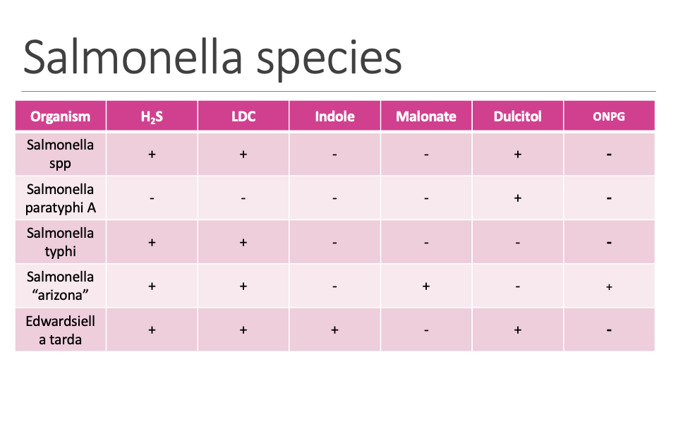
80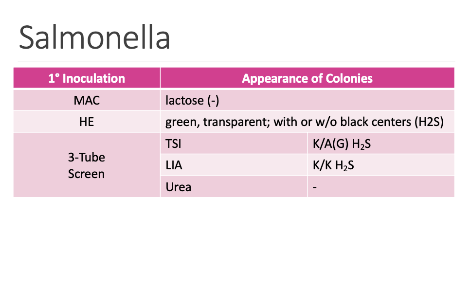
81
Q: How does non-typhoidal Salmonella infection typically resolve?
A: It is self-limiting and resolves in ≤ 1 week.
82
Q: What type of infection is typhoid (enteric) fever?
A: A serious blood infection.
83
Q: How does typhoid fever present in young children?
A: It is milder, but can be debilitating.
84
Q: What is the mortality rate of untreated typhoid fever?
A: > 10%
85
Q: What is the mortality rate of treated typhoid fever?
A: ≤ 1%, therefore treatment is necessary.
86
Q: When is antimicrobial treatment required for typhoid fever?
A: If invasive (in blood), treatment with antimicrobials is needed.
87
Q: What are the culture findings in typhoid fever during the first 2 weeks?
A: Blood (+), stool/ urine (−).
88
Q: What are the culture findings in typhoid fever after 2 weeks?
A: Blood (−), stool/ urine (+).
89
Q: How does paratyphoid fever compare to typhoid fever?
A: It has similar symptoms as typhoid fever.
90
Q: Does uncomplicated salmonellosis require treatment?
A: No, treatment is not needed.
91
Q: What are the reservoirs for Salmonella?
A: Animals (chicken, reptiles) and water.
92
Q: How is Salmonella commonly transmitted to humans?
A: Ingestion of poultry, chicken, pork, or dairy.
93
Q: What type of pathogen is Shigella?
A: A primary intestinal pathogen.
94
Q: How is Shigella food poisoning transmitted?
A: By direct fecal contamination.
95
Q: What foods are commonly associated with Shigella ingestion?
A: Egg salad and vegetables.
96
Q: What are the motility and Gram stain characteristics of Shigella?
A: Non-motile Gram-negative rod (GNR) that grow well on MAC
97
Q: How does Shigella grow on MacConkey (MAC) agar?
A: It grows well on MAC.
98
Q: Describe the biochemical profile of Shigella.
A: Biochemically inert:
LDC (−)
LAC (−)
CIT (−)
H₂S (−)
MAL (−)
LDC (−)
LAC (−)
CIT (−)
H₂S (−)
MAL (−)
99
Q: How many subgroups does Shigella have? Q: What antigen are Shigella serotypes based on?
4:
Shigella dysenteriae (enterotoxin, neurotoxin)
Shigella flexneri (most common worldwide)
Shigella boydii
Shigella sonnei (most common in US)
- serotypes are based on somatic O Ag
Shigella dysenteriae (enterotoxin, neurotoxin)
Shigella flexneri (most common worldwide)
Shigella boydii
Shigella sonnei (most common in US)
- serotypes are based on somatic O Ag
100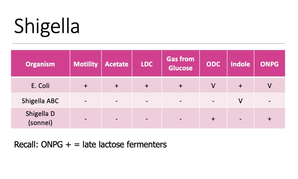
101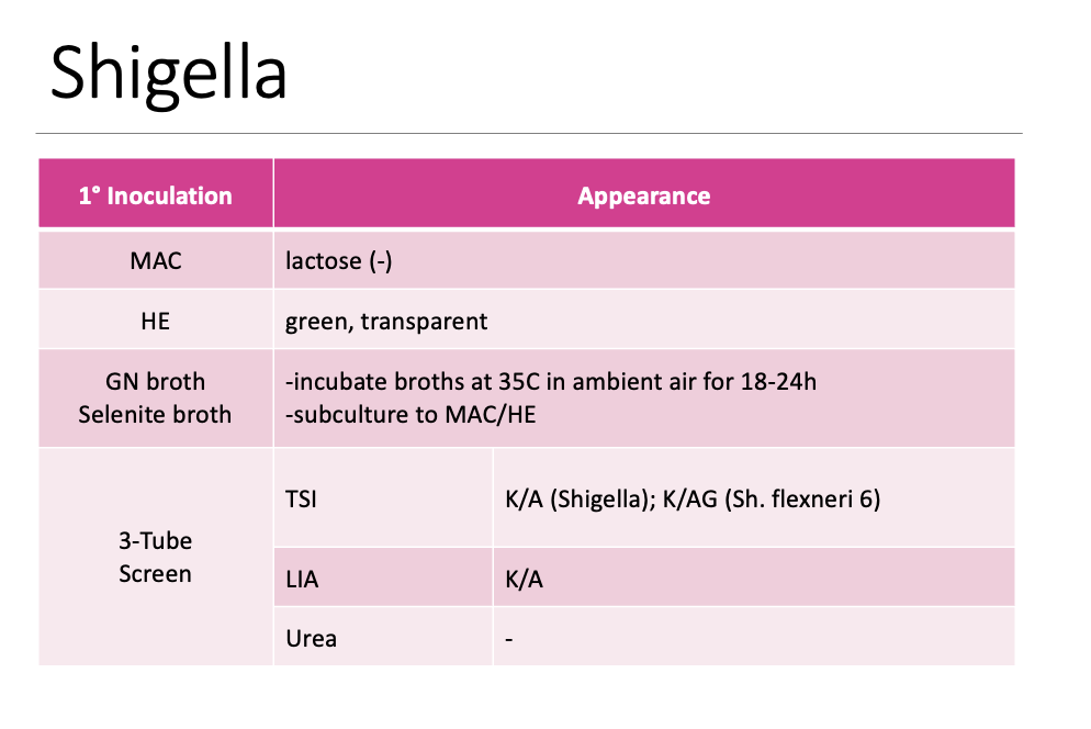
102
Q: What type of pathogen are Yersinia enterocolitica and Yersinia pseudotuberculosis?
A: Primary intestinal pathogens.
103
Q: What are the Gram stain and oxygen characteristics of Yersinia species?
A: Gram-negative rods (GNR), facultative anaerobes.
104
Q: What unusual staining characteristic may be seen with Yersinia species?
A: Bipolar staining (not always present).
105
Q: What are the lactose fermentation and MR reactions of Yersinia species?
A: LAC (−), MR (+).
106
Q: What temperature range can Yersinia species grow in?
A: 4°C–43°C.
107
Q: What is the primary reservoir for Yersinia species?
A: Swine.
108
Q: Which Yersinia species is most common?
A: Yersinia enterocolitica.
109
Q: How are Yersinia infections transmitted?
A: Fecal-oral transmission.
110
Q: Why can Yersinia be associated with transfusion reactions?
A: The organism likes colder temperatures and can be recovered from donor blood units, leading to transfusion reactions.
111
Q: On what medium and at what temperature is Yersinia cultured for isolation?
A: CIN agar incubated at room temperature (RT).
112
Q: How do Yersinia colonies appear on CIN agar?
A: They ferment mannitol, absorb neutral red dye, and produce clear colonies with a pink center.
113
Q: Which organism can produce colonies similar to Yersinia on CIN agar?
A: Aeromonas.
114
Q: What is the ONPG reaction of Yersinia and what does it indicate?
A: ONPG positive (+), indicating a delayed lactose fermenter.
115
Q: How does motility of Yersinia species change with temperature?
A: All species except Yersinia pestis are non-motile at 35–37°C but motile at 25°C.
116
Q: How does Yersinia enterocolitica appear on TSI and MAC agar?
A: Sucrose positive (A/A on TSI) but clear on MAC.
117
Q: What are the biochemical characteristics of Yersinia enterocolitica?
A:
Urea: +
Motility (22°C): +
Motility (35°C): −
Sucrose: +
Ornithine: +
Urea: +
Motility (22°C): +
Motility (35°C): −
Sucrose: +
Ornithine: +
118
Q: What are the biochemical characteristics of Yersinia pestis?
A:
Urea: −
Motility (22°C): −
Motility (35°C): −
Sucrose: −
Ornithine: −
Urea: −
Motility (22°C): −
Motility (35°C): −
Sucrose: −
Ornithine: −
119
Q: What are the biochemical characteristics of Yersinia pseudotuberculosis?
A:
Urea: +
Motility (22°C): +
Motility (35°C): −
Sucrose: −
Ornithine: −
Urea: +
Motility (22°C): +
Motility (35°C): −
Sucrose: −
Ornithine: −
120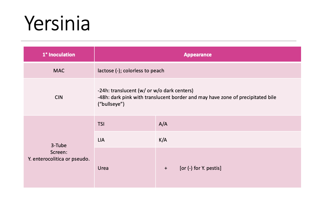
121
Q: How is Shiga toxin detected?
A: By BD MAX Enteric Bacterial Panel as a PCR target.
122
Q: What is the first step if Shiga toxin PCR is positive?
A: Inoculate stool into GN broth.
123
Q: What is done after GN broth incubation when PCR is positive for Shiga toxin?
A: After 16–24 hours incubation, send GN broth to LACDPH.
124
Q: Which organism is a Shiga toxin producer mentioned on this slide?
A: E. coli O157:H7
125
Q: What agar is used to recover Shiga toxin–producing E. coli O157:H7?
A: Sorbitol MacConkey agar (SMAC).
126
Q: How does E. coli O157:H7 appear on SMAC agar?
A: It does not ferment sorbitol and produces colorless colonies.
127
Q: What does EHEC stand for?
A: Enterohemorrhagic E. coli.
128
Q: What toxin types are associated with EHEC?
A: Serotoxigenic (STEC) and verotoxigenic (VTEC).
129
Q: What conditions are caused by EHEC?
A: Hemorrhagic diarrhea, colitis, and hemolytic uremic syndrome (HUS).
130
Q: How is hemolytic uremic syndrome (HUS) characterized?
A: Hemolytic anemia and low platelet count, leading to kidney failure and death.
131
Q: What are the two most common EHEC serotypes associated with diarrhea or HUS?
A: O157:H7 and O157:NM (nonmotile).
132
Q: What does ETEC stand for?
Q: What toxins are produced by ETEC?
Q: What type of diarrhea is caused by ETEC and how common is it in the U.S.?
Q: What is ETEC a common cause of?
Q: What toxins are produced by ETEC?
Q: What type of diarrhea is caused by ETEC and how common is it in the U.S.?
Q: What is ETEC a common cause of?
A: Enterotoxigenic E. coli.
A: Heat-labile enterotoxin (LT) and heat-stable enterotoxin (ST).
A: Mild watery diarrhea; uncommon in the U.S..
A: Common cause of traveler’s diarrhea
A: Heat-labile enterotoxin (LT) and heat-stable enterotoxin (ST).
A: Mild watery diarrhea; uncommon in the U.S..
A: Common cause of traveler’s diarrhea
133
Q: What does EPEC stand for?
Q: How does EPEC cause disease?
Q: What symptoms are associated with EPEC infection?
Q: In which population is EPEC typically seen?
Q: How does EPEC cause disease?
Q: What symptoms are associated with EPEC infection?
Q: In which population is EPEC typically seen?
A: Enteropathogenic E. coli.
A: It attaches to intestinal cells and typically does not produce exotoxin.
A: Prolonged, non-bloody diarrhea, vomiting, and fever.
A: Infants and children.
A: It attaches to intestinal cells and typically does not produce exotoxin.
A: Prolonged, non-bloody diarrhea, vomiting, and fever.
A: Infants and children.
134
Q: What does EIEC stand for?
Q: How does EIEC cause diarrhea and how common is it in the U.S.?
Q: How does EIEC cause diarrhea and how common is it in the U.S.?
A: Enteroinvasive E. coli.
A: Causes watery or bloody diarrhea via direct invasion of colonic epithelial cells; rare in the U.S.
A: Causes watery or bloody diarrhea via direct invasion of colonic epithelial cells; rare in the U.S.
135
Q: What does EAEC stand for?
Q: What are the stool characteristics of EAEC infection?
Q: What characteristic growth pattern is seen with EAEC?
Q: What are the stool characteristics of EAEC infection?
Q: What characteristic growth pattern is seen with EAEC?
A: Enteroaggregative E. coli.
A: Non-bloody stool that does not contain WBCs.
A: Clumps of bacteria form on the cell surface.
A: Non-bloody stool that does not contain WBCs.
A: Clumps of bacteria form on the cell surface.
136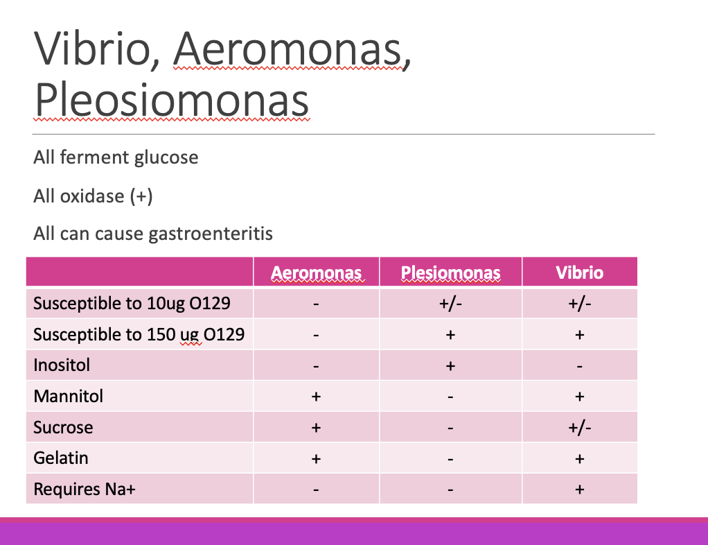
137
Q: What is the general morphology of Vibrio species?
A: Small Gram-negative rods, some are curved or comma-shaped.
138
Q: Where are Vibrio species naturally found?
A: Seawater.
139
Q: What are the typical biochemical and motility characteristics of Vibrio species?
A: Oxidase positive, catalase positive, and motile.
140
Q: What growth requirement do most Vibrio species have?
A: They are halophilic and require salt for growth.
141
Q: Which Vibrio species is a notable exception to the halophilic requirement?
A: Vibrio cholerae.
142
Q: How do Vibrio species grow in no-salt media?
A: They do not grow in no-salt media (e.g., MRVP tube).
143
Q: Why can Vibrio species grow on MacConkey (MAC) agar?
A: The salt content of MAC is sufficient to support growth of halophilic vibrios.
144
Q: Are Vibrio species considered part of normal flora?
A: No, they are not normal flora.
145
Q: How are Vibrio infections commonly acquired?
A:
Ingestion of contaminated or poorly cooked seafood
Ingestion of seawater or contaminated water (can lead to respiratory illness if it enters the lungs, e.g., drowning)
Wound infections after trauma while swimming or working in contaminated water, or exposure to marine animals (spikes, bites)
Ingestion of contaminated or poorly cooked seafood
Ingestion of seawater or contaminated water (can lead to respiratory illness if it enters the lungs, e.g., drowning)
Wound infections after trauma while swimming or working in contaminated water, or exposure to marine animals (spikes, bites)
146
Q: Why are early collection, prompt incubation, and suitable media important for Vibrio recovery?
A: They are important to ensure recovery of the organism.
147
Q: What selective media are used to recover Vibrio species?
A:
Alkaline Peptone Water (pH 8.4–8.5) with high salt (6.5% NaCl)
TCBS agar
Alkaline Peptone Water (pH 8.4–8.5) with high salt (6.5% NaCl)
TCBS agar
148
Q: What must be done if Vibrio is isolated?
A: Notify County Public Health.
149
Q: What does TCBS agar indicate for Vibrio species?
A:
Yellow = sucrose fermenters
Blue-green = non-sucrose fermenters
Yellow = sucrose fermenters
Blue-green = non-sucrose fermenters
150
Q: What is the next step after growth on TCBS agar when Vibrio is suspected?
A: Perform rule-out oxidase (+) colonies and isolate suspected colonies from TCBS to BAP.
151
Q: Which organisms produce yellow colonies (sucrose fermenters) on TCBS?
A:
V. cholerae
V. fluvialis
V. alginolyticus
Sucrose-fermenting Proteus spp
Enterococci
V. cholerae
V. fluvialis
V. alginolyticus
Sucrose-fermenting Proteus spp
Enterococci
152
Q: Which organisms produce blue-green colonies (non-sucrose fermenters) on TCBS?
A:
V. parahaemolyticus
V. mimicus
V. hollisae
Coliforms, Proteus spp, Enterococci (usually small and translucent)
Pseudomonas spp and Aeromonas spp
V. parahaemolyticus
V. mimicus
V. hollisae
Coliforms, Proteus spp, Enterococci (usually small and translucent)
Pseudomonas spp and Aeromonas spp
153
Q: Which Vibrio cholerae serogroups can produce toxin and cause epidemic cholera?
A: Serogroup O1 and O139 (O139 in Asia only)
154
Q: Which Vibrio cholerae biotype caused the seventh cholera pandemic in 1961?
A: Vibrio cholerae biotype El Tor (El Tor)
155
Q: What type of illness do Vibrio cholerae strains that do NOT produce toxin cause?
A: Mild gastroenteritis
156
Q: What causes the characteristic “rice water stool” in cholera?
A: Enterotoxin choleragen (cholera toxin, ctx)
157
Q: What are the key biochemical characteristics of Vibrio cholerae?
A: Sucrose (+),
Oxidase (+),
O129 Sensitive,
6.5% NaCl (+),
Glucose fermenter (+)
Oxidase (+),
O129 Sensitive,
6.5% NaCl (+),
Glucose fermenter (+)
158
Q: What is the O129 disk used for in identifying Vibrio cholerae?
A: It is a vibriostatic agent; most Vibrios are susceptible
159
Q: How does O129 help distinguish Vibrio cholerae from Aeromonas?
A: Vibrio cholerae is O129 sensitive, while Aeromonas is resistant (R)
160
Q: What is the recommended treatment approach for Vibrio cholerae infection?
A: Antibiotics plus ancillary rehydration therapy
161
Q: Which antibiotics are shown to reduce the duration and amount of diarrhea in cholera?
A: Tetracycline or doxycycline
162
Q: Which Vibrios are sucrose negative, and where is Vibrio parahaemolyticus found?
A: Sucrose-negative Vibrios: Vibrio parahaemolyticus and Vibrio vulnificus; V. parahaemolyticus is found in contaminated seafood
163
Q: What type of illness does Vibrio parahaemolyticus usually cause and how long does it last?
A: Mild to moderate infection that is self-limiting for 2–3 days
164
Q: What severe disease is Vibrio vulnificus associated with and what is the fatality rate?
A: Fatal septicemia with 60–70% mortality
165
Q: Describe the infection progression and common sources associated with Vibrio vulnificus.
A: Wound infection → septicemia → death; found near coastal H₂O and estuaries and associated with oysters
166
Q: Which Vibrios are sucrose positive and what illnesses are they associated with?
Vibrio alginolyticus
Vibrio fluvalis
Vibrio fluvalis
167
Q: What types of infections are associated with Vibrio alginolyticus?
acute gastroenteritis
Superficial wound, necrotizing fasciitis, conjunctivitis
Soft tissue infections especially ear
Superficial wound, necrotizing fasciitis, conjunctivitis
Soft tissue infections especially ear
168
Q: What types of infections are associated with Vibrio fluvalis
?
?
Gastroenteritis
169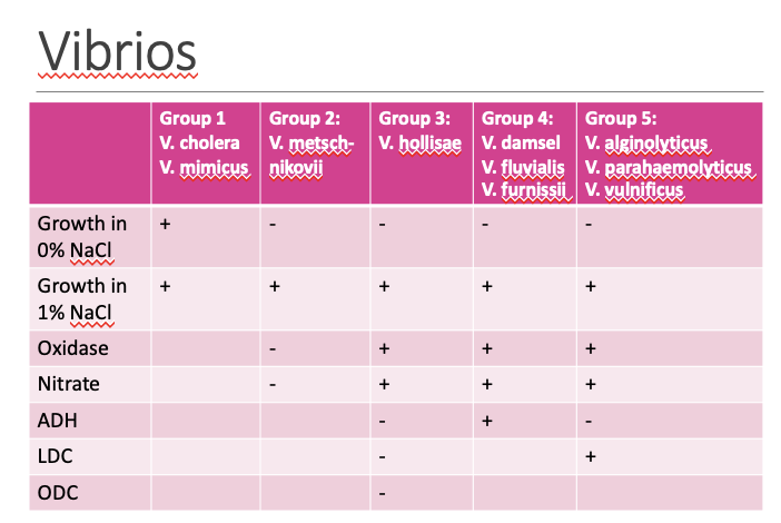
170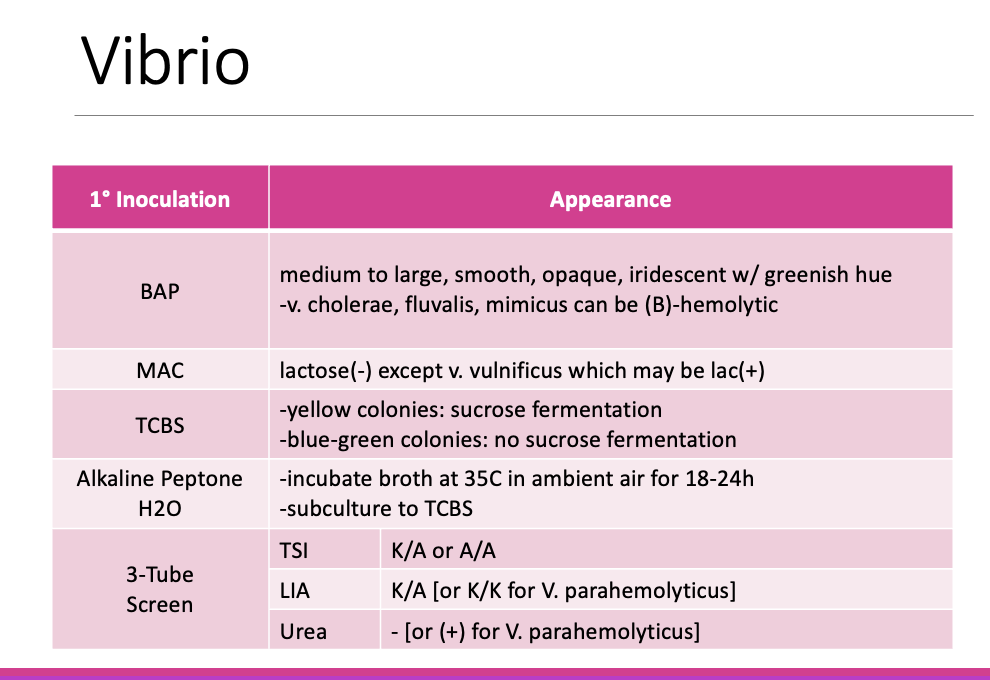
171
Q: What are the general characteristics and common associations of Aeromonas spp.?
A: Most commonly associated with diarrheal disease or wound infections acquired near or in water; most strains are beta-hemolytic on 5% BAP, oxidase positive, and ferment glucose
172
Q: Which Aeromonas species is the most common?
A: Aeromonas hydrophila
173
Q: What habitats are Aeromonas species found in?
A: Aquatic environments including groundwater/drinking water, clean or polluted lakes or rivers, and marine environments such as brackish water and water with low saline content
174
Q: How do Aeromonas species cause human infections and what are common sources?
A: Similar to Vibrio—infection via ingestion of contaminated water, exposure to marine animals, or wounds in sea water; sources include fresh produce, meat (beef, poultry, pork), dairy (raw milk, ice cream), and the gut of medicinal leeches (Aeromonas veronii biovar sobria)
175
Q: What animal infections are associated with Aeromonas species?
A: Severe infections in fish causing economic loss, and infections in frogs, pigs, cattle, birds, and marine animals
176
Q: What are the features of Aeromonas gastroenteritis causing acute watery diarrhea?
A: Acute watery diarrhea with no RBCs/WBCs in stool, abdominal pain, sometimes fever, nausea, vomiting; self-limiting infection, though children may need fluid replacement therapy; associated with Aeromonas caviae
177
Q: What characterizes cholera-like disease caused by Aeromonas species?
A: Dysenteric disease resembling Shigella with abdominal pain, fever, nausea, and bloody stool with mucus and PMNs; associated with A. veronii biovar sobria
178
Q: What severe complications are associated with Aeromonas infections and which species are involved?
A: Severe infections with complications such as HUS and kidney disease, associated with A. hydrophila and A. veronii biovar sobria
179
Q: What characterizes extraintestinal Aeromonas wound infections?
A: Traumatic injury in or around water leading to wound infections, including cellulitis and myonecrosis (destruction of muscle tissue);
infections are typically polymicrobial, but A. hydrophila is most common;
medicinal leeches used on wound sites postoperatively are associated with Aeromonas veronii biovar sobria
infections are typically polymicrobial, but A. hydrophila is most common;
medicinal leeches used on wound sites postoperatively are associated with Aeromonas veronii biovar sobria
180
Q: Which patients are at risk for Aeromonas septicemia and what is the fatality rate?
A: Patients with liver disease and hematological malignancies; 30–50% fatalities
181
Q: What are the key laboratory characteristics of clinically relevant Aeromonas species?
A: Beta-hemolytic, oxidase positive, spot indole positive, O129 resistant, no growth in 6.5% NaCl, INO (−), MAN (+), SUC (+), GEL (+)
182
Q: How can Aeromonas species be differentiated from Yersinia enterocolitica on CIN agar?
A: They may resemble each other on CIN, so an oxidase test is needed: Aeromonas = oxidase positive,
Yersinia enterocolitica = oxidase negative
Yersinia enterocolitica = oxidase negative
183
Q: How should Aeromonas be identified at the complex level?
A: As belonging to the Aeromonas hydrophila complex, Aeromonas caviae complex, or Aeromonas veronii biovar sobria
184
Aeromonas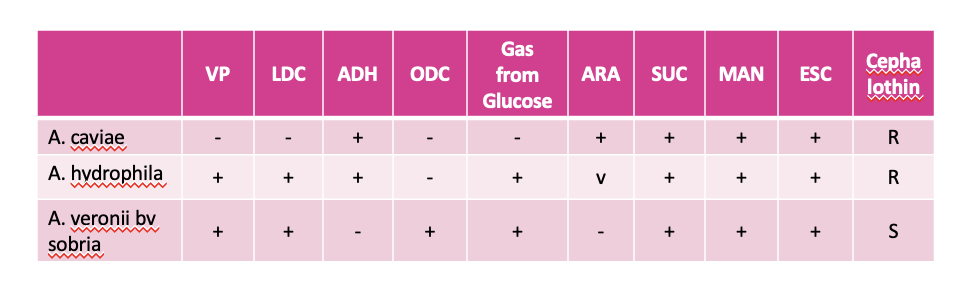
185
Q: How is Plesiomonas infection typically acquired and what illness does it cause?
A: Infection occurs primarily through ingestion of contaminated water or unwashed food, causing mild gastroenteritis
186
Q: What are the key laboratory characteristics of Plesiomonas, and what is its only species?
A: Similar to Aeromonas hydrophila; oxidase positive, glucose fermenter; only species is Plesiomonas shigelloides
187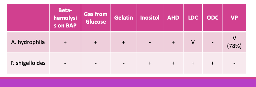
188
Q: What are the key laboratory characteristics of Edwardsiella tarda?
A: H₂S positive, indole positive (helps distinguish from Salmonella)
189
Q: What are the three-tube screen results for Edwardsiella tarda?
A: TSI: K/AG+,
LIA: K/K+,
Urea: negative
LIA: K/K+,
Urea: negative
190
Q: What type of disease can Edwardsiella tarda cause and why must it be distinguished from Salmonella?
A: Can cause GI disease and needs to be distinguished from Salmonella
191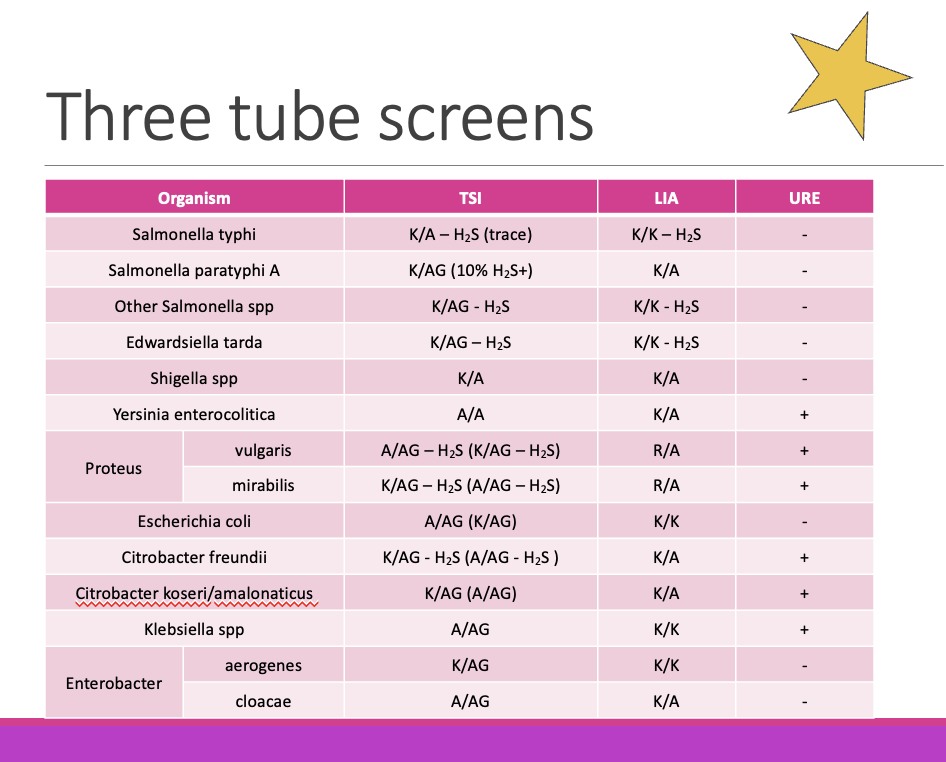
192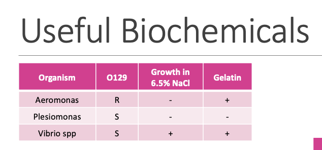
193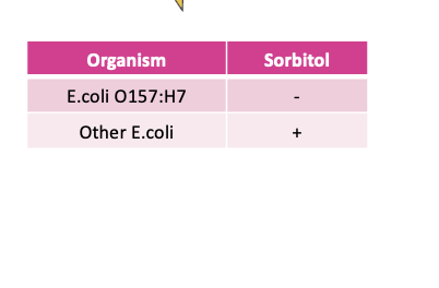
194
195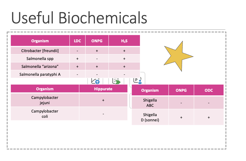
196
Q: Why have many laboratories switched to molecular diagnostics for detection of bacterial enteric pathogens?
A: Molecular methods are sensitive and provide faster turnaround time
197
Q: What are the pros and cons of molecular diagnostic methods?
Pro: Sensitive, faster turnaround time
Con: More expensive, cannot distinguish viable vs nonviable organisms, limits susceptibility testing
Con: More expensive, cannot distinguish viable vs nonviable organisms, limits susceptibility testing
198
Q: What pathogens are detected by the BD MAX™ System and what method does it use?
A: Uses PCR to detect common pathogens including Salmonella, Shigella, Campylobacter, and Shiga toxin
199
Q: What is the advantage of the BioFire® FilmArray® Gastrointestinal (GI) Panel?
A: Can detect more agents of GI disease, including viruses and parasites