Blood Culture
Study these flashcardsFree · spaced repetition · iPhone, Android, Mac & web
All cards
1
What is the purpose of site preparation during blood specimen collection?
To ensure the blood specimen is not contaminated with normal skin flora.
2
What should be done to disinfect the skin before venipuncture?
Defat the skin with 70% alcohol wipe, apply iodine tincture or chlorhexidine and allow to dry
3
What is the most important factor in blood culture collection and why?
Blood volume; higher blood volume increases probability that lab will recover the organism, making total volume more important than number of specimens.
4
Based on the patients age, what are the recommended volumes for blood culture?
Adults: optimally 2 sets of cultures using 10-20 mL per culture; infants and children: 1-5 mL per culture (volume drawn can be dependent on patient's weight).
5
When should blood cultures be collected and how many sets are recommended?
Collect blood before antibiotics are given to patient is most optimal; 2-3 culture sets (1 set = 1 aerobic & 1 anaerobic bottle) are sufficient if volume is adequate and may vary depending on cases (i.e. if blood is collected pre or post antibiotic).
6
How should blood cultures be timed, transported, and incubated?
• Generally 2 or 3 sets of blood cultures are collected an hour apart to account for intermittent sepsis;
• transport blood cultures at room temperature;
• incubate bottles at 35° to 37°C.
• transport blood cultures at room temperature;
• incubate bottles at 35° to 37°C.
7
Why must blood specimens for culture be treated with an anticoagulant?
Organisms are difficult to recover from clotted specimens, so blood must be treated with an anticoagulant.
8
What is sodium polyanethol sulfonate (SPS) and what does it do?
Sodium polyanethol sulfonate (SPS) is an anticoagulant that is anti-phagocyte and anti-complement, inhibits some antibiotics, and inhibits all Peptostreptococcus anaerobius and some strains of Neisseria species, Gardnerella vaginalis, and Streptococcus milliformis.
9
Which anticoagulants are inappropriate for blood cultures?
• Citrate (blue top),
• heparin (green top),
• oxalate (gray top),
• EDTA (ethylenediaminetetraacetic acid) (lavender top).
• heparin (green top),
• oxalate (gray top),
• EDTA (ethylenediaminetetraacetic acid) (lavender top).
10
How does dilution affect blood culture recovery and what ratios are used?
Dilution of blood to medium is a factor; traditionally a 1:10 ratio of blood to medium is used, but enhanced recovery occurs with a 1:5 ratio in conventional blood cultures. (commercial/automated systems specify their own appropriate dilutions)
11
What do blood culture media typically contain and what broths are used?
Blood culture media typically contain nutrient broth and anticoagulant; varying broths include trypticase soy broth, brain-heart infusion, peptone-supplemented broth, thioglycollate broth, and Columbia or Brucella broth.
12
How are conventional (manual) blood cultures incubated and monitored for growth?
Conventional blood cultures are incubated in a stationary incubator and bottles are visually examined for signs of growth such as hemolysis, gas, clumping, and turbidity.
13
How are conventional blood cultures worked up and how long are they held?
Bottles are periodically blind subcultured to CHOC at 24 and 48 hours, cultures are signed out at 7 days, and specific rule-outs such as Brucella are held for 21 days.
14
What is the BD BBL Septi-Chek system and how is it structured?
It's a biphasic (both liquid and solid media), self-contained subculture system with a conventional blood bottle attached to a paddle (chamber with slide coated with agar).
15
What media is used and how is growth detected in BD BBL Septi-Chek?
The agar paddle contains CHOC, MAC, and malt agar; it is periodically bathed in the blood-broth mixture, examined for growth daily, subcultured at 24 and 48 hours, and a larger volume of broth is subcultured.
16
What is the Isolator/Isostat blood culture system and what is it used for?
Isolator/Isostat is a lysis centrifugation system that is good for intracellular, fastidious, or slow growing organisms.
17
What type of media and anticoagulants are used in the Isolator/Isostat system?
Instead of regular blood culture media, blood is collected in media containing saponin which lyses RBC & WBC, and SPS & EDTA are used as anticoagulants.
18
How is the blood specimen processed in the Isolator/Isostat system after collection?
The tube is centrifuged, the supernatant is discarded, and the sediment is vortexed and placed on appropriate solid agar.
19
What are the main advantages of automated blood culture systems?
Instrumentation allows for continuous blood monitoring, is more hands-off, increases sensitivity, and reduces the need to blind subculture.
20
How do automated blood culture instruments detect positive growth?
Each instrument system uses different methodologies and algorithms to flag for positive blood culture growth.
21
How does the BacT/Alert system detect positive blood cultures?
It uses colorimetric detection of CO₂ by detecting an increase in CO₂ via generation of free hydrogen ions, which cause a color change in the sensor.
22
What media and anticoagulant are used in the BacT/Alert blood culture system?
It uses TSB (tryptic soy broth) and SPS (sodium polyanethol sulfonate).
23
How does the BACTEC FX system detect positive blood cultures?
Uses fluorescent light detection of CO₂; increased CO₂ generates free hydrogen ions causing a decrease in pH, which in turn increases fluorescent output of the sensor, utilizing an LED sensor read by a photodetector.
24
What media does BACTEC FX use and where is this instrument used?
It uses a soybean casein digest broth and is the instrument used in the UCLA.
25
How does the VersaTREK blood culture system detect microbial growth?
Detects pressure changes; decreased pressure indicates O₂ consumption, while increased pressure indicates production of CO₂, H₂, and N₂.
26
What media are used in VersaTREK aerobic and anaerobic bottles?
The aerobic bottle uses soycasein-peptone broth, and the anaerobic bottle uses protease-peptone broth.
27
How should positive blood cultures be prioritized and worked up?
Positive blood cultures are high priority and should be processed as quickly as possible, with identification and antimicrobial susceptibility tests performed.
28
What is the first step in working up a positive blood culture and why is it done?
A Gram stain smear is the first step; it determines the morphotype of the organism and guides setup of appropriate media/plates for the morphotype seen.
29
How should positive blood cultures be subcultured based on Gram stain morphotype?
GPC in clusters → BAP; GPC in pairs/chains → BAP/CHOC; GNR → BAP/MAC; PLGPR/GPR → BAP/CHOC; small GNR/GNCB → BAP/CHOC/MAC.
30
What steps are taken if no bacteria are seen on Gram stain from a positive blood culture?
Repeat Gram stain one more time and confirm with Acridine Orange (fluorescent stain) to visualize small or faintly staining organisms.
31
What follow-up actions are required when no bacteria are seen on Gram stain?
Perform blind subculture on BAP/CHOC/THIO and hold for 72 hours; if anaerobic bottle, also subculture to Brucella Blood Agar Plate and incubate anaerobically; return blood culture bottle to instrument within 3 hours.
32
Are all instrument-flagged positive blood cultures true positives?
No; not all bottles flagged positive by the instrument are true positive.
33
What l factors can cause an instrument false positive blood culture result?
An increase in metabolically active cells that produce CO₂, such as leukocytosis or leukemia, which typically produce linear or flat graphs rather than the logarithmic graph seen with true bacterial growth.
34
What technical factors can lead to an instrument-positive blood culture?
Overfilling blood culture bottles or obstruction of the instrument's ability to read the bottle, including labels obscuring the detector or a bottle defect.
35
What is the BCID system and how is it used on positive blood cultures?
BCID systems provide rapid molecular blood culture diagnostics by testing the direct blood specimen from the bottle, including Luminex Verigene, BioFire FilmArray, and Accelerate Pheno System (can perform sensi on GNR).
36
What are the main advantages of BCID testing?
BCID provides rapid turnaround time for identification (about 2 hours), allowing more targeted antibiotic therapy or rule-out of potential contaminants such as CoNS or SA.
37
What are the limitations and drawbacks of BCID systems?
BCID systems can be costly, may detect non-viable nucleic acid (FDA class 2 recall due to blood culture media contaminated with DNA on non-viable organism), and are limited by the target organism available on the panel.
38
What BCID system does the UCLA Bacteriology Lab currently use?
The cobas® eplex system developed by GenMark.
39
How are BCID panels selected and what panels are available?
One of three panels is selected based on the Gram stain result: BCID-GP (gram positive panel), BCID-GN (gram negative panel), or BCID-FP (fungal panel).
40
What additional clinically important information can the BCID system detect?
Can also detect resistance markers such as mecA, vanA, and CTX-M.
41
Why is preliminary identification from blood cultures important and how was it performed before BCID systems?
Early identification of the organism is of utmost importance; prior to BCID, conventional biochemicals were set up directly from blood cultures to shorten time to identification.
42
How is preliminary identification performed for gram-positive cocci in clusters versus pairs/chains?
• GPCCL: perform tube coagulase by inoculating blood directly into rabbit plasma tube; if SA, perform PBP2a for MRSA.
• GPCPr/Ch: use vancomycin 30 mcg and P disk, 6.5% NaCl, and bile-esculin slant by placing Va and P disks on BAP already plated with blood and inoculating blood directly onto 6.5% NaCl tube and bile-esculin slant.
• GPCPr/Ch: use vancomycin 30 mcg and P disk, 6.5% NaCl, and bile-esculin slant by placing Va and P disks on BAP already plated with blood and inoculating blood directly onto 6.5% NaCl tube and bile-esculin slant.
43
How is preliminary identification performed for small gram-positive rods and gram-negative rods?
• Small GPR: inoculate blood directly onto TSI slant and stab the butt.
• GNR: perform TSI, API20E, and preliminary Kirby-Bauer disk diffusion using the same TSI procedure, inoculating a few drops of blood into 5 mL saline for API20E, and inoculating MH with 4 drops of blood, streaking and applying antibiotics per usual MH protocol.
• GNR: perform TSI, API20E, and preliminary Kirby-Bauer disk diffusion using the same TSI procedure, inoculating a few drops of blood into 5 mL saline for API20E, and inoculating MH with 4 drops of blood, streaking and applying antibiotics per usual MH protocol.
44
Which organisms are always treated as true pathogens in blood cultures, even if only 1 of multiple sets is positive?
• Gram-negative rods
• Enterococcus species
• Staphylococcus aureus
• Streptococcus pneumoniae
• Haemophilus influenzae
• Streptococcus pyogenes
• Streptococcus agalactiae
• Large colonies beta-hemolytic Streptococci
• Listeria monocytogenes
• Enterococcus species
• Staphylococcus aureus
• Streptococcus pneumoniae
• Haemophilus influenzae
• Streptococcus pyogenes
• Streptococcus agalactiae
• Large colonies beta-hemolytic Streptococci
• Listeria monocytogenes
45
What is the required laboratory workup for these commonly recovered pathogens?
Perform full species-level identification and antimicrobial susceptibility testing.
46
Which organisms are considered probable skin contaminants when isolated from only 1 of multiple blood culture sets?
• Viridans group Streptococcus
• Coagulase negative Staphylococcus, not lugdunensis
• Corynebacterium species, not jeikeium
• Bacillus species, not anthracis
• Propionibacterium (Cutibacterium) acnes
• Coagulase negative Staphylococcus, not lugdunensis
• Corynebacterium species, not jeikeium
• Bacillus species, not anthracis
• Propionibacterium (Cutibacterium) acnes
47
When is full identification and susceptibility testing required for probable skin contaminants?
Perform full identification and susceptibility testing if recovered from multiple blood culture sets; exception: perform full ID and susceptibility on all isolates from patients ≤ 3 months old regardless of number of positive sets.
48
What is the clinically relevant Listeria species and how can it be mistaken in the lab?
It is a facultative intracellular pathogen (lives in phagocytes) and its colony morphology and biochemical reactions can be confused with group B streptococci.
49
How is listeriosis transmitted and what foods are commonly implicated?
Transmission occurs via contaminated food, especially unpasteurized milk and dairy, with outbreaks linked to soft cheese, ice cream, and milk.
50
Which patients are at highest risk for listeriosis and what are the typical outcomes?
High-risk patients include
• Pregnant women and fetuses - Nonspecific, mild to moderate symptoms but may lead to sepsis and meningitis
• Neonates - Listeria can cross placenta, acquire infection "in utero" resulting in abortion, stillbirth, premature birth, septicemia, or meningitis
• Elderly and immunocompromised (advanced malignancy, renal transplant, diabetes) - mainly causes meningitis and septicemia
• Pregnant women and fetuses - Nonspecific, mild to moderate symptoms but may lead to sepsis and meningitis
• Neonates - Listeria can cross placenta, acquire infection "in utero" resulting in abortion, stillbirth, premature birth, septicemia, or meningitis
• Elderly and immunocompromised (advanced malignancy, renal transplant, diabetes) - mainly causes meningitis and septicemia
51
What is the carrier rate of Listeria?
Carrier rate is 1-5%.
52
What are the Gram stain characteristics of Listeria monocytogenes?
• Gram stain: Small "ditpheroid-like", coccobacillary, non-sporeforming GPR. (Pairs may be mistaken for Streptococci)
53
What is the colonial morphology of Listeria monocytogenes?
On BAP: small translucent gray colonies with a narrow zone of beta-hemolysis; on McBride agar: colonies appear blue-green.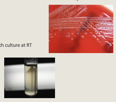
54
What are the key biochemical characteristics of Listeria monocytogenes?
Biochemicals:
• Catalase (+)
• Motile: Optimal at 25°C (Tumbling motility on wet mount from 2-6 hour broth culture at RT and Umbrella motility on semisolid agar at RT)
• CAMP (+)
• Hippurate (+)
• Esculin (+)
• Catalase (+)
• Motile: Optimal at 25°C (Tumbling motility on wet mount from 2-6 hour broth culture at RT and Umbrella motility on semisolid agar at RT)
• CAMP (+)
• Hippurate (+)
• Esculin (+)
55
What is the Anton (ocular) test for Listeria monocytogenes and what does it demonstrate?
The Anton (ocular) test assesses pathogenicity/virulence; inoculation of the organism into a rabbit's eye causes purulent conjunctivitis.
56
What is the purpose of the cold enrichment test and which organisms does it help recover?
Cold enrichment enhances recovery of Listeria and Yersinia and is used for epidemiological studies of contaminated specimens.
57
How is cold enrichment performed for Listeria monocytogenes?
Although optimum growth is 30-37 °C, Listeria can grow at 4 °C; inoculated UVM-Listeria Selective Enrichment Broth is refrigerated at 4 °C for 3 months, subcultured weekly for 4 weeks, then monthly for the next 2 months.
58
What is the principle of the hippurate hydrolysis test?
Sodium hippurate hydrolysis produces glycine, which is detected by ninhydrin; in the deaminated glycine-ninhydrin reaction, ammonia reacts with residual and reduced ninhydrin to form a blue-purple condensation complex.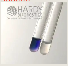
59
How is the hippurate hydrolysis test performed and interpreted?
Procedure:
1. Add 0.1mL sterile water to plastic test tube (glass test tubes may result in false negative)
2. Emulsify 1-3 colonies (more colonies are needed for smaller organisms such as Gardnerella vaginalis)
3. Drop hippurate disk in mixture
4. Incubate for 2 hours at 35-37C
5. Add 2 drops of ninhydrin
6. Mix and reincubate for 30 minutes
7. Interpretation
Positive: Blue-purple color
Negative: No change or light gray
***(Run positive and negative QC to correlate color change (GBS + and GAS -)
1. Add 0.1mL sterile water to plastic test tube (glass test tubes may result in false negative)
2. Emulsify 1-3 colonies (more colonies are needed for smaller organisms such as Gardnerella vaginalis)
3. Drop hippurate disk in mixture
4. Incubate for 2 hours at 35-37C
5. Add 2 drops of ninhydrin
6. Mix and reincubate for 30 minutes
7. Interpretation
Positive: Blue-purple color
Negative: No change or light gray
***(Run positive and negative QC to correlate color change (GBS + and GAS -)
60
Which organisms are hippurate positive?
Campylobacter jejuni, Group B Streptococcus, Listeria monocytogenes, and Gardnerella vaginalis.
61
What is the CAMP test and what is its principle?
CAMP (Christie, Atkins, and Munch-Peterson) test detects production of a diffusible extracellular hemolytic protein (CAMP factor) by GBS and Listeria monocytogenes that acts synergistically with beta-lysin of Staphylococcus aureus to enhance lysis of RBCs.
62
How is the CAMP test performed?
Streak the organism to be tested perpendicular to a streak of Staphylococcus aureus ATCC 25923 on BAP within 2 mm and incubate overnight at 35-37 °C.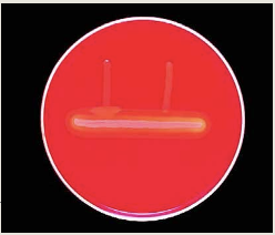
63
How are CAMP test results interpreted and what is an important limitation?
• Positive: enhanced zone of hemolysis adjacent to where the two streak lines meet (arrowhead may appear but not always);
• negative: no enhancement;
***Some strains of GAS may have a positive CAMP reaction, test should be limited to colonies characteristic of GBS (small zone of hemolysis)
• negative: no enhancement;
***Some strains of GAS may have a positive CAMP reaction, test should be limited to colonies characteristic of GBS (small zone of hemolysis)
64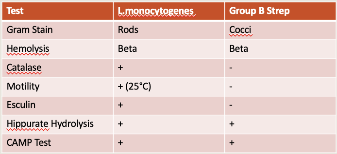
Listeria monocytogenes
65
What is Erysipelothrix rhusiopathiae and what animals does it primarily infect?
Erysipelothrix rhusiopathiae is primarily an animal pathogen that infects a wide variety of animals, especially swine.
66
What diseases are caused by Erysipelothrix rhusiopathiae in animals and humans?
• In swine it causes erysipelas, acquired by ingestion of contaminated food and water with multiplication in tonsils and gut leading to bloodstream spread;
• In humans it causes erysipeloid, a localized cutaneous infection with red skin lesions that may disseminate and cause bacteremia and endocarditis.
• In humans it causes erysipeloid, a localized cutaneous infection with red skin lesions that may disseminate and cause bacteremia and endocarditis.
67
How do humans acquire Erysipelothrix rhusiopathiae infection and who is at risk?
Humans become infected through contact with animals or animal products; infection is occupational, commonly affecting butchers, fishermen, and veterinarians.
68
What specimens should be collected for suspected Erysipelothrix rhusiopathiae infection?
For skin lesions, collect skin biopsies; for sepsis, collect blood cultures.
69
What are the Gram stain characteristics of Erysipelothrix rhusiopathiae?
• Gram Stain: short slender GPR , may be slightly pleomorphic,
• easily decolorizes
• GS performed from solid media may appear filamentous
• easily decolorizes
• GS performed from solid media may appear filamentous
70
What is the colonial morphology of Erysipelothrix rhusiopathiae on blood agar?
• Alpha or non-hemolytic on BAP
• Smooth colonies - small convex circular
• Rough colonies - larger, matte with fimbriated edge
• Smooth colonies - small convex circular
• Rough colonies - larger, matte with fimbriated edge
71
What are the key biochemical characteristics of Erysipelothrix rhusiopathiae?
Biochemicals:
• Catalase (-)
• Produce H2S in TSI
• Non-motile
• Catalase (-)
• Produce H2S in TSI
• Non-motile
72
What types of infections are caused by Corynebacterium jeikeium and in which patients are they most common?
Corynebacterium jeikeium causes nosocomial infections in immunocompromised patients, including bacteremia and bacterial endocarditis, and is associated with indwelling devices.
73
What clinical syndromes can Corynebacterium jeikeium cause and what is its antibiotic susceptibility pattern?
• It can cause community and nosocomially acquired bacteremia, sepsis, valve endocarditis, meningitis, osteomyelitis, and tissue infections;
• It is highly resistant to most gram-positive antibiotics but is sensitive to vancomycin.
• It is highly resistant to most gram-positive antibiotics but is sensitive to vancomycin.
74
What are the Gram stain characteristics of Corynebacterium jeikeium?
Small coccobacillary pleomorphic gram-positive rods (PLGPR).
75
What is the colonial morphology of Corynebacterium jeikeium?
• Small pinpoint gray-to-white, non-hemolytic colonies;
• Lipophilic and will not grow on CHOC.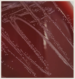
• Lipophilic and will not grow on CHOC.
76
What are the key biochemical characteristics of Corynebacterium jeikeium?
Biochemicals:
•Catalase (+)
•Motility (-)
•Urea (-)
•Nitrate (-)
•Glucose (+)
•Catalase (+)
•Motility (-)
•Urea (-)
•Nitrate (-)
•Glucose (+)
77
What is the normal habitat of viridans group Streptococcus?
Normal respiratory and genitourinary tract flora.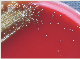
78
Why is viridans group Streptococcus clinically important in endocarditis?
It is a common cause of subacute endocarditis, such as damaged valves after dental surgery involving infected gums, reproductive or urinary (genitourinary tract) surgery or operations on the gastrointestinal tract (e.g. GI endoscopy, sigmoidoscopy or in those with artificial heart valves.
79
What proportion of endocarditis cases is caused by viridans group Streptococcus and what other infections can it cause?
Causes approximately 30-40% of endocarditis cases and may also cause dental cavities, wound infections, and brain abscesses.
80
Which species groups comprise the viridans group Streptococcus?
• Streptococcus mitis/oralis group
• Streptococcus sanguinis group
• Streptococcus salivarius group
• Streptococcus mutans group
• Streptococcus sanguinis group
• Streptococcus salivarius group
• Streptococcus mutans group
81
Which additional Streptococcus species fall under the viridans group umbrella and why are they clinically important?
Streptococcus anginosus (milleri) group, which is associated with abscesses and other pyogenic infections, and Streptococcus gallolyticus (formerly Streptococcus bovis), where Streptococcus gallolyticus subsp. gallolyticus bacteremia is linked to the presence of colonic cancer.
82
When is full species-level identification required for viridans group Streptococcus?
Perform full identification using API Strep on critical/sterile sites or when isolated from multiple blood culture bottles
83
When is limited biochemical identification sufficient for viridans group Streptococcus?
When not from critical sites or multiple blood cultures, biochemical identification as viridans group Streptococcus versus Streptococcus anginosus group is sufficient.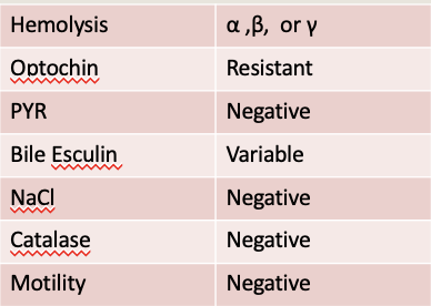
84
Why is Streptococcus agalactiae (Group B Streptococcus) a major concern in neonates?
It is a major cause of neonatal and perinatal bacteremia and meningitis, Neonates acquire it during passage through birth canal (may acquire it "in-utero" before delivery)
85
Where is Streptococcus agalactiae normally found in adults?
It is part of the normal vaginal flora and rectum and is usually asymptomatic.
86
What infections can Streptococcus agalactiae cause in adults and who is most at risk?
May also cause variety of infections in adults, including UTI, skin and soft tissue infections, postpartum fever, and sepsis. Usually in patients with risk factors such as catheters, diabetes, or advanced age.
87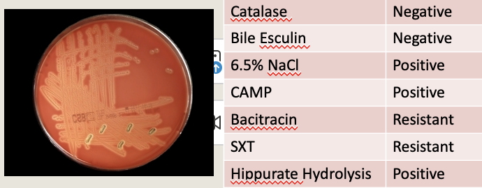
Streptococcus agalactiae
88
What infections are most commonly caused by Enterococcus species?
Enterococcus species mostly cause UTIs.
89
How does Enterococcus cause bacteremia and which patients are typically affected?
Enterococcus enter blood stream from UTI, abdominal or pelvic abscess, wounds, and IV devices; patients are usually elderly, immunocompromised, or have underlying diseases.
90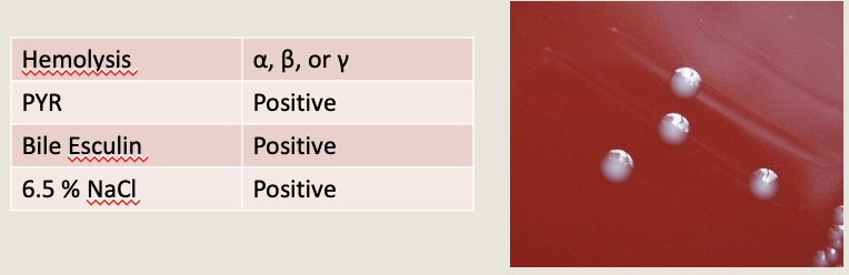
What are the key laboratory characteristics of Enterococcus species?
91
Why is speciation of Enterococcus species especially important in blood cultures?
Speciation is more important in blood cultures because different Enterococcus species can have different antimicrobial susceptibility patterns.
92
How are Enterococcus species fully identified in the lab?
Fully ID with API Strep
93
How do Enterococcus faecalis and Enterococcus faecium differ in hemolysis and susceptibility patterns?
• Enterococcus faecalis - Gamma hemolytic (usually)
- Ampicillin S, Synercid R
• Enterococcus faecium - Alpha hemolytic
- Ampicillin R, Synercid S
- Ampicillin S, Synercid R
• Enterococcus faecium - Alpha hemolytic
- Ampicillin R, Synercid S
94
Which Enterococcus species have intrinsically elevated vancomycin results?
Enterococcus gallinarum and Enterococcus casseliflavus/flavescens have intrinsically elevated vancomycin results.
95
Why is Streptococcus pneumoniae clinically significant?
It is a major cause of community-acquired pneumonia and a leading cause of bacteremia and sepsis in adults and children.
96
How does Streptococcus pneumoniae present differently in children versus adults?
• In children, it commonly causes Otitis media(middle ear infection) + pneumonia leading to bacteremia;
• In adults, pneumonia may progress to bacteremia.
• In adults, pneumonia may progress to bacteremia.
97
Which populations are most affected by severe Streptococcus pneumoniae infections and what complications can occur?
It is a common cause of meningitis in adults, infants, and toddlers (Meningitis as a result of bacteremia), with most serious infections occurring in infants <3 years old and adults >65 years old.
98
What is the main virulence factor of Streptococcus pneumoniae?
Virulence is attributable to polysaccharide capsule (resists intracellular killing)
99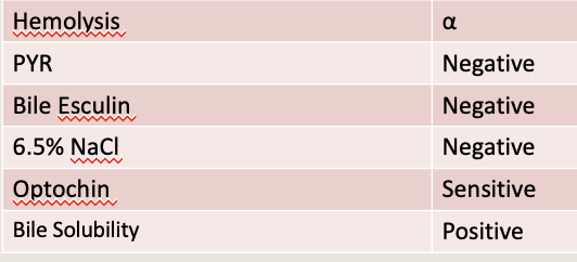
What are the key laboratory identification characteristics of Streptococcus pneumoniae?
100
Why is Staphylococcus epidermidis clinically significant in endocarditis and bacteremia?
It is the most common agent of prosthetic valve endocarditis and the most common agent of bacteremia in immunocompromised patients.
101
How does Staphylococcus epidermidis commonly cause bloodstream infections?
It reaches the blood via vascular access devices such as catheters and is the most common isolate of IV catheters causing Catheter-Related Bloodstream Infection (CRBI).
102
How are catheter-related infections due to Staphylococcus epidermidis evaluated in the lab?
IV catheters are the source of bacteremia; catheter tips can be submitted for culture by rolling the catheter tip on a blood agar plate and reporting identification with colony count.
103
What other device-related infections are commonly caused by Staphylococcus epidermidis?
It is also the most common cause of infection of CSF shunts and CSF catheters.
104
Why is Staphylococcus lugdunensis clinically important?
It causes endocarditis, bacteremia, and abscesses.
105
What are the colony characteristics of Staphylococcus lugdunensis?
Beta-hemolytic with white colonies (Must be differentiated from other coagulase negative Staphylococcus)
106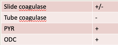
How does Staphylococcus lugdunensis differ from other coagulase-negative Staphylococci Biochemicals?
107
What reagents are required for the rapid ODC (ornithine decarboxylase) test?
0.5 mL Moeller decarboxylase broth base and 0.5 mL Moeller decarboxylase broth with ornithine.
108
How is the rapid ODC test set up and incubated?
• Inoculate both broths with colonies from an overnight culture to create a heavy suspension,
• overlay suspensions with mineral oil to prevent surface oxidation leading to false positives,
• incubate for 5 hours in a 35 °C heat block.
• overlay suspensions with mineral oil to prevent surface oxidation leading to false positives,
• incubate for 5 hours in a 35 °C heat block.
109
How are rapid ODC test results interpreted?
Interpretation:
• POS: broth with ornithine = purple, plain broth = yellow
• NEG: both remain yellow
• INVALID: base broth is purple
• POS: broth with ornithine = purple, plain broth = yellow
• NEG: both remain yellow
• INVALID: base broth is purple
110
What are HACEK organisms and what infection are they classically associated with?
HACEK organisms are fastidious gram-negative coccobacilli associated with infective endocarditis.
111
Which organisms are included in the HACEK group?
• Haemophilus species
• Aggregatibacter species
• Cardiobacterium hominis
• Eikenella corrodens
• Kingella spp.
• Aggregatibacter species
• Cardiobacterium hominis
• Eikenella corrodens
• Kingella spp.
112
What special growth requirements do HACEK organisms have?
They require media supplemented with hemin (X factor) and/or NAD (V factor) and enriched incubation with 5-10% CO₂.
113
On which media do HACEK organisms not grow well?
They do not grow on traditional enteric media such as MAC.
114
What is the expected incubation time for HACEK organisms in blood cultures?
Up to 14 days with conventional blood culture methods and about 5 days in continuous monitoring systems.
115
How are HACEK organisms fully identified in the laboratory?
Full identification is performed using the RAPID NH panel.
116
Why is Haemophilus influenzae type b (Hib) clinically significant?
Hib is the most pathogenic type of H. influenzae, is one of six polysaccharide capsule types, and has an available vaccine.
117
Where is Haemophilus influenzae normally found and what infections can it cause?
It is part of the normal oral flora and can cause epiglottitis (resulting in obstruction), otitis media, sinusitis, bronchitis, and pneumonia.
118
What is the role of Haemophilus influenzae in meningitis?
• In the pre-vaccine era, H. influenzae was the leading cause of bacteremia and meningitis in children ages 1 month-2yrs;
• nasopharyngeal colonization and otitis media can lead to meningitis; adult meningitis is uncommon;
• meningitis is frequent and presents early in children.
• nasopharyngeal colonization and otitis media can lead to meningitis; adult meningitis is uncommon;
• meningitis is frequent and presents early in children.
119
How does Haemophilus influenzae cause bacteremia and who is at increased risk?
• Increased risk of invasive disease if not vaccinated
• Nasopharyngeal colonization / respiratory infection/otitis media -> enters blood -> meninges
• Can be found in elderly with underlying diseases (head trauma or neurosurgery)
• Nasopharyngeal colonization / respiratory infection/otitis media -> enters blood -> meninges
• Can be found in elderly with underlying diseases (head trauma or neurosurgery)
120
What are the Gram stain characteristics of Haemophilus influenzae?
Small pleomorphic gram-negative coccobacilli.
121
What is the colony morphology of Haemophilus influenzae?
On CHOC: smooth, round, tan colonies (some strains mucoid); will not grow on BAP or MAC but will grow on horse or rabbit BAP.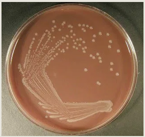
122
What are the key biochemical and growth requirements of Haemophilus influenzae?
Biochemical:
• Oxidase and catalase variable
• Require both X and V factor
• Growth only around XV disk (not X, V)
• Growth on Rabbit BAP, no hemolysis
• Satellite Test (+) on BAP
• Satellite Test (-) on Nutrient Agar
• Beta-lactamase positive (30-40%)
• Oxidase and catalase variable
• Require both X and V factor
• Growth only around XV disk (not X, V)
• Growth on Rabbit BAP, no hemolysis
• Satellite Test (+) on BAP
• Satellite Test (-) on Nutrient Agar
• Beta-lactamase positive (30-40%)
123
How can Haemophilus influenzae be fully identified if needed?
Full identification can be performed using the RAPID NH panel.
124
What organisms are included under non-fastidious gram-negative rods?
Enterobacterales and non-fermenting gram-negative rods.
125
What is the clinical significance of Enterobacterales?
They can be present in any type of infectious disease, and Salmonella typhi causes typhoid fever.
126
Which non-fermenting gram-negative rod is most commonly isolated and who does it affect?
Pseudomonas aeruginosa is the most commonly isolated non-fermenter and infections occur primarily in immunocompromised patients.
127
How does Pseudomonas aeruginosa cause bloodstream and cardiac infections?
It can spread from the GI tract or respiratory tract to the blood and infect heart valves and prosthetic heart valves, especially in IV drug users who inject drugs contaminated with PSA from the environment.
128
Where are Pediococcus/Leuconostoc found and who do they infect?
They are found on human mucous membranes and in the environment; infections are rare but usually occur in immunocompromised patients.
129
What infections can Pediococcus/Leuconostoc cause?
They can cause endocarditis, bacteremia, osteomyelitis, and wound infections.
130
How do Pediococcus and Leuconostoc differ on Gram stain?
Gram Stain:
• Pediococcus = GPCCl
• Leuconostoc = GPCPrCh
• Pediococcus = GPCCl
• Leuconostoc = GPCPrCh
131
What is the colony morphology of Pediococcus/Leuconostoc and what do they resemble?
They are alpha-hemolytic and resemble viridans group streptococci and enterococci.
132
What key biochemical features help identify Pediococcus/Leuconostoc?
Biochemical:
• Catalase (-)
• Vancomycin resistant - helps differentiate from most Str. viridans and Enterococci
• Catalase (-)
• Vancomycin resistant - helps differentiate from most Str. viridans and Enterococci
133
What are Abiotrophia/Granulicatella also known as and what do they require for growth?
They are nutritionally variant streptococci (NVS) and require pyridoxal (vitamin B6) for growth.
134
Where are Abiotrophia/Granulicatella normally found?
Upper respiratory tract, urogenital tract, and gastrointestinal tract.
135
What infections can Abiotrophia/Granulicatella cause?
Endocarditis, postpartum sepsis, and many other systemic infections.
136
What is the Gram stain morphology of Abiotrophia/Granulicatella?
Gram-positive cocci in pairs and chains (GPCPrCh).
137
How do Abiotrophia/Granulicatella grow on routine media?
Colonial morphology:
• BAP: No Growth
• CHOC: Viridans Strep-like (small colonies with greening zone)
• BAP: No Growth
• CHOC: Viridans Strep-like (small colonies with greening zone)
138
What key biochemical features help identify Abiotrophia/Granulicatella?
Biochemical:
• Catalase (-)
• Satellite Test (+) (on BAP)
• Full ID with API Strep, if needed. Otherwise sufficient to ID as NVS
• Catalase (-)
• Satellite Test (+) (on BAP)
• Full ID with API Strep, if needed. Otherwise sufficient to ID as NVS
139
What organisms are identified using the Satellite Test?
Haemophilus species and nutritionally variant streptococci (NVS).
140
What is X factor and its properties in the Satellite Test?
Hemin; heat stable; supplied by blood agar.
141
What is V factor and its source in the Satellite Test?
V factor: NAD, heat-labile (supplied by yeast, potato extract and certain bacteria (e.g. Staphyloccci) - Bacteria that needs V factor will form colonies near SA
142
How does the Satellite Test work on blood agar (BAP)?
X factor present in blood agar, strains that need both X and V factors will statellite SA
143
How does the Satellite Test work on nutrient agar?
• X factor not present (no blood)
• Organisms that satellite around Staphylococcus only requires V factor
• Organisms that satellite around Staphylococcus only requires V factor
144
What key observation defines a positive Satellite Test?
Growth of colonies adjacent to Staphylococcus aureus streak.
145
What is the procedure for the Satellite Test?
Procedure:
• Streak Haemophilus or suspected NVS across the surface of an agar plate (e.g., BAP).
• Streak Staphylococcus aureus ATCC 25923 perpendicular to the initial streak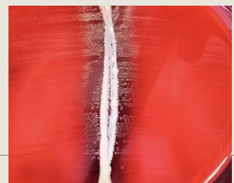
• Streak Haemophilus or suspected NVS across the surface of an agar plate (e.g., BAP).
• Streak Staphylococcus aureus ATCC 25923 perpendicular to the initial streak
146
What incubation conditions are required for the Satellite Test and how is it interpreted?
Incubate overnight in CO2
• Positive: faint growth or tiny colonies seen near the SA streak
• Negative: no growth around SA streak
• Positive: faint growth or tiny colonies seen near the SA streak
• Negative: no growth around SA streak
147
What disease is caused by Brucella species and what biosafety level is required?
Brucellosis (undulant fever); infectious pathogen that requires the use of biological safety level 3 precautions for all manipulations of specimens and cultures
148
What is the clinical significance of Brucella species?
A zoonotic disease where domestic animals serve as reservoirs and an important veterinary pathogen.
149
How is Brucella transmitted to humans?
By ingestion of unpasteurized contaminated milk or cheese, iInhalation (aerosolization-primary mechanism of transmission in laboratories), or direct contact via skin abrasions and mucous membranes (including the conjunctiva)
150
Which occupational and exposure risk factors are associated with Brucella infection?
Handling infected animals (dairy farmers, slaughterhouse workers, meat inspectors, veterinarians), ingestion of contaminated animal products (especially imported unpasteurized goat cheese in the U.S.), and handling Brucella cultures (laboratorians).
151
Why has the incidence of Brucella decreased in the United States?
Aggressive vaccination in cattle =↓ risk of infection, ↓ #s in US after 1975
152
What is the clinical presentation of Brucella infection?
Ranges from asymptomatic infection to serious debilitating disease, and may present as systemic or localized infection.
153
What symptoms are associated with brucellosis?
Intermittent fevers, chills, weakness malaise, body aches, sweating, headache, and weight loss (Lymphadenopathy and splenomegaly are common)
154
What complications can occur with brucellosis?
Arthritis, spondylitis, and endocarditis.
155
How common is death from Brucella infection, and what is it usually associated with?
Death is uncommon and is most often associated with endocarditis.
156
What clinical specimens are used for culture and isolation of Brucella?
Blood and bone marrow aspirates.
157
Which commercial blood culture systems can recover Brucella?
BacT/Alert, BACTEC, and ESP (Biphasic Castenada bottles-ok)
158
How long should cultures be incubated for suspected Brucella?
Incubation: growing-incubate at least 21 days (New automated Blood culture systems: 10 days)
159
What is the Gram stain morphology of Brucella species?
Gram Stain: Small gram negative coccobacilli or short rods arranged singly or in pairs and short chains. "Fine sand" appearance (Usually poorly stained (resists counterstain)-stain with safranin for ~3 minutes)
160
What is the colonial morphology of Brucella?
• After 48 hr of incubation colonies appear small, convex, smooth, translucent, nonhemolytic
• After 4-5 days colonies are 2-7mm, spheroidal, moist, slightly opalescent, slightly yellow or bluish-white; may become brownish with age
• After 4-5 days colonies are 2-7mm, spheroidal, moist, slightly opalescent, slightly yellow or bluish-white; may become brownish with age
161
What are other characteristics of Brucella species?
• Facultative, intracellular organism
• Aerobic to capnophilic, no growth under strict anaerobic conditions
• Oxidase (+), urea +, non-motile
• Aerobic to capnophilic, no growth under strict anaerobic conditions
• Oxidase (+), urea +, non-motile
162
What are the safety category for Brucella species in blood cultures?
Category B Select Agent of Bioterrorism
163
What is the gram stain for Brucella species?
Gram Stain: Small GNCB
• Usually positive in blood bottles at/after 3 days of incubation
• UCLA protocol: tape all cultures positive for GNCB on 3rd day
• Usually positive in blood bottles at/after 3 days of incubation
• UCLA protocol: tape all cultures positive for GNCB on 3rd day
164
What is the colony morphology for Brucella species and what do you do if unable to rule-out?
Colony: Poorly growing at 24 hours on BAP CHOC, NG on MAC (May require CO2 for initial growth)
Send to public health, if unable to rule-out:
Grows on BAP without need to satellite
• Oxidase +
• Catalase +
• Urea +
Send to public health, if unable to rule-out:
Grows on BAP without need to satellite
• Oxidase +
• Catalase +
• Urea +
165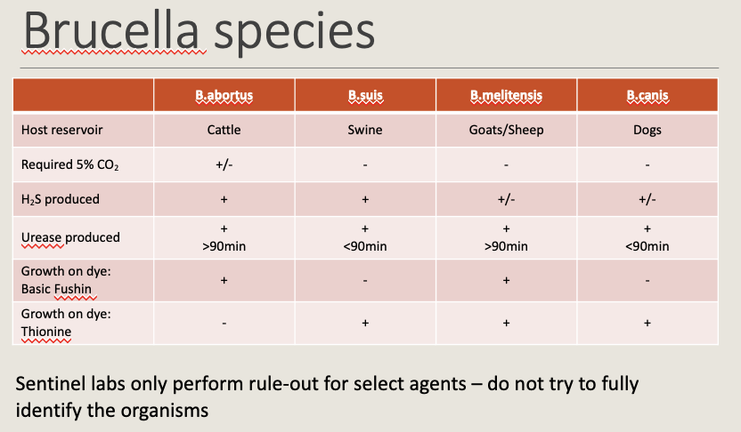
166
What are some culture negative bloodstream infection?
• Coxiella burnettii
• Chlamydophila species
• Rickettsia species
• Tropheryma whipplei (Whipple's disease)
• Chlamydophila species
• Rickettsia species
• Tropheryma whipplei (Whipple's disease)