Body Fluids
Study these flashcardsFree · spaced repetition · iPhone, Android, Mac & web
All cards
1
What are three key functions of CSF?
• Protect the brain & spinal cord from sudden pressure changes
• Maintain a stable chemical environment for the brain and spinal cord
• Remove the waste products of cerebral metabolism
• Maintain a stable chemical environment for the brain and spinal cord
• Remove the waste products of cerebral metabolism
2
What infectious condition is an indication for lumbar puncture?
Meningitis (bacterial, fungal or viral).
3
What types of hemorrhage are indications for lumbar puncture?
Subarachnoid or intracerebral hemorrhage.
4
What type of disease is an indication for lumbar puncture?
Neurologic disease.
5
What type of tumor is an indication for lumbar puncture?
Primary or metastatic tumor (including leukemia).
6
Besides disease, what other indication for lumbar puncture exists?
Some radiographic techniques.
7
What is the first gross examination factor of CSF and what indicates normal CSF?
Color is the first gross examination factor and colorless indicates a normal CSF.
8
What does bloody CSF suggest?
1. Subarachnoid hemorrhage
2. Intracranial hemorrhage
3. Infarct
4. Traumatic tap
2. Intracranial hemorrhage
3. Infarct
4. Traumatic tap
9
What is another color finding in CSF besides bloody?
Xanthochromia
10
What can cause xanthochromia?
1. Subarachnoid hemorrhage
2. Intracranial hemorrhage
3. Infarct
4. Traumatic tap
2. Intracranial hemorrhage
3. Infarct
4. Traumatic tap
11
What is the second gross examination factor of CSF and what is considered normal?
Clarity and Clear is normal.
12
What does cloudy (turbid) CSF indicate?
1. Pleocytosis (WBCs > 200/microL)
2. Microorganisms
3. RBCs > 400/microL
4. Increased protein
2. Microorganisms
3. RBCs > 400/microL
4. Increased protein
13
What is the third gross examination factor of CSF and what is considered normal CSF?
Clot formation, and no clot is normal.
14
What are causes of clot formation in CSF?
1. Total protein sharply elevated
2. Very bloody traumatic tap
2. Very bloody traumatic tap
15
What finding suggests a traumatic tap?
Traumatic tap shows progressive clearing from 1st tube to subsequent tubes.
16
What is seen after centrifugation of a bloody CSF in a traumatic tap vs a pathologic bleed?
After centrifugation of a bloody CSF, the supernatant is clear in a traumatic tap and a pathologic bleed is usually xanthochromic.
17
What about clot presence distinguishes a tramatic tap vs a pathologic bleed?
A clot may be seen in a bloody traumatic tap, whereas a pathologic bleed is rarely associated with a clot formation.
18
Should differentials be performed on all CSFs regardless of cell count and why are diffs from wet mount not recommended?
Yes, and not recommended because one can’t be certain of all cell types present; cell concentration provides a larger number of cells for diff.
19
What advantage does a stained smear offer?
A stained smear offers a permanent record for CSF cellular evaluation (especially beneficial in neoplastic conditions)
20
What hematopoietic cells may be found in CSF?
Lymphocytes, monocytes/macrophage, granulocytes, and rbcs.
21
How do these cells compare morphologically to those in blood or hematopoietic tissue?
They have same morphologic characteristics as in peripheral blood or hematopoietic tissue.
22
What are some artifactual changes seen on cytocentrifuge smears?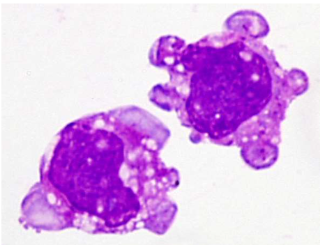
• Cytoplasmic or nuclear holes
• Cytoplasmic tailing
• Paranuclear zone in mononuclear cells
• Nuclear chromatin spreading in cells near the periphery of the smear
• Cytoplasmic tailing
• Paranuclear zone in mononuclear cells
• Nuclear chromatin spreading in cells near the periphery of the smear
23
In normal CSF Diff: What is the predominant cell type in adult CSF and its range?
Lymphocytes - Range 40-99%.
24
What is the monocyte range in adults?
Range 3-45%.
25
What is the neutrophil range in adults?
Range 0-6%.
26
In normal CSF Diff: What is the lymphocyte range in neonates?
Range 5-35%.
27
What is the predominant cell type in neonates and its range?
Monocytes - Range 50-90%.
28
What is the neutrophil range in neonates?
Range 0-8%.
29
In CSF, what is the normal WBC count for < 1 year?
0-30/microL
30
In CSF, what is the normal WBC count for 1-4 years?
0-20/microL
31
In CSF, what is the normal WBC count for 5 years to puberty?
1-10/microL
32
What is the normal WBC count for adults?
0-5/microL
33
In adults, what is the predominant CSF cell type?
Lymphocytes.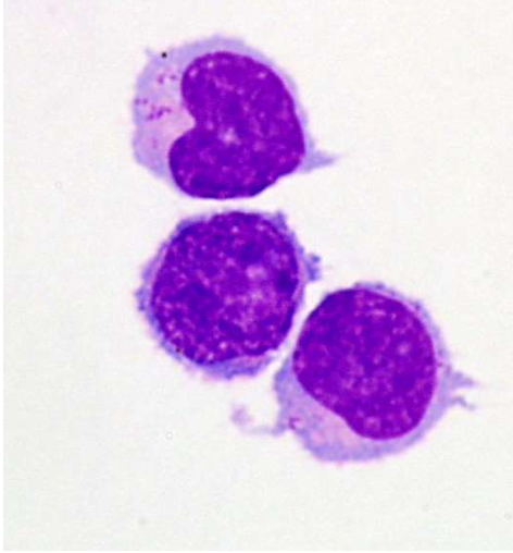
34
Which lymphocyte types are not normal in CSF?
Reactive, atypical, plasmacytoid lymphocytes and plasma cells are not normal.
35
With what are reactive lymphocytes usually associated?
Viral infections.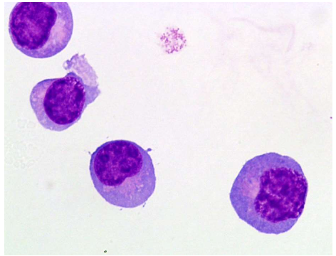
36
What is the predominant cell type in newborns?
Monocytes/macrophages.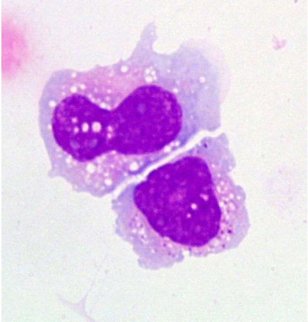
37
Is pure monocytic pleocytosis in CSF commonly seen?
Rarely seen (usually increased monocytes are present with neutrophils and lymphocytes-mixed cell reaction)
38
With what conditions is a mixed cell reaction most commonly associated?
Chronic bacterial meningitis, TB or fungal or viral meningitis and asceptic meningeal reaction.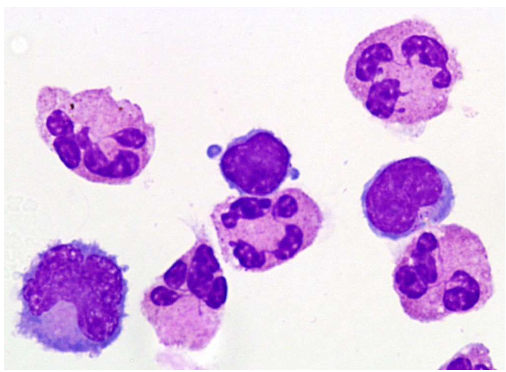
39
How does a macrophage that is not very actively phagocytic appear?
Morphologically similar to the monocyte.
40
What may active macrophages engulf?
A variety of material including other macrophages, rbcs, wbcs, microorganisms, pigment and lipids.
41
Is macrophage vacuolization commonly seen and what happens when vacuoles fuse?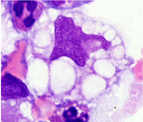
Yes, they are commonly seen. Fusion of vacuoles may push the nucleus to the periphery forming a signet ring shape. 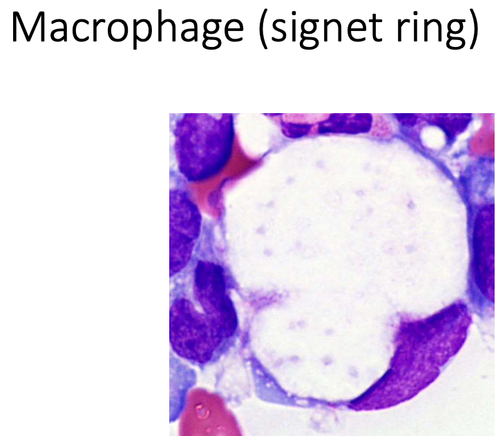
42
What must macrophage vacuolization be differentiated from?
Tumor cells.
43
What may suggest a pathologic bleed or previous traumatic tap?
Several erythrophagocytes may suggest a pathologic bleed (or previous traumatic tap).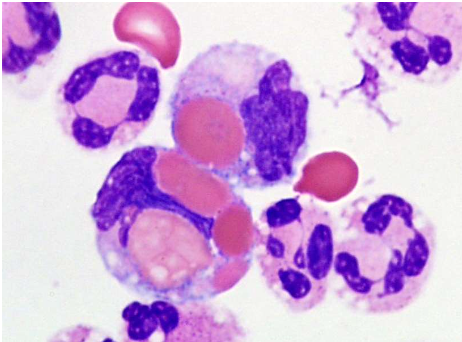
44
When may RBC ingestion occur after hemorrhage?
RBC ingestion may occur within a few hours after hemorrhage.
45
What are siderophages?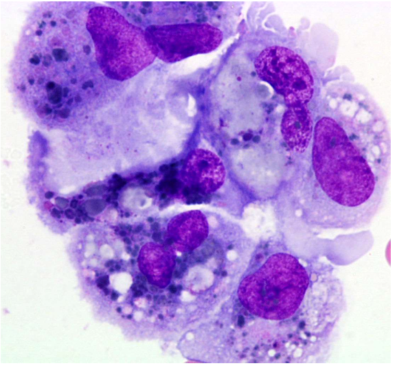
Hemosiderin laden macrophages (blue-black or dark brown granules).
46
What are siderophages a good indication of?
Hemorrhage.
47
How long may siderophages persist within the CSF?
2-8 weeks.
48
From what must siderophages be distinguished and how?
May need to be distinguished from melanoma cells (perform iron stain).
49
How often are hematoidin crystals seen and when may hematoidin crystals be found post hemorrhage?
Rarely seen and 10-14 days post hemorrhage.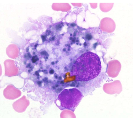
50
What is a leukophage?
Macrophage with ingested wbcs.
51
When do leukophages occur?
Occur in the clearing processes in connection with cell necrosis, surgical procedures, hemorrhage, and lymphocytic and granulocytic inflammation.
52
What percentage of neutrophils is considered normal?
<10 % considered normal.
53
When is finding neutrophils significant?
If CSF wbc count is increased, finding is significant.
54
With what conditions is neutrophilic pleocytosis associated?
Acute bacterial meningitis, fungal or TB or viral meningitis, previous lumbar puncture, cerebral infarct, CNS hemorrhage, and trauma.
55
When may pyknotic neutrophils be seen?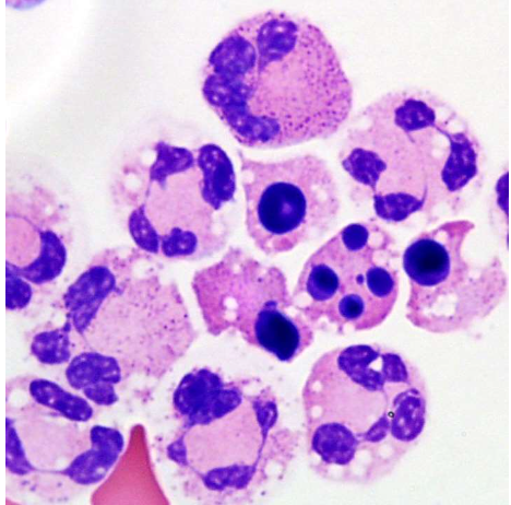
May be seen in bacterial meningitis.
56
What may pyknotic neutrophils be mistaken for?
May be mistaken for nrbcs.
57
How usual is finding eosinophils in normal CSF?
Very unusual finding in normal CSF.
58
When may eosinophilic pleocytosis be seen?
Parasitic infections; may be seen in response to tumors, subacute meningitis and traumatic puncture.
59
What is the diagnostic significance of basophilic pleocytosis?
No diagnostic significance.
60
Where has basophilic pleocytosis been seen?
Has been seen in cases of CML with CNS involvement.
61
What lining cells may pass into the CSF?
Cells lining the CSF space (ependymal or choroid plexus cells) may pass into the CSF.
62
Is it easy to differentiate choroid plexus and ependymal cells?
Difficult to differentiate between choroid plexus and ependymal cells.
63
When may lining cells be seen?
May be seen following trauma, surgery, infarcts or by sample obtained by ventricular or cisternal puncture.
64
What are the physical characteristics of lining cells? 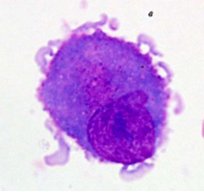
• Delicate chromatin
• Nuclei size of small lymphocyte
• Abundant cytoplasm & grey-blue
• Usually seen in clusters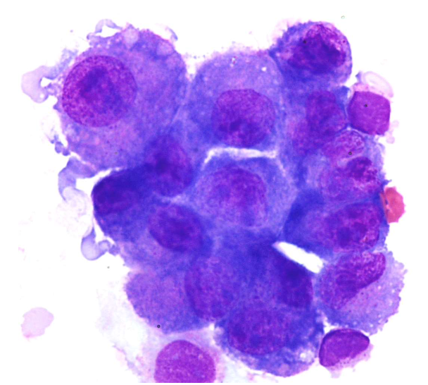
• Nuclei size of small lymphocyte
• Abundant cytoplasm & grey-blue
• Usually seen in clusters
65
What are the 3 general sources of tumor or malignant cells in CSF?
Leukemia/lymphoma (hematopoietic origin); Primary brain tumor; Metastatic carcinomas.
66
Which tumor is most common?
Leukemia/Lymphoma
67
Which Leukemia/Lymphoma is most common?
ALL is the most common.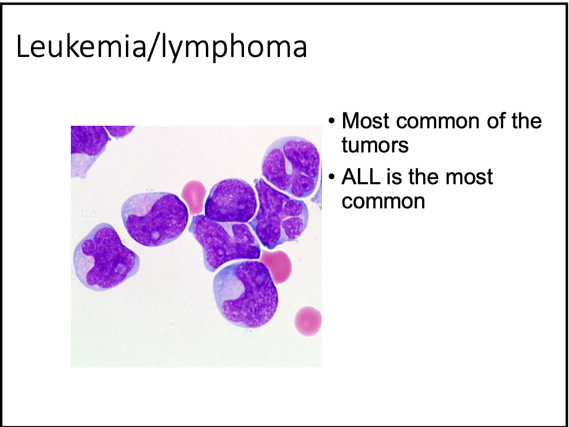
68
Name examples of primary brain tumors.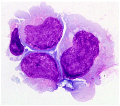
Medulloblastoma; Retinoblastoma; Neuroblastoma; Meningiomas.
69
Name common metastatic tumors for CSF.
Lung; Breast; Melanoma.
70
Name other cells rarely seen in CSF.
LE cell; Cartilage cells & bone marrow elements (accidental puncture of the vertebral body).
71
What are the 3 basic types of serous fluids?
Pleural (thoracentesis or chest fluid); Pericardial (pericardiocentesis fluid); Peritoneal (ascites, abdominal paracentesis fluid).
72
How much fluid is normally present and how is it derived?
Normally there is only a small amount of fluid (derived by plasma ultrafiltration, separating the membranes which form their respective “cavities”).
73
When do cavities become potential cavities?
These cavities are only potential cavities which become so in the presence of disease causing accumulation of fluid.
74
What is accumulation of fluid called?
Effusion.
75
What are the indications of Paracentesis?
Diagnostic evaluation; Relief of symptoms (e.g. shortness of breath); Infusion of drugs; Drainage of hemothorax or empyema.
76
How should serous fluids be collected and why?
Should be collected in sterile EDTA tubes because serous fluids frequently clot.
77
Why should serous fluids be examine promptly?
Specimens should be examined promptly becasue rapid cell deterioration may occur
78
What causes transudates and with what are they associated?
Caused by disease(s) outside of body cavity; Associated with CHF, cirrhosis, and nephrotic syndrome.
79
What causes exudates and with what are they associated?
Caused by disease(s) involving the organ or surface of the body cavity; Associated with infection TB, inflammation, malignancies, pancreatitis, trauma and infarction.
80
What is a normal serous fluid appearance?
Normal appearance is clear & pale yellow.
81
What does bloody sorous fluid indicate?
hemorrhagic fluid or traumatic tap.
82
What does milky white serous fluid indicate?
may be chylous (caused by malignancy, trauma or lymphatic obstruction) or pseduo chylous effusion (RA, TB or any other chronic effusion of any cause).
83
What does green serous fluid result from?
results from bile contamination (perforated gall bladder, perforated duodenal ulcer or pancreatitis).
84
Are total wbc and rbc counts useful to differentiate transudates from exudates?
The total wbc and rbc counts are of limitied value in differentiating transudates from exudates.
85
What may be more helpful in differentiating transudates from exudates?
The WBC differential may be more helpful.
86
List lymphocyte origin cells in serous fluid.
Small lymphocytes; Large lymphocytes; Reactive lymphocytes; Plasmacytoid lymphocytes; Plasma cells.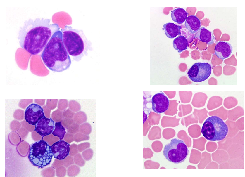
87
Where are mature lymphs seen?
Mature lymphs are seen in varying numbers in almost all effusions.
88
What does lymphocytosis >50% suggest?
Lymphocytosis >50% suggest TB, carcinoma, or lymphoproliferative disorder (leukemia or lymphoma).
89
When may less pronounced lymphocytosis be seen?
Less pronounced lymphocytosis may be seen in CHF and cirrhosis.
90
With what are reactive, atypical, plasmacytoid lymphs associated?
Associated with Ag-Ab type disorders.
91
In what conditions may plasma cells be seen?
Plasma cells may be seen in RA, TB, malignancy, and other conditions associated with lymphocytosis.
92
What is markedly increased in purulent effusions?
Marked increase in segs (>50%) is seen in purulent effusions.
93
What may be suspected in pleural fluids if segs are markedly increased (>50)?
bacterial pneumonia & pulmonary infarct may be suspected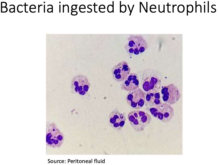
94
What may be suspected in peritoneal fluids if segs are markedly increased (>50) ?
chemical peritonitis and pancreatitis may be suspected
95
How common are immature neutrophils in CSF?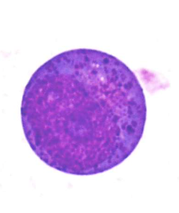
Less commonly seen.
96
When may they be seen?
May be seen in cases where increase in those same cells in the peripheral blood.
97
How common is peritoneal eosinophilia vs pleural fluid eosinophilia?
Peritoneal eosinophilia is less common than pleural fluid eosinophilia
98
When is peritoneal eosinophilia seen?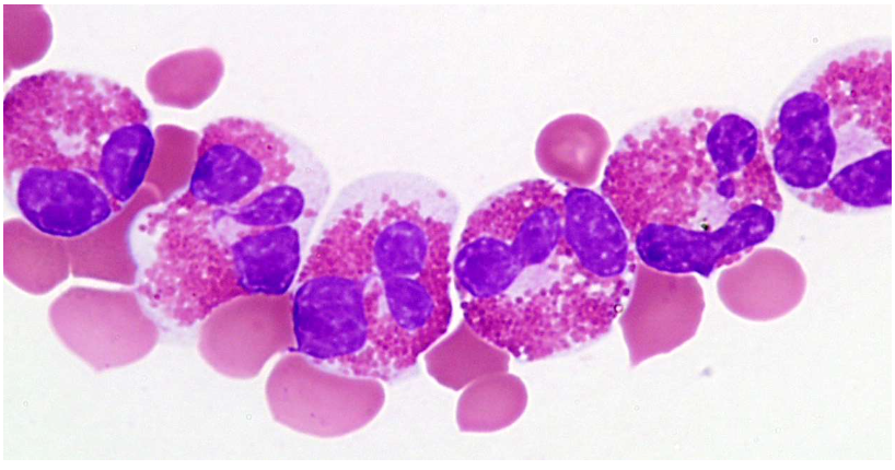
Seen in malignant ascites, ruptured hydatid cyst, CHF, chronic peritoneal dialysis and vasculitis
99
Describe pleural eosinophilia.
Pleural eosinophilia is nonspecific; May or may not accompany blood eosinophilia
100
What are mast cells and their significance?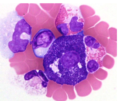
Mast cells (tissue basophils) are non-specific and found in a variety of conditions
101
Describe the morphology of Monocytes/Macrophages
Morphology quite variable (typical blood monocyte to macrophage)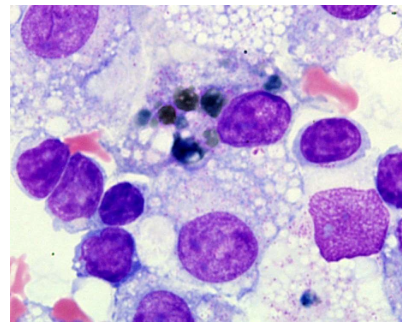
102
List macrophage phagocytic findings.
Erythrophagocytosis; Siderophage; Neutrophage (macrophage with engulfed neutrophil may be found following any cause of neutrophilia)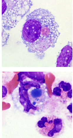
103
Where do mesothelial cells line and how are they shed?
Normally line the pleural, pericardial & peritoneal surfaces; May be shed individually or in clusters
104
When found in pairs, what feature may mesothelial cells show?
When found in pairs or clusters frequently have coupled cell borders with “windows” between many of the cells
105
What is the size of mesothelial cells?
Size 12-20 um in diameter
106
What is the N:C ratio of mesothelial cells?
Low N:C ratio (less than one half)
107
Describe the nucleus and the cytoplasm of mesothelial cells.
Nucleus often eccentric & round to ovoid shape (may have nucleoli). Cytoplasm is light to dark blue 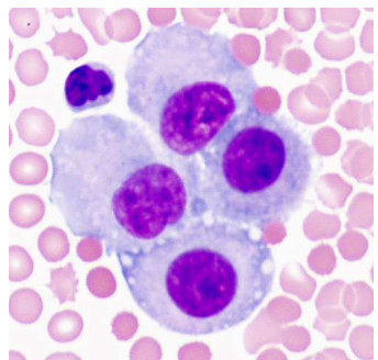
108
What perinuclear feature may be present?
May have perinuclear zone of pallor (fried egg appearance)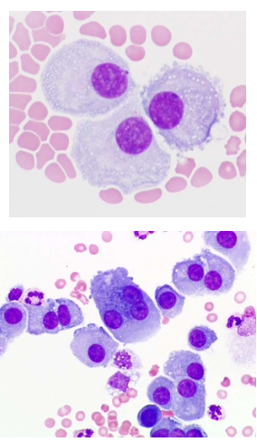
109
What other features may mesothelial cells have?
May have cytoplasmic vacuoles & capable of phagocytosis (resemble macrophage)
110
What changes may be seen in mesothelial cells and what can cause potential confusion?
May show reactive changes; May be mistaken for malignant cells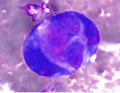
111
Which malignancies are most commonly seen at UCLA and who often has effusions?
Most commonly seen at UCLA are disseminated lung & breast cancer; Effusions are common in patients with lymphoma, mesothelioma, various sarcomas, ovarian and gastrointestinal neoplasms
112
What are some characteristics of malignant cells?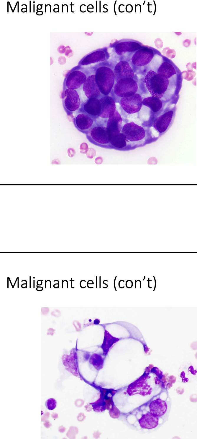
Cells may be larger than normal cells; May exhibit clumping; May show molding or a mosiac pattern; May possess macronucleoli; May be vacuolated; and cells may look “foreign”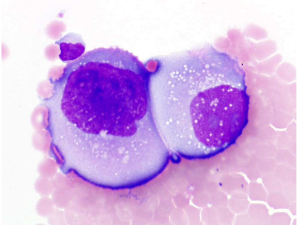
113
Where may LE cells be found and what characterizes them?
Spontaneous lupus erythematosus cells may be found; Characteristically an intact neutrophil contains a large, homogenous, pink inclusion (degenerated nucleus)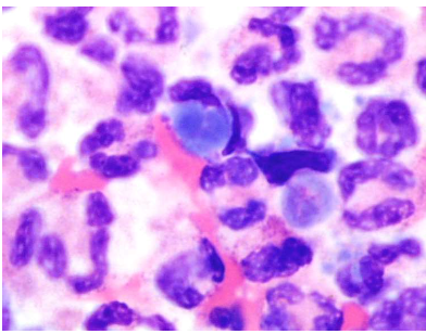
114
What is the function and lining of synovial fluid?
To lubricate the joint space and transport nutrients to the articular cartilage; Lined by synovial cells (capable of protein synthesis and phagocytosis)
115
What are some indications for synovial fluid aspiration?
Effusion of unknown etiology; Possible infection; Release pain or allow increased mobility; Instillation of drugs
116
What specimen collection tubes are recommended for synovial fluid?
EDTA recommended for microscopic examination; Plain, sterile tube recommended for crystal analysis
117
Describe the appearance, viscoid nature and WBC count of synovial fluid
Pale yellow and clear; Usually viscoid and WBC count 0-200/microL
118
What are the normal synovial fluid differentials?
Neutrophils 0-25%; Lymphocytes 0-78%; Monocytes 0-71%; Macrophages 0-26%; Synovial cells 0-12%
119
What percent of neutrophils suggests septic arthritis and what should be done?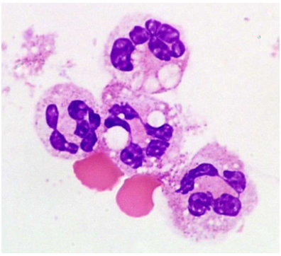
More than 80% suggestive of septic arthritis (regardless of total wbc count); Gram stain should be performed when neutrophil percent is markedly increased
120
What do synovial cells resemble and what is their specificity?
Resemble mesothelial cells; Presence is expected and has no diagnostic specificity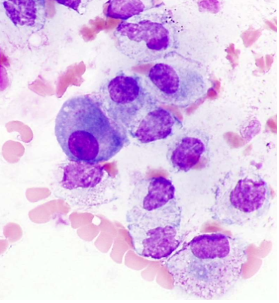
121
Where may LE cells be seen and in what context?
May be seen from patients with RA as well as in lupus erythematosus; May found in absence of peripheral blood LE formation
122
How should crystal examination be performed on synovial fluid?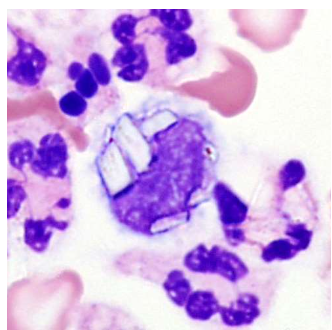
Should be performed routinely using polarized light; Should be performed on plain, sterile tube
123
How common is involvement of synovial fluid by malignant cells?
Involvement of synovial fluid is rare
124
How are counts performed on synovial fluid and what does CAP recommend?
Manual using a hemacytometer; CAP recommends counting both sides of the hemacytometer
125
What about electronic counters (synovial fluid)?
Electronic cell counters; newer models are FDA approved for fluids
126
What method is used for cell concentration and what are its attributes?
Many labs use the cytocentrifuge method; Rapid, easy to use and may process up to 12 specimens simultaneously; Recovery is satisfactory; Some artifactual morphologic changes
127
Case Study #1: History; CSF from a 48 year old woman complaints of stiff neck and double vision 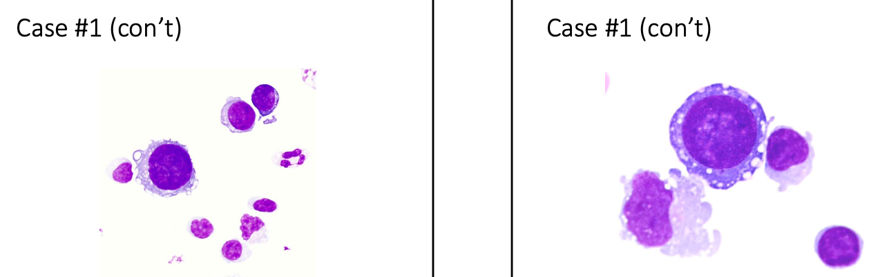
Diagnosis: Metastatic breast carcinoma
128
Case Study #2: History; Hip fluid from a 76 year old woman. Color of the fluid was green-blue.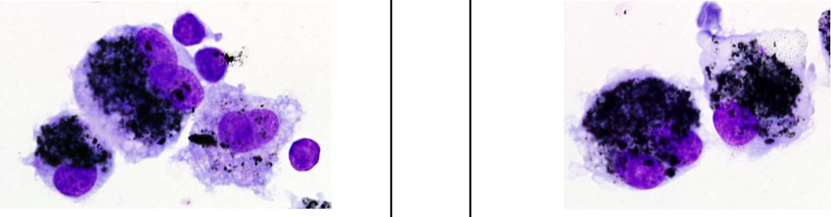
Diagnosis:
• A cytoprep smear of the fluid was stained for non-specific esterase-the cells stained postive
• The macrophages ingested pieces of the woman’s hip replacement.
• The color of the fluid was consistent with cobalt chrome.
• A cytoprep smear of the fluid was stained for non-specific esterase-the cells stained postive
• The macrophages ingested pieces of the woman’s hip replacement.
• The color of the fluid was consistent with cobalt chrome.
129
Case Study #3: History; An elderly woman admitted with dehydration and a pleural effusion.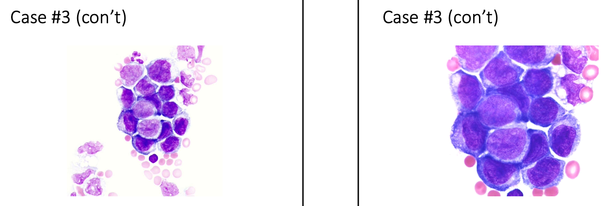
Diagnosis: Gastric carcinoma
130
Case Study #4: History: Pleural fluid from a 68 year old male with shortness of breath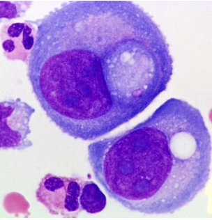
Diagnosis: Adenocarcinoma-Lung