Auto Chemistry Rotation
Study these flashcardsFree · spaced repetition · iPhone, Android, Mac & web
All cards
1
What specimen types are run at UCLA Chemistry?
Plasma, Serum, Urine, CSF, and Other Body Fluids.
2
What are examples of "Other Body Fluids" run at UCLA Chemistry?
Pancreatic cyst, synovial, pleural, chest, ascites, peritoneal, abdominal, feces, peri-nephric, peri-cardial, and PTH aspirates.
3
What is done to all specimens before running them on the analyzer?
All specimens are spun down before running on the analyzer.
4
What do the different Cobas Racks (Non-TLA) colors represent?
Green and grey = Serum, plasma, pancreatic cyst; Yellow and grey = Urine, feces lytes; Red = Stat (usually serum/plasma only); Pink and grey = Body Fluids; Brown and grey = CSF; Blue and grey = False bottom tubes; Solid grey = Samples aliquotted from the MPA.
5
What methods are used on the C8000?
Ion Selective Electrode, Photometric, and ECL (Electrochemiluminescent)
6
What type of measurement does the ISE unit perform?
The ISE unit performs indirect measurement of electromotive force (EMF) in millivolts between ion-selective electrodes and the reference electrode.
7
What does "indirect measurement" mean in the context of the ISE unit?
It means that all samples are diluted at a 1:31 ratio.
8
How are EMF values converted to mmol/L values in the ISE unit?
They are converted by a calculation algorithm that uses the EMF data together with data from a 2-point calibration with two primary calibrators.
9
How is drift between consecutive ISE measurements offset?
An ISE measurement before and after each routine sample measurement is used to offset the drift
10
Which electrolytes are measured by the ISE unit?
Sodium, potassium, and chloride.
11
How is the concentration of sodium, potassium, and chloride determined in the ISE unit?
It is calculated from the EMF of the specific electrode.
12
What equation is used to calculate electrolyte concentrations in the ISE unit?
The equation derived from the Nernst Equation.
13
What types of tests use photometric principles on the cobas c modules?
All tests that use absorbance measurements for result calculation, including clinical chemistry and immunological tests.
14
What are the two fundamental types of photometric test principles used by the instrument?
Endpoint and rate assays
15
How do endpoint assays work in photometric testing?
In endpoint assays, measurements are taken by the photometer at specific measuring points. If measurements are taken after the reactions are completed, the intensity of the colored (or turbidimetric) product is an indicator of the sample component's concentration. These are called endpoint assays.
16
How do rate assays work in photometric testing?
With rate assays, measurements are taken as the reaction proceeds. In this case, the rate of the reaction is proportional to the sample component's concentration or activity being analyzed.
17
What happens during the Electrochemiluminescent (ECL) process?
ECL is a process in which highly reactive species are generated from stable precursors at the surface of an electrode. These highly reactive species react with one another, producing light. The ECL processes are known to occur with numerous molecules including compounds of ruthenium, osmium, rhenium, or other elements.
18
What is the basis of ECL immunology test development?
The development of ECL immunology tests is based on the use of a ruthenium(II)-tris(bipyridyl) complex and tripropylamine (TPA).
19
When is the final chemiluminescent product formed in ECL testing, and how is light emitted?
The final chemiluminescent product is formed during the detection step. The chemiluminescent reactions that lead to the emission of light from the ruthenium complex are initiated electrically rather than chemically.
20
How is the ECL chemiluminescent reaction initiated and what is its advantage?
It is achieved by applying a voltage to the immunological complexes (including the ruthenium complex) that are attached to streptavidin-coated microbeads. The advantage of electrically initiating the chemiluminescent reaction is that the entire reaction can be precisely controlled.
21
What is the ISE module composed of, what does it measure, and what type of measurement does it use?
The ISE module is composed of two ISE (ion selective electrode) units, measures Na (sodium), K (potassium), and Cl (chloride), and uses indirect measurement.
22
What are the main functions and features of the c702 chemistry module at UCLA?
The c702 is the main chemistry module at UCLA used for clinical chemistry and homogeneous immunoassays, with a throughput of up to 2,000 tests per hour.
23
What additional features ensure accuracy and efficiency in the c702 module?
It ensures specimen integrity via serum indices, clot and liquid level detection, uses contact-free ultrasonic mixing, and has 2 sample probes and 4 reagent probes.
24
What are the main functions and testing focuses of the c502 module at UCLA?
The c502 performs clinical chemistry and homogeneous immunoassays. At UCLA, it focuses on chemistries related to TDM (therapeutic drug monitoring) and DAU (drugs of abuse).
25
What are the performance and quality features of the c502 module?
The c502 has a throughput of up to 600 tests per hour, ensures specimen integrity via serum indices, clot and liquid level detection, and uses contact-free ultrasonic mixing.
26
What type of tests and technology does the e801 module use?
The e801 performs heterogeneous (separation step) immunoassays using ElectroChemiLuminescence (ECL) technology.
27
What are the performance and quality features of the e801 module?
The e801 has a throughput of up to 300 tests per hour, uses carryover-free disposable tips, and includes clot and air bubble detection.
28
What is the purpose of calibration?
The purpose of calibration is to maintain assurance of accuracy and repeatability of measurements.
29
How is a machine calibrated?
In order to calibrate a machine, known standards are measured and plotted against a measured (response) value, such as absorbance.
30
What does calibration convert during measurement?
When we measure something, we convert an absorbance value to something meaningful, such as analyte concentration or enzyme activity.
31
What law is associated with calibration and absorbance?
Beer-Lambert Law.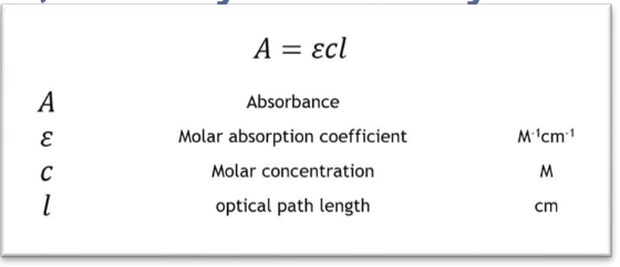
32
When are calibrations performed?
Calibrations are performed when loading new lots of reagent, after major maintenance, and when troubleshooting QC.
33
What is a lot calibration (lot cal)?
A lot cal is a calibration curve for a specific lot of reagent.
34
What is a reagent calibration (reagent cal)?
A reagent cal is a calibration curve for a specific reagent pack.
35
What calibrator is used for blank calibration?
The calibrator of the lowest analyte concentration is used for calibration, such as Calibrator 1 (for example, H₂O).
36
How is the calibration curve adapted in blank calibration?
The calibration curve is adapted without changing the K-factor.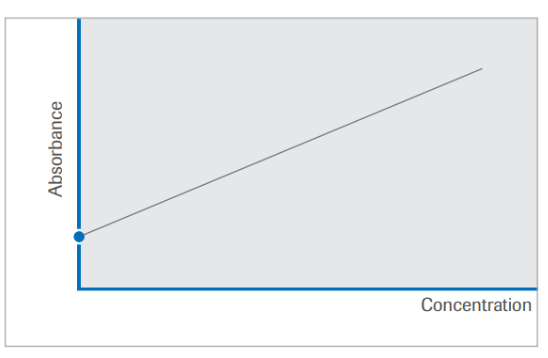
37
What calibrators are used for 2-point calibration?
Two calibrators are used: Calibrator 1, for example H₂O, and a second calibrator, for example C.f.a.s.
38
How is the calibration curve adapted in 2-point calibration?
The calibration curve is adapted by recalculating the K-factor.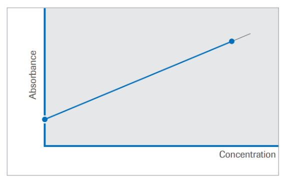
39
What calibrators are used for full calibration?
All calibrators are used for this nonlinear calibration, meaning all calibrators defined for this test.
40
How is the calibration curve adapted in full calibration?
The calibration curve is adapted by recalculating all K-factors.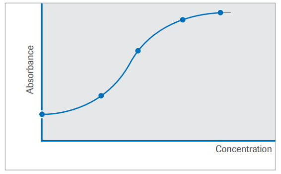
41
How do we ensure our test results are correct and reliable?
Quality Control (QC) validates whether our instrument is working properly and gives us confidence that our test results are correct.
42
When is QC generally performed?
QC is run every 24 hours, when a new reagent is loaded, after preventative maintenance, and after calibration.
43
How often is QC run for less stable tests?
Less stable tests have their QC run more frequently.
44
What is QC material designed to do?
QC material is meant to mimic a patient sample, so its matrix is ideally made from serum, urine, or spinal fluid.
45
What can each QC material test?
Each QC material can test multiple analytes.
46
What types of controls should QC material include?
Ideally, QC material will have a normal control and an abnormal control or span an analyte’s decision points.
47
What should be done before running QC material?
It should be well mixed and at the appropriate temperature before running.
48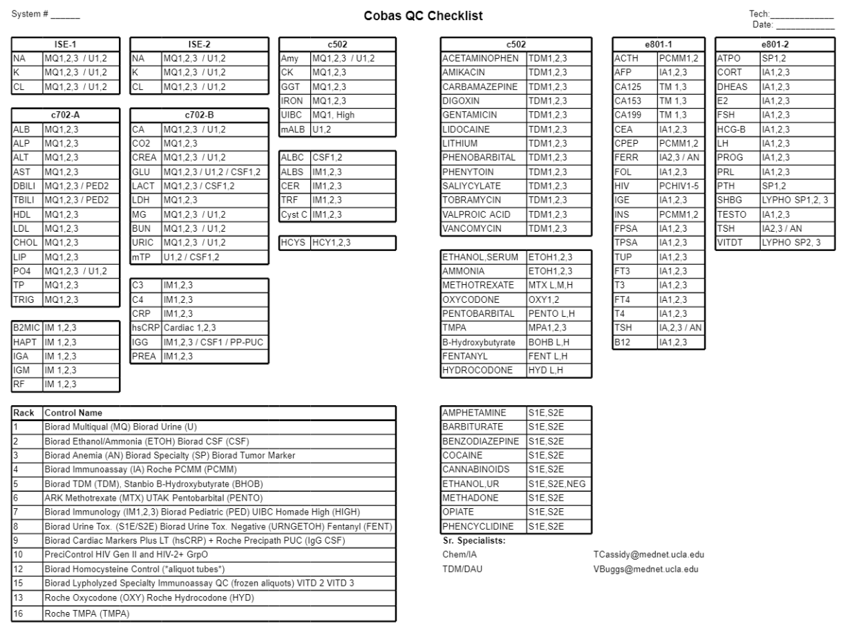
49
How is a QC range ideally established?
QC material is run around 20 times, once a day and by different people, to account for natural variation that occurs.
50
How is the QC range calculated after collecting data points?
The QC range is established by calculating the mean and standard deviation.
51
What does the mean represent in QC statistics?
The mean is an estimate of the analyte’s true value.
52
What does the standard deviation (SD) represent in QC statistics?
SD is a measure of precision.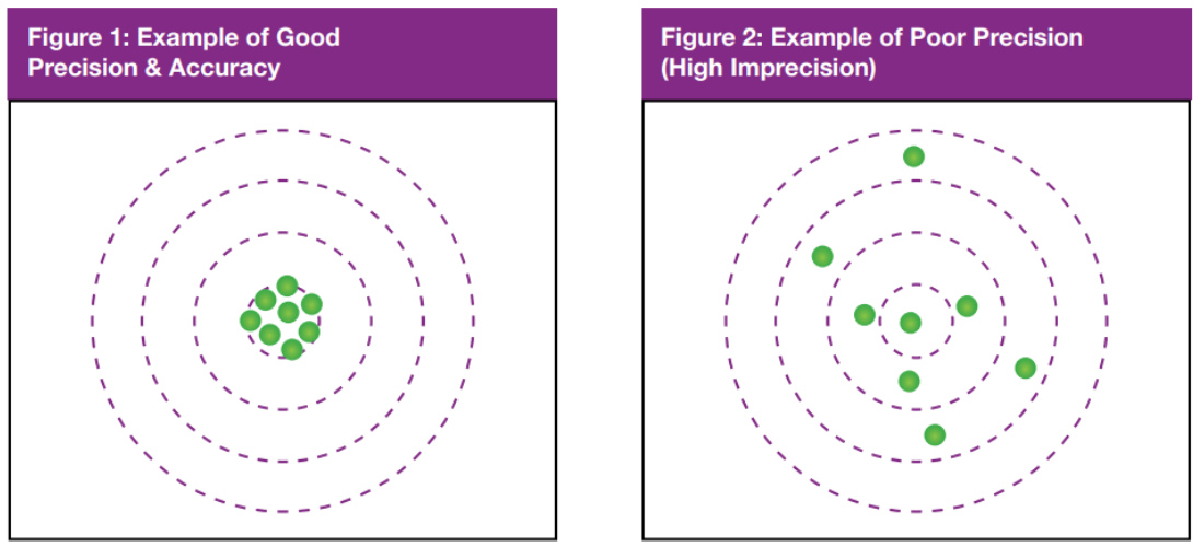
53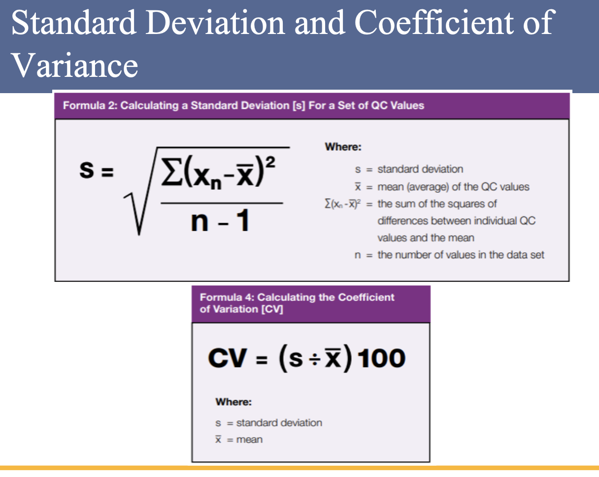
54
How is the QC range calculated?
Once the mean and SD are known, the QC range is calculated as mean (±) 2SD.
QC range calculation Example:
If the mean is 50 and the SD is 5, then the QC range is 40 to 60.
QC range calculation Example:
If the mean is 50 and the SD is 5, then the QC range is 40 to 60.
55
What percentage of QC runs should fall within 2SD?
95% of QC runs should be within 2SD.
56
How many basic rules are in the Westgard scheme, and what are they?
There are six basic rules: 1-3s, 2-2s, R-4s, 1-2s, 4-1s, and 10-x. These can be used individually or in combination (multi-rule) to evaluate the quality of analytical runs.
57
What are two main reasons for applying Westgard Rules?
They reduce false rejections made when applying just the 1-2s rule for run rejection and increase error detection more than provided when applying just the 1-3s rule.
58
Which Westgard Rules detect random and systematic errors?
1-3s and R-4s detect random error, while 2-2s, 4-1s, and 10-x detect systematic error.
59
What does the 1-3s rule identify in Westgard Rules?
The 1-3s rule identifies unacceptable random error or possibly the beginning of a large systematic error. Any QC result outside ±3SD violates this rule. Since only 0.3% or 3 out of 1000 points will fall outside the ±3SD limit, any value outside of ±3SD is usually considered to be associated with a significant error condition.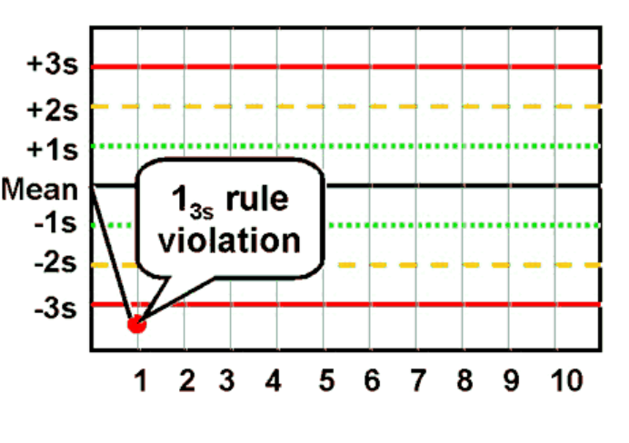
60
Why might a value outside ±3SD not always be concerning?
While a value outside ±3SD may be statistically significant, it may not be biologically or medically relevant because modern laboratory instruments are often more precise than what is needed medically.
61
What is the 1-2s rule in Westgard Rules and how is it applied?
The 1-2s rule is usually a warning rule violated when a single control observation is outside the ±2SD limit. It was originally designed as a warning rule for manual application of the Westgard rules. If one control measurement within a run exceeds the mean ±2SD, evaluate other controls in the run and in previous runs before accepting the run and reporting results.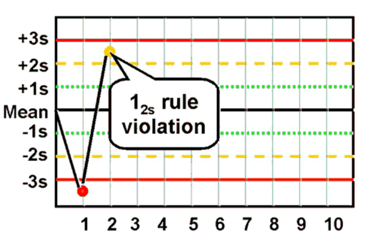
62
Why should laboratories not reject analytical runs based only on the 1-2s rule?
Some laboratories incorrectly consider any QC value outside ±2SD to be out of control and reject valid runs. An analytical run should not be rejected if a single QC value is outside ±2SD but within ±3SD, since about 4.5% of valid QC values naturally fall between ±2SD and ±3SD. Universally rejecting values outside ±2SD leads to rejecting good runs too frequently, and with computer-based Westgard applications, the 1-2s rule is usually unnecessary.
63
What happens when the 1-2s rule is used alone in quality control testing?
Using the 1-2s rule alone causes frequent rejection of valid runs because it fails to allow for valid points between 2SD and 3SD.
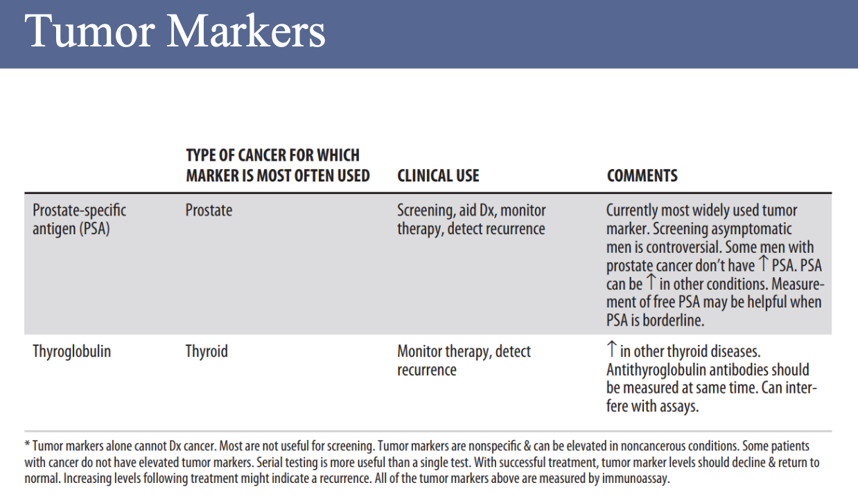
64
What are the consequences of using the 1-2s rule alone according to Dr. Westgard?
It may falsely reject 5% of all analytical runs when using one level of control, 10% when using two levels, and 14% when using three levels. These false rejections lead to unnecessary repeats of patient samples, wasted labor and materials, and delayed patient results.
65
What does the 2-2s rule detect and when is it violated?
The 2-2s rule detects systematic error only. It is violated when two consecutive QC results are greater than 2SD and on the same side of the mean.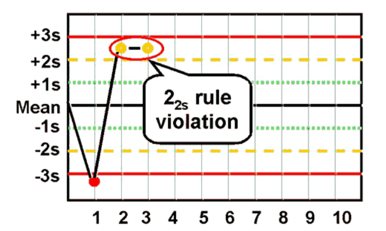
66
How is the 2-2s rule applied within and across runs?
Within a run, the rule is violated when two control values in the same run are >2SD on the same side of the mean, indicating systematic error affecting the entire analytical curve. Across runs, the rule is violated when a single level of control is >2SD from the mean in one run and again >2SD on the same side of the mean in the next run, indicating systematic error affecting only a single portion of the analytical curve.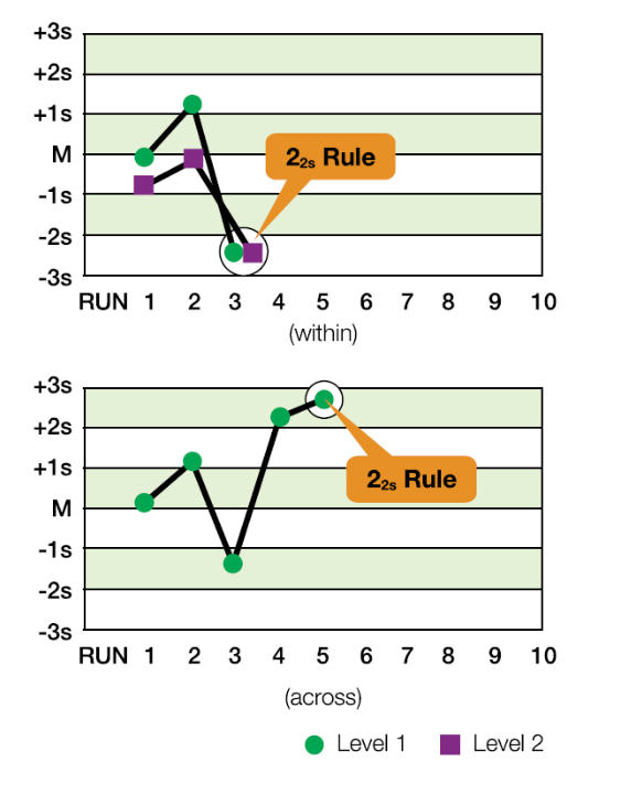
67
What type of error does the R-4s rule identify, and how is it applied?
The R-4s rule identifies random error and is applied only within the current run. It is violated when there is at least a 4SD difference between control values within a single run.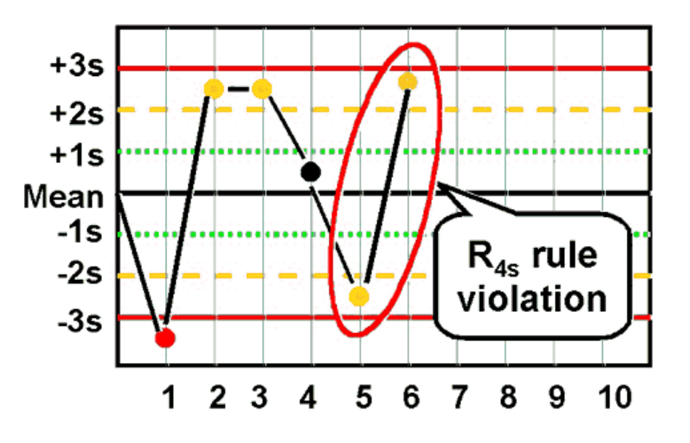
68
Give an example of a violation of the R-4s rule.
If Level 1 is +2.8SD above the mean and Level 2 is -1.3SD below the mean, the total difference is greater than 4SD (+2.8SD - (-1.3SD) = 4.1SD), indicating a violation of the R-4s rule.
69
When are the 3-1s and 4-1s rules violated in Westgard Rules?
These rules are violated when three or four consecutive results are greater than 1SD and on the same side of the mean.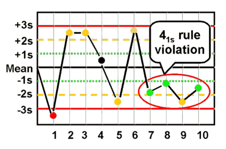
70
How are the 3-1s and 4-1s rules applied and what do violations indicate?
They are applied within a control level or across control levels. Violations within a control level indicate systematic bias in a single area of the method curve, while violations across control levels indicate systematic error over a broader range of concentrations.
71
When are the N-x rules violated in Westgard Rules?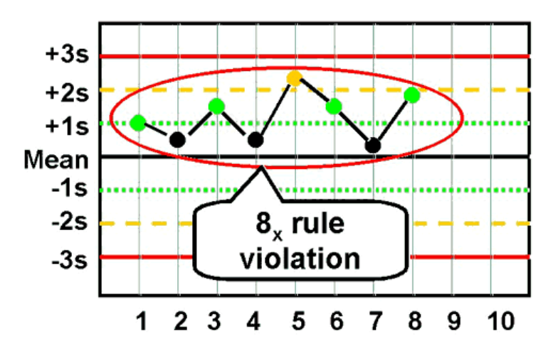
N-x rules are violated when there are 7, 8, 9, 10, or 12 control results on the same side of the mean.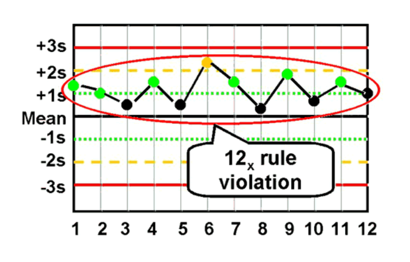
72
How are N-x rules applied and what do violations indicate?
They are applied within a control level or across control levels. Violations within a control level indicate systematic bias in a single area of the method curve, while violations across control levels indicate systematic error over a broader range of concentrations.
73
Which N-x rule is more sensitive to analytical bias and why?
The 7-x control rule is far more sensitive to analytical bias than the 12-x rule because the chance of finding seven consecutive control observations on one side of the mean is much higher than finding twelve.
74
What is the purpose of a Levey-Jennings Chart?
A Levey-Jennings Chart gives a visual representation of QC data and helps visualize systematic errors.
75
What does a Levey-Jennings Chart display?
It graphs QC value runs, showing run-to-run or day-to-day performance.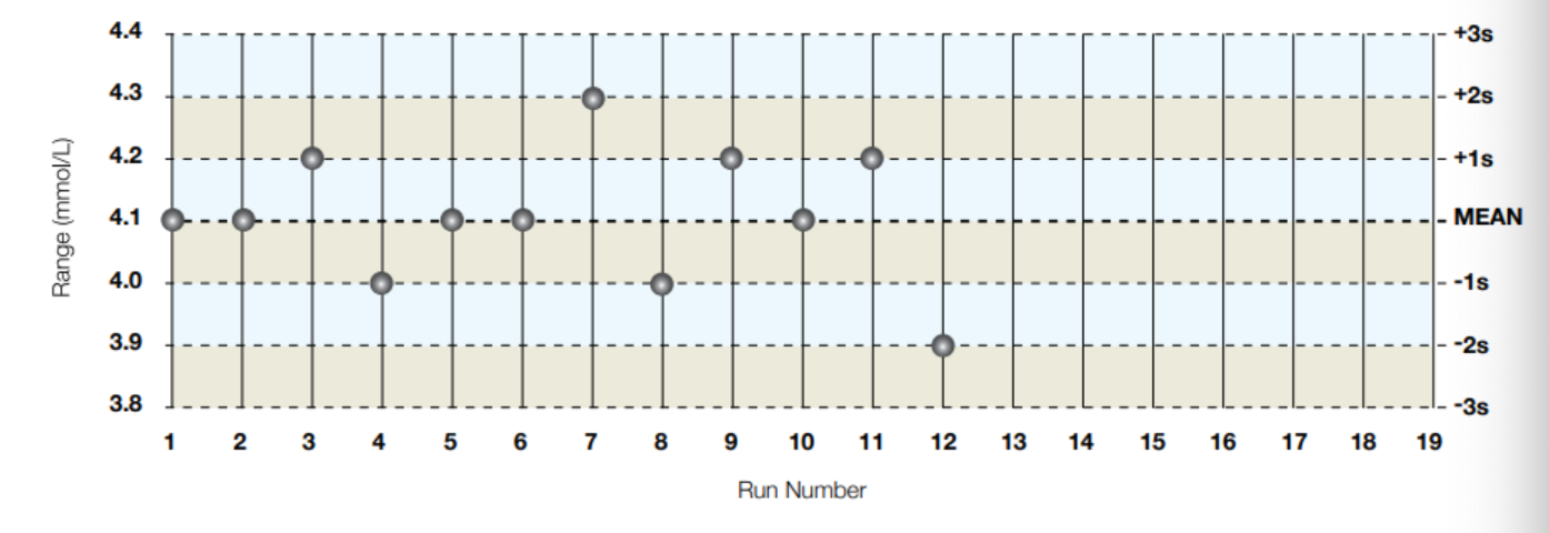
76
What are systematic errors and what causes them?
Systematic errors can be gradual (trend) or abrupt (shift) and are built in due to an issue such as bad calibration, failing hardware, or old QC.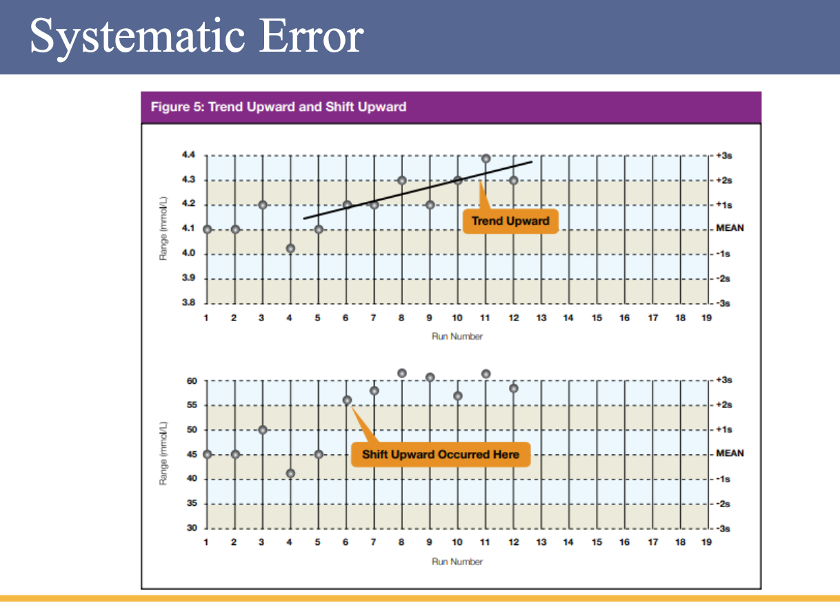
77
What are random errors and what causes them?
Random errors are due to natural variability.
78
What is the analytical measuring range?
The analytical measuring range is the range of an analyte that a method can measure without dilution or concentration.
79
What is the clinical measuring range?
The clinical measuring range is the range of an analyte expanded with dilution.
80
What do LoB, LoD, and LoQ represent?
Limit of Blank (LoB), Limit of Detection (LoD), and Limit of Quantitation (LoQ) describe the smallest concentration of a measurand that can be reliably measured by an analytical procedure.
81
What is the Limit of Blank (LoB)?
LoB is the highest apparent analyte concentration expected to be found when replicates of a blank sample containing no analyte are tested.
82
What is the Limit of Detection (LoD)?
LoD is the lowest analyte concentration likely to be reliably distinguished from the LoB and at which detection is feasible.
83
What is the Limit of Quantitation (LoQ)?
LoQ is the lowest concentration at which the analyte can not only be reliably detected but at which some predefined goals for bias and imprecision are met.
84
What is the purpose of dilution?
Dilution is the process of decreasing the amount of analyte until it is measurable; the purpose is to decrease the analyte concentration so it is within the analytical measuring range (AMR).
85
How are dilutions performed?
Some dilutions are done by the machine, while others are performed manually.
86
What is a diluent?
A diluent is a substance used to dilute an analyte.
87
What is the dilution factor?
The dilution factor is the number by which you multiply the measured diluted value to obtain the original value.
88
What is a serial dilution?
A serial dilution is a series of dilutions that can be done to save sample or to dilute something with a high concentration.
89
What is the purpose of the dilution factor calculation?
It determines the relationship between the total volume, sample volume, and diluent volume in a dilution process.
90
What is the equation for chemical dilutions that should not be confused with dilution factor calculations?
C1V1 = C2V2.
91
What are the symbols used in dilution factor calculations?
Dilution factor = F, Total volume = T, Diluent volume = D, Sample volume = S.
92
What formulas are used to calculate sample and diluent volumes?
(T / F) = S and T – S = D.
93
What is the purpose of performing a serial dilution?
A serial dilution is performed when the concentration of an analyte is still too high (above the AMR), allowing further dilution using an already diluted sample.
94
What does the phrase “Garbage in, garbage out” mean in relation to specimen quality?
It means that poor-quality specimens will produce poor or unreliable test results.
95
What factors can affect test results and specimen quality?
Age (stability), exposure to air (capped / uncapped), delay in centrifugation, recentrifugation, indices (HIL), pH, volume, clot, draw times, order of draw, light, IV contamination, temperature, and tourniquet time.
96
Why are indices important in testing?
Indices greatly affect test results and must be checked before releasing results.
97
How can indices impact test outcomes?
Depending on the test, indices can cause results to be either higher or lower.
98
What types of interference can indices cause?
Indices can cause chemical or spectral interference depending on the assay.
99
What is hemolysis?
Hemolysis is the destruction of red blood cells, which releases intracellular components such as hemoglobin.
100
What are the in vivo causes of hemolysis?
Biochemical, immunological, physical, or chemical causes.
101
What is an in vitro cause of hemolysis?
Incorrect sample processing.
102
Which tests are affected by hemolysis?
LDH, AST, K, and Folate.
103
What conditions or factors can cause elevated icterus?
It can be elevated in liver disease, biliary cirrhosis, alcoholism, or as a response to many drugs.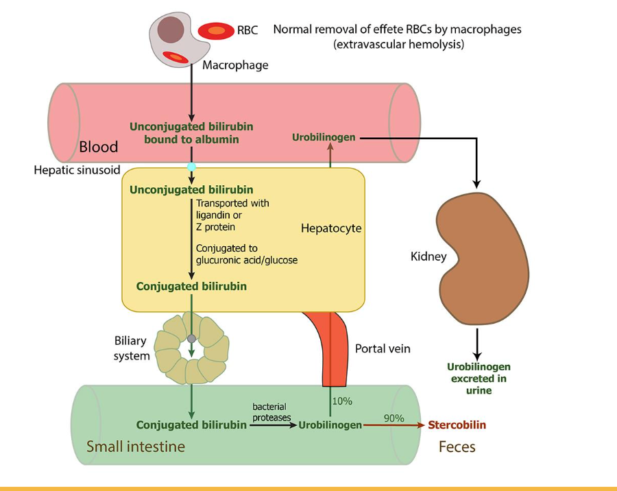
104
What is lipemia and what causes it?
Lipemia refers to the turbidity of a sample caused by the accumulation of lipoprotein particles, with chylomicron being the largest contributor. It can be removed by airfuge.
105
When is lipemia usually observed?
Lipemia is usually seen if the patient was not fasting or if the patient is receiving lipids.
106
What are other possible causes of lipemia?
Other causes include diabetes mellitus, ethanol use, chronic renal failure, and pancreatitis.
107
How do lipoproteins affect sampling in lipemic samples?
Lipoproteins cause a volume depletion effect by reducing the available water in the sample volume.
108
How does lipemia affect analytes dissolved in plasma or serum?
Most analytes, such as sodium (Na), are dissolved in the aqueous phase of plasma or serum. Lipoproteins displace water, leading to an apparent decrease in the concentration of these analytes.
109
How does lipemia affect lipid-soluble analytes?
For analytes that are lipid soluble, lipemia causes an apparent increase in concentration.
110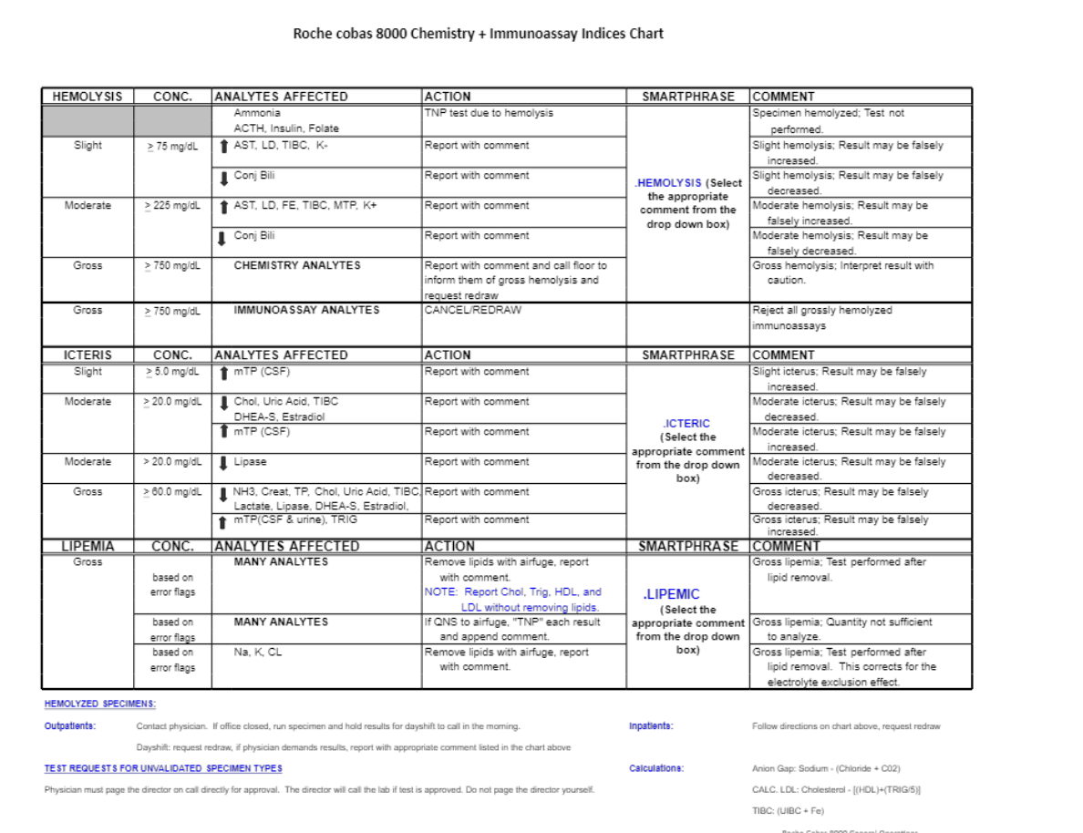
111
What is the purpose of different panels in laboratory testing?
Different panels provide information about the body’s chemical balance, metabolism, and body functions.
112
Why are panels useful in ordering tests?
They provide an easier and more organized way of ordering multiple related tests.
113
What tests are included in the Basic Metabolic Panel (BMP)?
Na, K, Cl, CO2, AGAP, Glu, Crea, BUN, and Ca.
114
What tests are included in the Hepatic Panel?
Tp, Alb, t.Bili, d.Bili, ALP, AST, and ALT.
115
What tests are included in the Comprehensive Metabolic Panel (CMP)?
The CMP includes all tests from the BMP plus the Hepatic Panel, but d.Bili (direct bilirubin) is not included.
116
What are electrolytes and how are they classified?
Electrolytes are ions capable of carrying an electric charge and are classified as anions or cations based on the type of charge they carry.
117
What functions do electrolytes help regulate?
Electrolytes help control nerve and muscle function and maintain the acid-base (pH) balance and water balance of the blood.
118
What is the function of sodium?
Sodium is the major extracellular cation that maintains the distribution of water and osmotic pressure.
119
What is the function of potassium?
Potassium is the major intracellular cation that regulates neuromuscular excitability, contraction of the heart, intracellular fluid (ICF) volume, and hydrogen ion (H⁺) concentration.
120
What is the function of chloride?
Chloride is the major extracellular anion that maintains osmolality, blood volume, and electric neutrality.
121
What is the function of total CO₂?
Total CO₂ is important for maintaining acid-base balance.
122
What is the formula for calculating the anion gap (AGAP)?
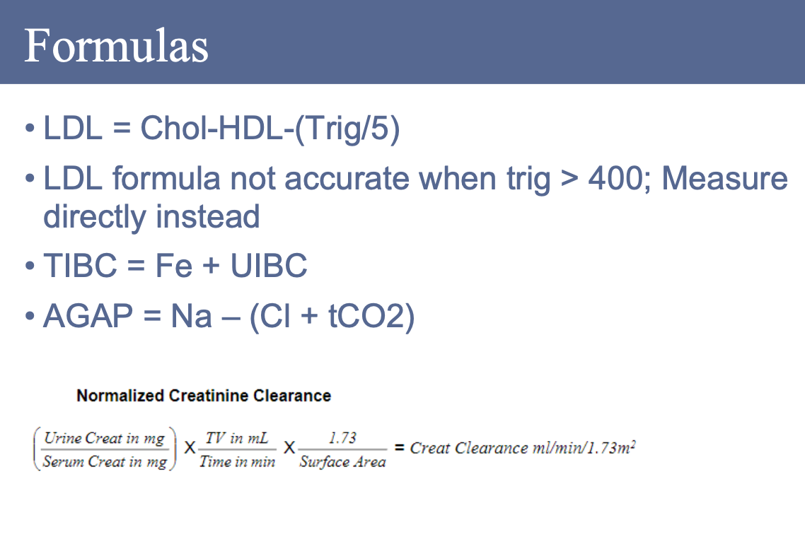
AGAP = Na – (Cl + CO₂).
123
What does the anion gap measure?
The anion gap measures the difference, or gap, between negatively charged and positively charged ions.
124
What does the anion gap take into account?
It takes into account the unmeasured anions and cations.
125
What conditions can cause an increased anion gap?
Diabetic acidosis, lactic acidosis, and renal failure.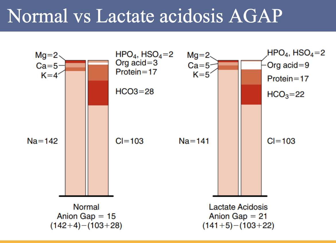
126
What can cause a low or negative anion gap?
A low or negative AGAP can be caused by low albumin or instrument issues.
127
What does an increase in Alkaline Phosphatase (ALP) indicate?
An increase in ALP indicates liver and bone disease.
128
What does an increase in Aspartate Aminotransferase (AST) indicate?
An increase in AST indicates liver disease.
129
What does an increase in Alanine Aminotransferase (ALT) indicate?
An increase in ALT indicates liver disease.
130
What does an increase in Gamma Glutamyl Transferase (GGT) indicate?
An increase in GGT indicates liver disease.
131
What does an increase in Lactate Dehydrogenase (LD) indicate?
An increase in LD indicates liver disease.
132
What does an increase in Creatine Kinase (CK) indicate?
An increase in CK indicates heart or muscle disease.
133
What does an increase in Amylase indicate?
An increase in Amylase indicates pancreatitis.
134
Where is AST primarily found in the body?
AST is widely distributed in human tissue, with the highest concentrations in cardiac tissue, liver, and skeletal muscle, and smaller amounts in the kidney, pancreas, and erythrocytes.
135
What are the main clinical uses of AST?
AST is mainly used to evaluate hepatocellular disorders and skeletal muscle involvement.
136
How do AST levels change during an acute myocardial infarction (AMI)?
In AMI, AST levels begin to rise within 6 to 8 hours, peak at 24 hours, and generally return to normal within 5 days.
137
Why is AST not useful for diagnosing AMI?
Because of its wide tissue distribution, AST levels are not specific enough to be useful in the diagnosis of AMI.
138
Where is ALT primarily found in the body?
ALT is distributed in many tissues but has comparatively high concentrations in the liver.
139
Why is ALT considered a liver-specific enzyme?
ALT is considered the more liver-specific enzyme of the transferases because of its high concentration in liver tissue.
140
What are the main clinical applications of ALT assays?
ALT assays are mainly used for evaluating hepatic disorders.
141
In which conditions are higher ALT elevations observed?
Higher elevations are found in hepatocellular disorders than in extrahepatic or intrahepatic obstructive disorders.
142
Where is ALP activity found in the body?
ALP activity is present on cell surfaces in most human tissues, with the highest concentrations in the intestine, liver, bone, spleen, placenta, and kidney.
143
In which conditions are ALP elevations most diagnostically significant?
Elevations of ALP are most significant in evaluating hepatobiliary and bone disorders.
144
When are ALP elevations more predominant in hepatobiliary disorders?
In hepatobiliary disorders, ALP elevations are more predominant in obstructive conditions than in hepatocellular disorders.
145
When are ALP elevations observed in bone disorders?
In bone disorders, ALP elevations are observed when there is involvement of osteoblasts.
146
Where is CK primarily found in the body?
CK is widely distributed in tissue, with the highest activities found in skeletal muscle, heart muscle, and brain tissue, and much smaller quantities in the bladder, placenta, gastrointestinal tract, thyroid, uterus, kidney, lung, prostate, spleen, liver, and pancreas.
147
Why are CK levels often elevated?
Because of the high concentrations of CK in muscle tissue, levels are frequently elevated in disorders of cardiac and skeletal muscle.
148
What conditions are associated with elevated CK levels?
Elevated CK levels are associated with acute myocardial infarction (AMI) and muscular dystrophy, particularly the Duchenne type.
149
Where is LDH primarily found in the body?
LDH is widely distributed, with high activities in the heart, liver, skeletal muscle, kidney, and erythrocytes, and lesser amounts in the lung, smooth muscle, and brain.
150
Why can LDH levels be elevated in many conditions?
Because LDH is active in numerous body tissues, it can be elevated in a variety of disorders.
151
What conditions are associated with increased LDH levels?
Increased LDH levels are found in cardiac, hepatic, skeletal muscle, and renal diseases, as well as in hematologic and neoplastic disorders.
152
In which conditions are the highest LDH levels seen?
The highest levels of total LDH are seen in pernicious anemia and hemolytic disorders.
153
Where is GGT activity primarily found in the body?
GGT activity is found primarily in the kidney, brain, prostate, pancreas, and liver.
154
What are the main clinical applications of GGT assays?
GGT assays are mainly used for evaluating liver and biliary system disorders.
155
Where is GGT located within the liver?
In the liver, GGT is located in the canaliculi of the hepatic cells and in the epithelial cells lining the biliary ductules.
156
Why is GGT considered one of the most sensitive enzyme assays?
Because GGT is elevated in virtually all hepatobiliary disorders, it is one of the most sensitive enzyme assays for detecting these conditions.
157
What are the major tissue sources of serum amylase?
The acinar cells of the pancreas and the salivary glands are the major tissue sources of serum amylase, with lesser concentrations found in skeletal muscle, the small intestine, and the fallopian tubes.
158
What is the main diagnostic significance of serum and urine amylase measurements?
Serum and urine amylase measurements are primarily used in the diagnosis of acute pancreatitis.
159
Where is lipase (LPS) primarily found in the body?
Lipase concentration is found primarily in the pancreas, but it is also present in the stomach and small intestine.
160
What is the main clinical use of serum lipase measurement?
Serum lipase measurement is used almost exclusively for the diagnosis of acute pancreatitis.
161
How does lipase measurement compare to amylase measurement?
Lipase measurement is similar to amylase in diagnosing acute pancreatitis but is considered more specific for pancreatic disorders than amylase measurement.
162
What does total bilirubin measure and when is it elevated?
Total bilirubin measures both conjugated and unconjugated bilirubin and is elevated in liver disease and hemolysis.
163
What special handling is required for bilirubin samples?
Bilirubin samples must be protected from light for accurate measurement because high levels can cause brain damage in newborns (kernicterus).
164
What is conjugated (direct) bilirubin?
Conjugated bilirubin, or direct bilirubin, is converted in the liver so it can be removed from the body and is water soluble.
165
What is unconjugated (indirect) bilirubin?
Unconjugated bilirubin, or indirect bilirubin, is water insoluble.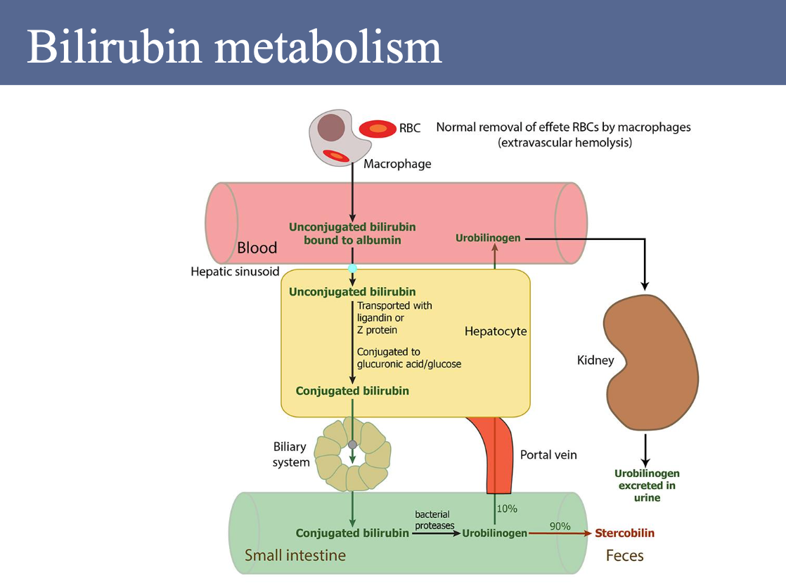
166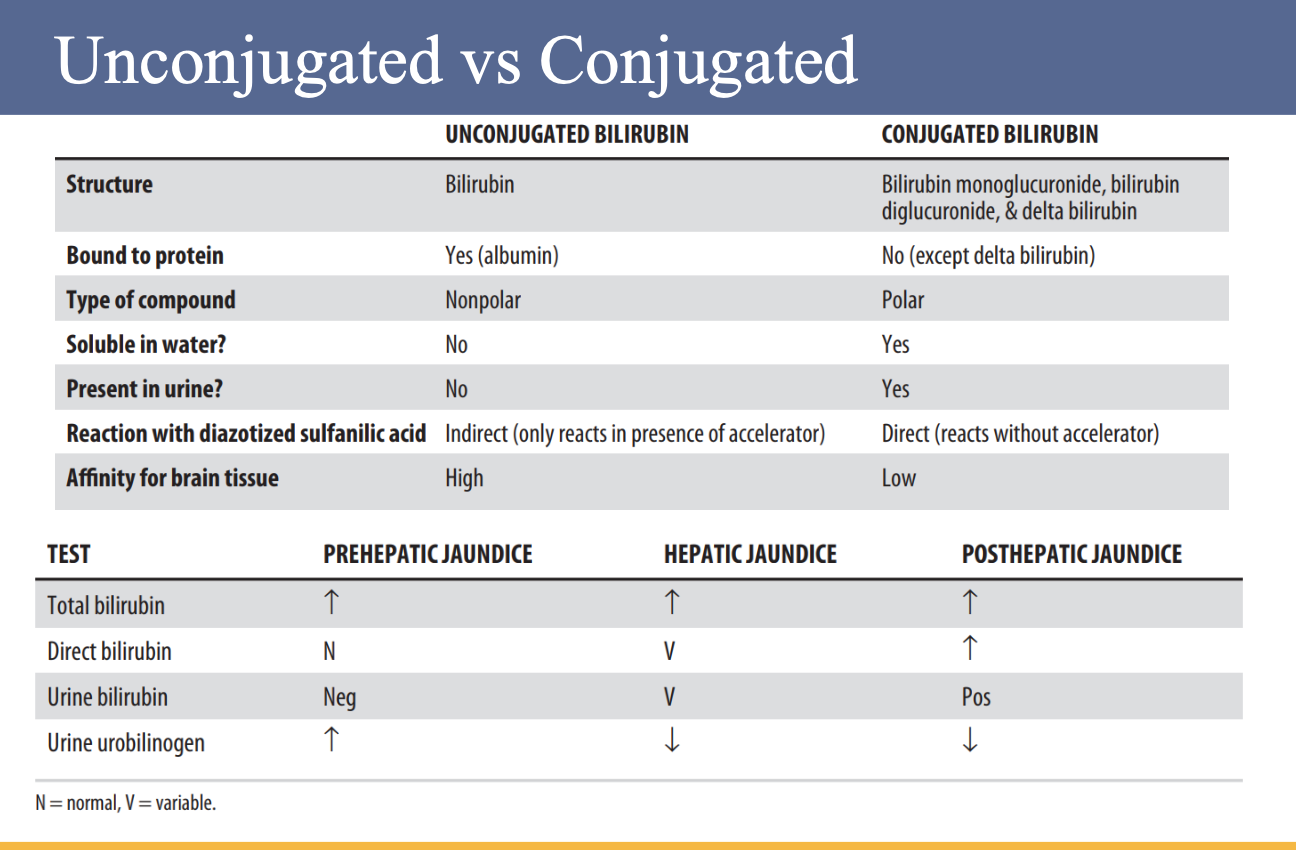
167
What is jaundice and what causes it?
Jaundice is the yellowing of the skin and sclera caused by abnormally elevated levels of bilirubin in the blood.
168
How is jaundice categorized?
Jaundice is categorized into three types: pre-hepatic, intra-hepatic, and post-hepatic.
169
What are the differences between medical and surgical jaundice?
Pre-hepatic and intra-hepatic jaundice are known as medical jaundice, while post-hepatic (or obstructive) jaundice is considered surgical jaundice.
170
What happens in pre-hepatic jaundice?
In pre-hepatic jaundice, excess production of bilirubin overwhelms the liver’s ability to conjugate and excrete it into the gut, leading to predominantly unconjugated hyperbilirubinemia.
171
What is the most common cause of pre-hepatic jaundice?
The most common cause is hemolytic anemia, which results in excess heme breakdown.
172
What causes intra-hepatic jaundice?
Intra-hepatic jaundice is caused by parenchymal liver disease that impairs the liver’s ability to conjugate or excrete bilirubin.
173
How does the type of bilirubin elevation vary in intra-hepatic jaundice?
The fraction of bilirubin that is elevated varies—viral hepatitis often causes predominantly unconjugated hyperbilirubinemia, while conjugated hyperbilirubinemia occurs with cholestasis from drugs or primary biliary cholangitis.
174
What causes post-hepatic (obstructive) jaundice?
Post-hepatic jaundice occurs when there is an impediment to the flow of bile due to a partial or complete obstruction of the extrahepatic biliary passage between the liver and duodenum.
175
Where can the obstruction occur in post-hepatic jaundice?
The obstruction can occur within the biliary ducts themselves or more distally within the pancreas.
176
What type of bilirubinemia is seen in post-hepatic jaundice?
Post-hepatic jaundice is characterized by predominantly conjugated hyperbilirubinemia.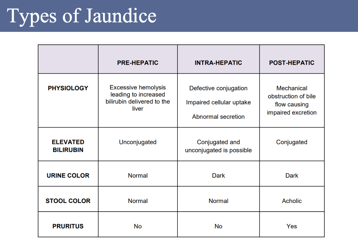
177
What are hormones and what do they do?
Hormones are chemical substances secreted by endocrine glands that travel through the blood and regulate the functions of tissues and organs elsewhere in the body.
178
What are the three types of hormones?
The three types of hormones are steroids, amines, and proteins/peptides.
179
What are examples of steroid hormones and what are they made from?
Steroid hormones are made from cholesterol and include cortisol, aldosterone, androgens (male hormones), estrogens (female hormones), and progesterone.
180
What are examples of amine hormones?
Amine hormones include thyroid hormones and adrenomedullary hormones.
181
What are examples of protein and peptide hormones?
Protein and peptide hormones include pituitary, parathyroid, and pancreatic hormones.
182
What is the function of TSH?
TSH is secreted by the pituitary gland and stimulates the production of T4 and T3.
183
What are the functions of T4 and T3?
T4 and T3 stimulate metabolic activity and increase oxygen consumption, heat production, heart rate, gastrointestinal movement, and brain activity. They also influence the growth and development of the musculoskeletal and nervous systems.
184
How is thyroid hormone production regulated?
When T4 and T3 levels are low, the hypothalamus releases TRH, which stimulates TSH release. When T4 and T3 levels are high, TRH release is inhibited.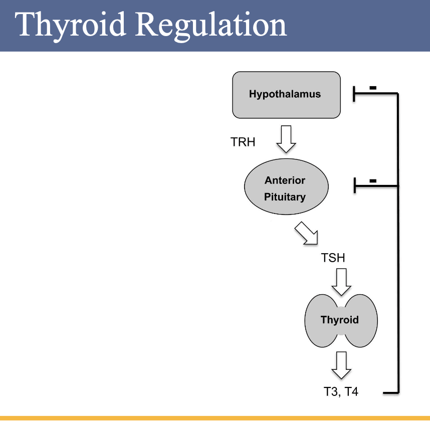
185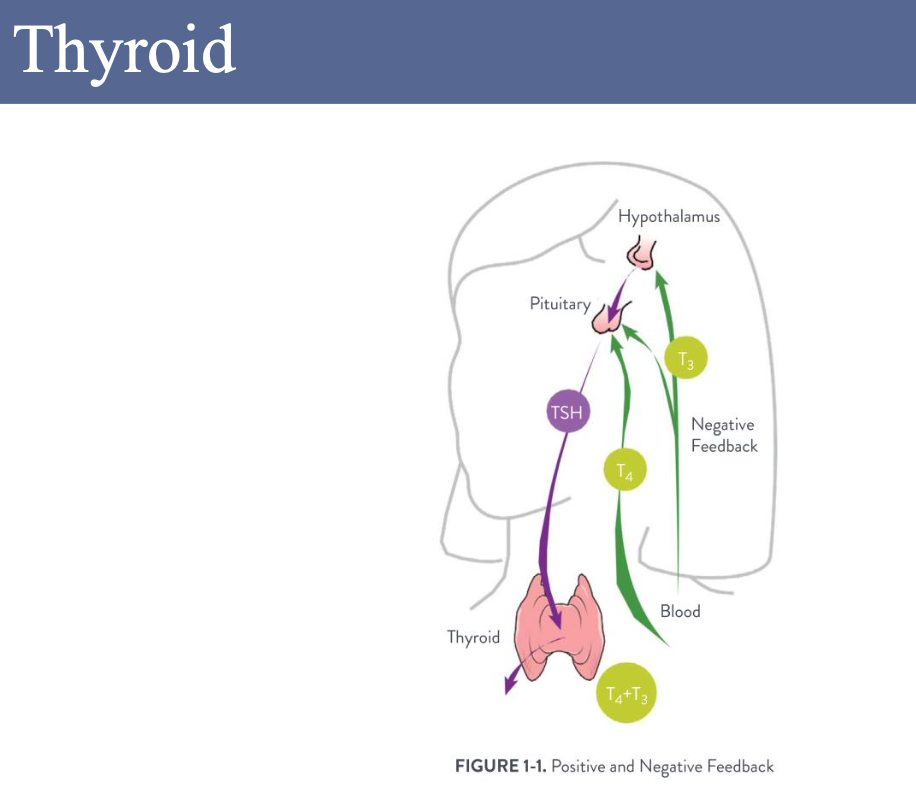
186
What is the function of TSH in the thyroid?
TSH regulates the production of T3 and T4 by the thyroid.
187
What is the function of T3 and T4?
T3 and T4 regulate growth and development.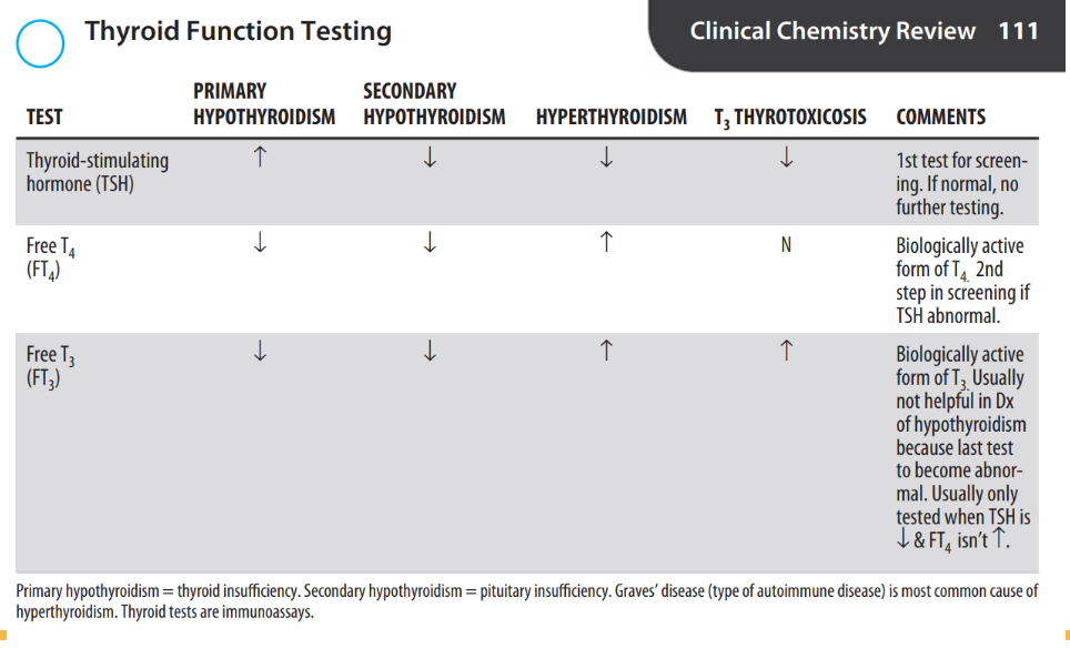
188
What is the purpose of the TSH test?
TSH is used to evaluate thyroid function.
189
What is the purpose of the T4 test?
T4 helps with diagnosis and determination of hyperthyroidism or hypothyroidism.
190
When is the T3 test ordered and what is its purpose?
T3 is ordered after abnormal TSH and T4 results and helps diagnose hypothyroidism.
191
What is the purpose of the aTPO test?
aTPO is used to diagnose and monitor autoimmune thyroid disease.
192
What is the purpose of the Tg test?
Tg is used as a tumor marker for thyroid cancer.
193
Why is tgAB measured?
tgAB is measured because it can interfere with Tg measurement.
194
What is the purpose of the T-Uptake test?
T-Uptake estimates protein binding of thyroid hormones
195
What is the function of ACTH?
ACTH regulates cortisol and androgens.
196
What is the function of FSH?
FSH regulates and stimulates the growth and function of the ovaries and testes. It works with LH to stimulate follicle growth and maturation in women and induces spermatogonium development in men.
197
What is the function of LH?
LH regulates and stimulates the growth and function of the ovaries and testes. In men, it stimulates testosterone production; in women, LH peaks mid-cycle to induce ovulation and the formation of the corpus luteum.
198
What is the function of PRL?
PRL regulates lactation.
199
What is the function of TSH?
TSH regulates the production of T3 and T4.
200
What is the function of ADH?
ADH regulates the reabsorption of water in the distal renal tubules.
201
What is the function of oxytocin?
Oxytocin regulates uterine contractions during childbirth and lactation.
202
What are the functions of thyroid hormones T4 and T3?
T4 and T3 regulate metabolism, growth, and development.
203
What is the function of calcitonin?
Calcitonin inhibits calcium resorption.
204
What hormone does the parathyroid gland produce and what does it regulate?
The parathyroid gland produces PTH, which regulates calcium and phosphate.
205
What are the main hormones produced by the adrenal cortex?
The adrenal cortex produces aldosterone and cortisol.
206
What is the function of aldosterone?
Aldosterone regulates the reabsorption of sodium (Na) in the renal tubules.
207
What is the function of cortisol?
Cortisol regulates carbohydrate, fat, and protein metabolism; maintains water and electrolyte balance; and suppresses inflammatory and allergic reactions.
208
What hormones are produced by the adrenal medulla?
The adrenal medulla produces epinephrine and norepinephrine.
209
What is the function of epinephrine and norepinephrine?
Epinephrine and norepinephrine regulate the “fight or flight” response by stimulating the sympathetic nervous system.
210
What hormones are produced by the ovaries and what are their functions?
The ovaries produce estrogen and progesterone. Estrogen regulates female reproductive organs and secondary sex characteristics, controls the menstrual cycle, and maintains pregnancy. Progesterone prepares the uterus for ovum implantation and maintains pregnancy.
211
What hormone is produced by the placenta and what is its function?
The placenta produces HCG, which regulates progesterone production by the corpus luteum during early pregnancy and supports the development of fetal gonads.
212
What hormone is produced by the testes and what is its function?
The testes produce testosterone, which is responsible for the development of male reproductive organs and secondary sex characteristics.
213
What is the function of insulin?
Insulin regulates carbohydrate metabolism and increases the movement of glucose into cells for metabolism.
214
What is the function of glucagon?
Glucagon regulates glycogenolysis, gluconeogenesis, and lipolysis.
215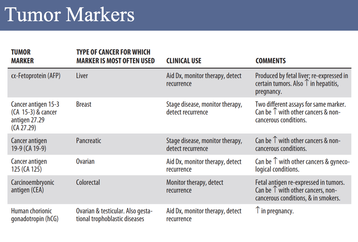
216
What is therapeutic drug monitoring (TDM)?
Therapeutic drug monitoring is the measurement of specific drugs and/or their metabolites at timed intervals to maintain a relatively constant concentration of the medication in the blood.
217
What is the goal of therapeutic drug monitoring?
The goal is to balance producing the desired therapeutic effect while avoiding adverse responses in the patient.
218
What is the therapeutic index?
The therapeutic index is the ratio between the toxic dose and the therapeutic (effective) dose of a medication.
219
What is a trough level and when is it measured?
The trough is the lowest concentration of a drug in the blood and is drawn before the next dose.
220
What is a peak level and when is it measured?
The peak is the highest concentration of a drug in the blood and is drawn about one hour or more after the dose, depending on the drug.
221
What is pharmacokinetics?
Pharmacokinetics is the study of what the body does to the drug.
222
What is pharmacodynamics?
Pharmacodynamics is the study of what the drug does to the body.
223
How are most drugs and their metabolites eliminated from the body?
Most drugs, especially water-soluble ones, are eliminated largely by the kidneys in urine.
224
What is the purpose of pharmacogenetic testing?
Pharmacogenetic testing provides information about a person’s genes to help health care providers choose the medicine and dosage that are the best fit for the individual.
225
What important factors should be checked before resulting in therapeutic drug monitoring (TDM)?
Before resulting, check for:
• Volume – Ensure there is enough sample.
• Clot – A clot can prevent accurate sampling and cause erroneous results, even if clot detection is missed by the machine.
• Previous history – Confirm whether a trough looks like a trough (low value) and a peak looks like a peak (high value).
• Volume – Ensure there is enough sample.
• Clot – A clot can prevent accurate sampling and cause erroneous results, even if clot detection is missed by the machine.
• Previous history – Confirm whether a trough looks like a trough (low value) and a peak looks like a peak (high value).
226
What are common issues with TDM sample timing?
Trough and peak samples may be ordered incorrectly, switched, or collected at the wrong time; if timing is uncertain, the order may need to be changed to “Random.”
227
What code is associated with this procedure?
.LABVCP
228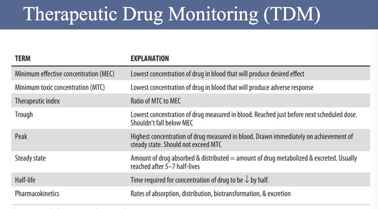
229
What drugs are included in the UCLA Drugs of Abuse (DAU) panel?
The panel includes amphetamine, barbiturates, benzodiazepine, cannabinoids, cocaine, methadone, opiates, oxycodone, ethanol, hydrocodone, and fentanyl.
230
What type of testing method is used for DAU tests?
DAU tests are semi-quantitative—the machine gives a numeric value that is converted to a positive or negative result based on the analyte’s cutoff.
231
What is the associated test code for Drugs of Abuse testing?
.LABDRUGDIL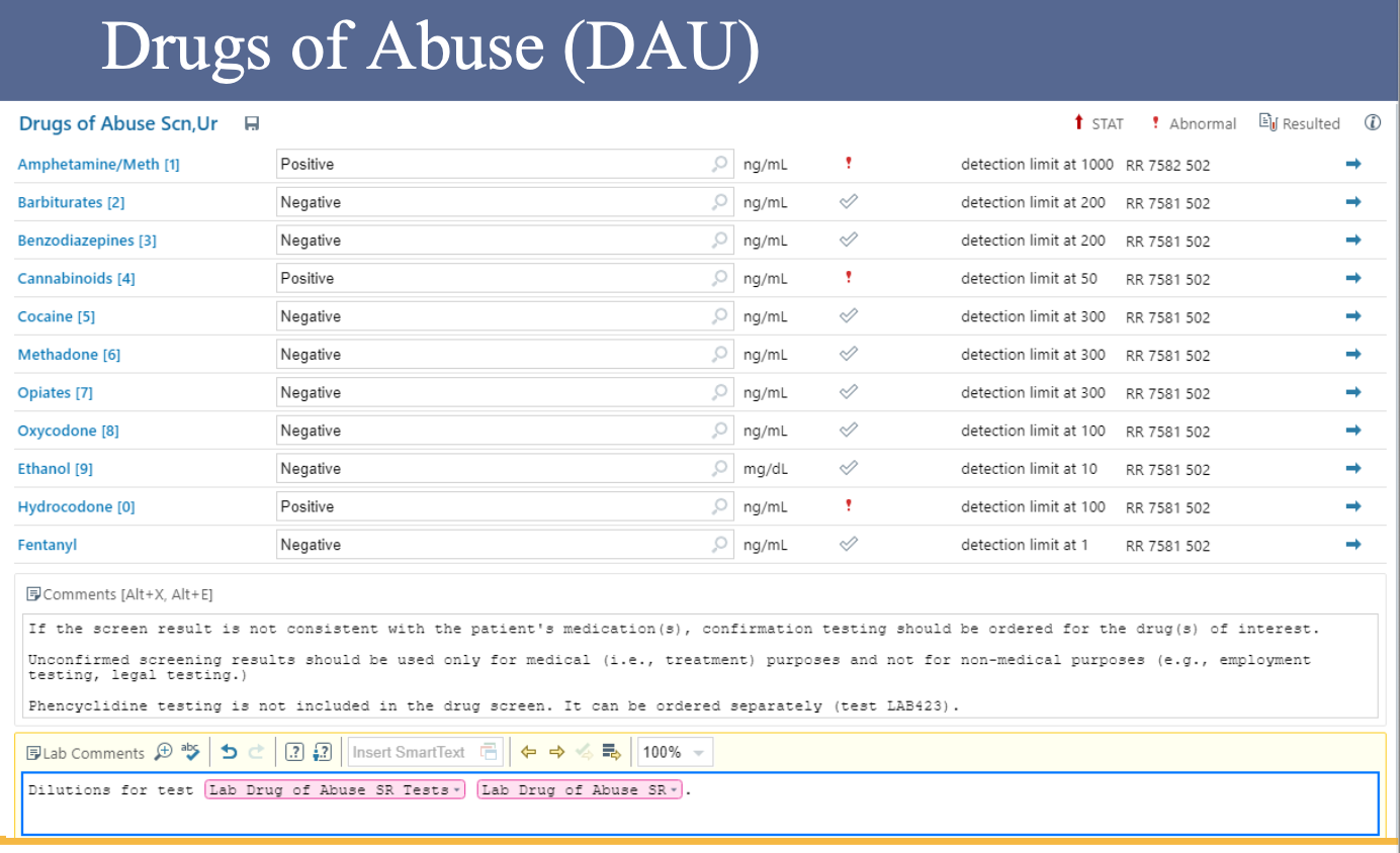
232
How can you determine if a sample is contaminated?
To assess possible contamination, look for a dilutional effect on other analytes. For example, an increase in sodium (Na) may suggest saline contamination. Checking other analytes such as creatinine and BUN for dilutional effects can support suspicion of IV saline contamination.
233
What should be done if results are unclear or contamination is uncertain?
If results are not clearly contaminated, UCLA’s SOP states that staff should not hint at contamination but may ask the RN or MD if the value is expected. The final decision is made by the MD.
234
What does it mean when a test is auto-verified?
A majority of tests are auto-verified, meaning they are automatically released unless an issue causes the result to be held, in which case the CLS must address it.
235
What should be done if an error code appears?
Pay attention to error codes—if there’s an error code, it’s most likely not safe to release the result.
236
What should you do when uncertain about a held result?
When in doubt, check the machine to see what it is doing.
237
What factors should be reviewed before releasing a result?
Check the test’s AMR (Analytical Measuring Range) and CRR (Clinical Reportable Range), perform a dilution if necessary, and review patient history to ensure the values make sense.
238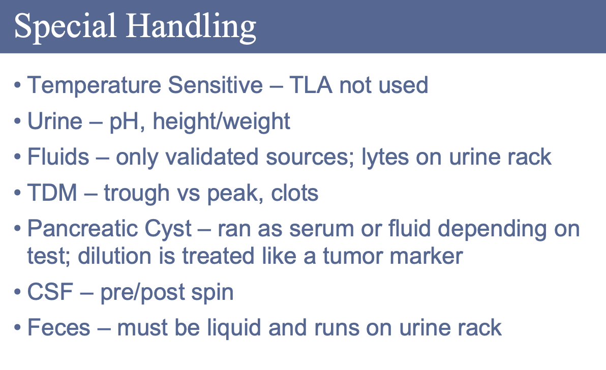
239
How can contamination occur in blood samples?
Contamination can occur when drawing from a line, if blood is drawn near the source of infusion, or due to an incorrect order of draw.
240
What are common causes and indicators of contamination?
Common causes include Total Parenteral Nutrition (TPN), which can increase glucose and potassium (K), or saline, which can increase sodium (Na) and chloride (Cl); EDTA contamination is also common.
241
What happens to test parameters when contamination occurs?
Some parameters may appear diluted when contamination occurs.
242
What is UCLA’s policy regarding contaminated samples?
At UCLA, if a sample is contaminated, all specimens drawn at the same time are considered contaminated, regardless of whether they match previous history or appear normal.
243