Lecture 3: Hemolytic Anemias Part 2
Study these flashcardsFree · spaced repetition · iPhone, Android, Mac & web
All cards
1
What are the two types of clinical trends in hemolysis?
Acute and Chronic
2
How is acute hemolysis described?
Sudden, episodic or paroxysmal, such as PNH, PCH, AHTR
3
How is chronic hemolysis described?
No anemia as long as BM can compensate, best example G6PD deficiency. Thalassemia Major is also chronic but so severe that BM can not compensate
4
What are the two development patterns of hemolysis?
Acquired and Inherited
5
What is acquired hemolysis?
Person is exposed to an agent that induces hemolysis, such as malaria
6
What is inherited hemolysis?
A mutant gene which causes hemolysis is passed on to offspring, such as thalassemia
7
What are the two categories of RBC defects?
Intrinsic (mostly inherited) and Extrinsic (mostly acquired)
8
What are intrinsic RBC defects?
Defects in RBC membrane, metabolism, Hb content (hemoglobinopathies)
9
What are extrinsic RBC defects?
Malaria, drugs, immune hemolysis, trauma, microangiopathic (MAHA: TTP, HUS, DIC), exception: PNH which is acquired intrinsic defect
10
What are the two sites of hemolysis?
Intravascular and Extravascular
11
What characterizes intravascular hemolysis?
RBC fragmentation (DIC, heart prosthetic valves)
12
What characterizes extravascular hemolysis?
Macrophage-mediated, in spleen, BM or liver
13
An elderly white woman is evaluated for worsening anemia, with a decrease of approximately 0.5 mg/dL of hemoglobin each week. The patient is pale, and her skin and eyes are slightly yellow. She complains of extreme fatigue and is unable to complete the tasks of daily living without napping in midmorning and midafternoon. She also tires with exertion, finding it difficult to climb even five stairs. Which of the features of this description points to a hemolytic cause for her anemia?
A. Pallor
B. Yellow skin and eyes
C. Need for naps
D. Tiredness on exertion
A. Pallor
B. Yellow skin and eyes
C. Need for naps
D. Tiredness on exertion
B. Yellow skin and eyes
14
What causes hemolytic anemias (HAs)?
Excessive macrophage-mediated hemolysis (extravascular(EV)) or fragmentation hemolysis (intravascular(IV))
15
EV: Main RBC abnormality formed?
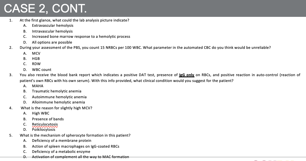
EV hemolysis occurs mainly in the spleen/liver macrophages. RBCs are partially phagocytized, leading to spherocyte formation (smaller, round, no central pallor).
16
IV: main RBC abnormality formed?
IV hemolysis occurs directly in the circulation due to mechanical trauma, complement, or toxins, resulting in schistocyte formation (fragmented RBCs).
17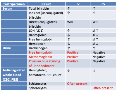
18
All Intrinsic RBC defects are inherited except?
Exception: PNH, intrinsic but acquired
19
A 5-year-old girl was seen by her physician several days before the current visit and was diagnosed with pneumonia. She was prescribed a standard course of antibiotics. Her mother has brought her to the physician again because the girl’s urine began to darken after the first visit and now is alarmingly dark. The girl has no history of anemia, and there is no family history of any hematologic disorder. The CBC shows a mild anemia, polychromasia, and a few schistocytes. This anemia could be categorized as:
A. Acquired, fragmentation
B. Acquired, macrophage mediated
C. Hereditary, fragmentation
D. Hereditary, macrophage mediated
A. Acquired, fragmentation
B. Acquired, macrophage mediated
C. Hereditary, fragmentation
D. Hereditary, macrophage mediated
A. Acquired, fragmentation
20
A patient has a personal and family history of a mild hemolytic anemia. The patient has consistently elevated levels of total and indirect serum bilirubin and urinary urobilinogen. The serum haptoglobin level is consistently decreased, whereas the reticulocyte count is elevated. The latter can be seen as polychromasia on the patient’s peripheral blood film. Spherocytes are also noted. Which one of the findings reported for this patient is inconsistent with a classical diagnosis of fragmentation hemolysis?
A. Elevated total and indirect serum bilirubin
B. Elevated urinary urobilinogen
C. Decreased haptoglobin
D. Spherocytes on the peripheral blood film
A. Elevated total and indirect serum bilirubin
B. Elevated urinary urobilinogen
C. Decreased haptoglobin
D. Spherocytes on the peripheral blood film
D. Spherocytes on the peripheral blood film
21
Hereditary Spherocytosis (HS) is the outcome of what kind of defect and what kind of mutation?
An intrinsic defect which is mutations in proteins which maintain vertical attachments
22
What type of genetic inheritance is HS mostly associated with?
Mostly autosomal dominant
23
HS is mostly due to mutations in which proteins?
ankyrin and spectrin
24
What happens to RBCs in HS?
RBCs lose surface area (low s/v ratio), lose deformability or some endure further membrane modifications forming microsherocytes. 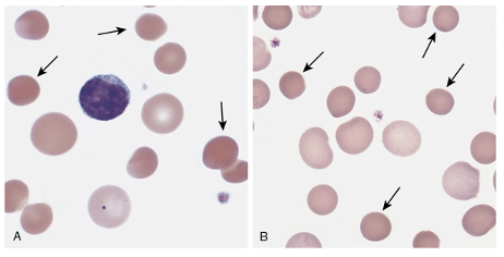
25
What happens when RBCs lose surface area and deformability?
They are destroyed by the spleen leading to EV hemolysis,
26
What is the triad seen in HS?
anemia, splenomegaly, jaundice
27
What is the hallmark finding on PBS in HS?
On PBS: Hallmark is spherocytosis (not specific), also increase Polychromasia, indicative of reticulocytosis (up to 10%).
28
What RBC indices are elevated in HS?
high MCHC (>36 mg/dL), high CHCM (>4%), which indicate hyperdense RBCs (used for HS screening)
29
What are the serum results for indirect bili, LDH, and hepatoglobin in HS?
Indirect bili ↑, LDH ↑, haptoglobin ↓
30
What findings are highly suggestive of HS?
Childhood hemolytic anemia, family history of similar abnormalities, uniform spherocytes on PBS
31
What makes the diagnosis of HS definitive?
Definitive diagnosis: family history of HS, splenomegaly, high MCHC, reticulocytosis, and spherocytes on the PBS
32
What additional tests can be performed if required for HS?
If required: osmotic fragility (OF) test and eosin-5’-maleimide (EMA) binding test
33
HS: Summary of Laboratory and Clinical Diagnosis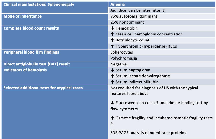
34
What defects cause hereditary elliptocytosis (HE)?
Defects in horizontal interactions
35
What percentage of HE cases are caused by spectrin gene mutations?
95% cases mutation in spectrin genes (α and β)
36
What happens to RBCs over time in HE?
RBCs become elliptical over time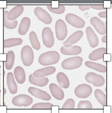
37
What may be seen on peripheral blood smear in HE?
On PBS: elliptocytes may be up to 100%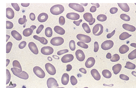
38
What is the severe form of HE and what causes RBC fragmentation?
Severe form of HE is HPP (Hereditary Pyropoikilocytosis) in which RBCs are fragmented on heating due to low stability (at 41-45°C)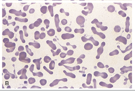
39
What does HPP look like before and after heating?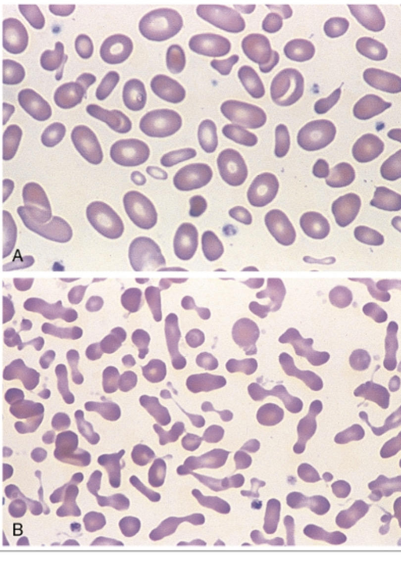
HPP: A-before heating, B-after heating
40
What are the RBC characteristics of HPP?
HPP: extreme poikilocytosis, MCV very low (50-65 fl), fragmentation, microspherocytosis, and elliptocytosis (similar to burns)
41
What is overhydrated hereditary stomatocytosis (intrinsic membrane defects)?
Overhydrated hereditary stomatocytosis: RBC membrane is highly permeable, deficiency in stomatin protein or mutation in RhAG protein has been reported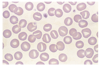
42
What is dehydrated hereditary stomatocytosis (hereditary xerocytosis)?
Dehydrated hereditary stomatocytosis or hereditary xerocytosis (Both intrinsic membrane defects): defect in membrane cation permeability causing RBCs to become dehydrated
43
A patient has anemia that has been worsening over the last several months. The hemoglobin level has been declining slowly, with a drop of 1.5 g/dL of hemoglobin over about 6 weeks. Polychromasia and anisocytosis are seen on the peripheral blood film, consistent with the elevated reticulocyte count and red cell distribution width (RDW). Serum levels of total bilirubin and indirect fractions are normal. The urinary urobilinogen level also is normal. When these findings are evaluated, the conclusion is drawn that the anemia does not have a hemolytic component. Based on the data given here, why was hemolysis ruled out as the cause of the anemia?
A. The decline in hemoglobin is too gradual to be associated with hemolysis.
B. The elevation of the reticulocyte count suggests a malignant cause.
C. Evidence of increased protoporphyrin catabolism is lacking.
D. Elevated RDW points to an anemia of decreased production.
A. The decline in hemoglobin is too gradual to be associated with hemolysis.
B. The elevation of the reticulocyte count suggests a malignant cause.
C. Evidence of increased protoporphyrin catabolism is lacking.
D. Elevated RDW points to an anemia of decreased production.
C. Evidence of increased protoporphyrin catabolism is lacking.
44
Which of the following sets of test results is typically expected with chronic fragmentation hemolysis?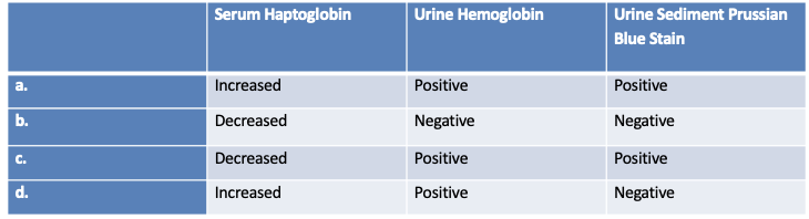
C. Decreased haptoglobin, Positive urine hemoglobin, Positive Prussian blue stain
45
What type of anemia is PNH (Paroxysmal nocturnal hemoglobinuria) and what defect does it involve?
Rare chronic IV hemolytic anemia, acquired membrane defect
46
What proteins are lacking on RBCs in PNH?
RBCs lack CD55 and CD59
47
What happens when CD55 and CD59 are absent on RBCs?
Absence of CD55 and CD59 renders RBCs susceptible to spontaneous lysis by complement.
48
What kind of RBC mosaicism is seen in PNH?
PNH RBCs display mosaicism: some RBCs are normal some abnormal, 3 RBC populations exist: type I, type II, type III
49
What characterizes type I RBCs in PNH?
Type I: normal level of CD55 and CD59, no lysis or little
50
What characterizes type II RBCs in PNH?
Type II: partial CD55 and CD59 deficiency, relatively resistant to lysis
51
What characterizes type III RBCs in PNH?
Type III: complete deficiency of CD55 and CD59, highly sensitive to lysis
52
What is the most common RBC types in PNH?
combination of I and III
53
What is the confirmatory test for PNH?
Confirmatory test: detection of CD55 and CD59 by flowcytometry
54
What is the role of the Hexose monophosphate shunt (HMS)?
The Hexose monophosphate shunt (HMP shunt), also known as the pentose phosphate pathway, has two main roles: to generate NADPH for reductive biosynthesis and protection against oxidative stress, and to produce pentose phosphates (like ribose-5-phosphate) as precursors for the synthesis of nucleotides and nucleic acids (RNA and DNA).
55
Why is G6PD critical to RBCs?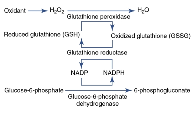
G6PD is critical to RBCs as it is the only means to produce NADPH
56
Can you tell what happens if G6PD is missing?
If G6PD is deficient:
- RBCs cannot make enough NADPH.
- Glutathione is not regenerated → oxidative stress damages hemoglobin.
- Hemoglobin denatures and precipitates as Heinz bodies (seen with supravital stain).
- The spleen removes these inclusions → “bite cells” on blood smear.
- Leads to episodic intravascular and extravascular hemolysis triggered by oxidative stress.
- RBCs cannot make enough NADPH.
- Glutathione is not regenerated → oxidative stress damages hemoglobin.
- Hemoglobin denatures and precipitates as Heinz bodies (seen with supravital stain).
- The spleen removes these inclusions → “bite cells” on blood smear.
- Leads to episodic intravascular and extravascular hemolysis triggered by oxidative stress.
57
Where is the G6PD enzyme encoded?
G6PD enzyme is encoded on X chromosome
58
What is the most common RBC enzyme defect?
G6PD deficiency is the most common RBC enzyme defect
59
Which population is protected against malaria by G6PD deficiency?
African males with G6PD deficiency (A- variant) are protected against plasmodium falciparum malaria
60
WHO classification of G6PD deficiency 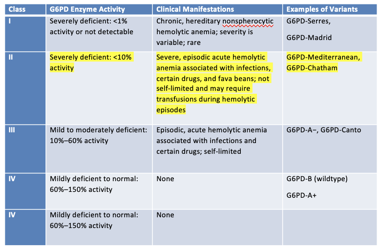
61
What are the triggers of acute hemolytic anemia in G6PD deficiency?
Triggers of acute hemolytic anemia in G6PD deficiency: Oxidizing drugs, infections, fava beans
62
What type of anemia is usually seen in G6PD deficiency?
Anemia, moderate to sever, usually normochromic/normocytic
63
What findings are seen with severe G6PD deficiency variants?
With severe variants: marked anisocytosis, poikilocytosis, spherocytosis, and schistocytosis
64
What RBC morphologies are associated with G6PD deficiency?
Bite cells (not specific), Heinze bodies (denatured Hb, only with supravital staining)
65
What is seen in reticulocytes during G6PD deficiency?
Profound reticulocytosis (up to 30%) (high MCV)
66
What are the serum and plasma findings in G6PD deficiency hemolysis?
Very low serum haptoglobin, high indirect bili, high LDH, increased plasma Hb (IV hemolysis)
67
A patient experiences an episode of acute intravascular hemolysis after taking primaquine for the first time. The physician suspects that the patient may have G6PD deficiency and orders an RBC G6PD assay 3 days after the hemolytic episode began. How will this affect the test result?
A. Absence of enzyme activity
B. False decrease in enzyme activity due to hemoglobinemia
C. False increase in enzyme activity due to reticulocytosis
D. No effect on enzyme activity
A. Absence of enzyme activity
B. False decrease in enzyme activity due to hemoglobinemia
C. False increase in enzyme activity due to reticulocytosis
D. No effect on enzyme activity
C. False increase in enzyme activity due to reticulocytosis
68
What is pyruvate kinase (PK) and what pathway is it part of?
Pyruvate kinase (PK) is a key enzyme of the glycolytic pathway.
69
What reaction does PK catalyze?
PK catalyzes the conversion of phosphoenolpyruvate to pyruvate forming ATP.
70
What happens to RBCs in PK deficiency?
PK deficiency causes premature destruction of RBCs — without PK, depletion of ATP results in lack of membrane integrity.
71
What is the clinical presentation of PK deficiency?
Clinical presentation: anemia, jaundice, splenomegaly & gallstones (chronic hemolysis).
72
How does PK deficiency present in neonates?
Neonates: severe anemia.
73
How does PK deficiency present in adults?
Adults: severe to compensated.
74
What are the CBC findings of PK deficiency?
75
What is the Biochemistry findings of PK deficiency?
76
What is the PBS findings of PK deficiency?
77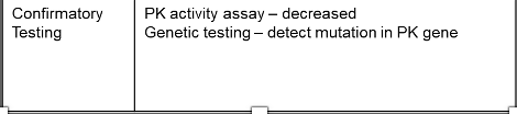
What are the confirmatory findings of PK deficiency?
78
What is immune hemolytic anemia (extrinsic defect)?
Conditions where red cells survival is shortened because of antibody-mediated mechanisms
79
What can some antibodies do in immune hemolytic anemia?
Some antibodies are able to activate complement
80
How are RBCs with antibodies or complement removed?
Removed by: Macrophages (extravascular), Complement mediated hemolysis (intravascular), Combination of extravascular and intravascular processes
81
How can immune hemolytic anemia be classified?
Classified as:
1. Autoimmune hemolytic anemia
2. Alloimmune hemolytic anemia
3. Drug-induced immune hemolytic anemia
1. Autoimmune hemolytic anemia
2. Alloimmune hemolytic anemia
3. Drug-induced immune hemolytic anemia
82
What type of hemolysis can IgM mediated hemolysis cause in immune hemolytic anemia?
IgM mediated hemolysis can result in both extravascular and intravascular hemolysis
83
What type of hemolysis is predominantly caused by IgG in immune hemolytic anemia?
IgG mediated hemolysis is predominantly extravascular --> RBCs removed by macrophages in the spleen/liver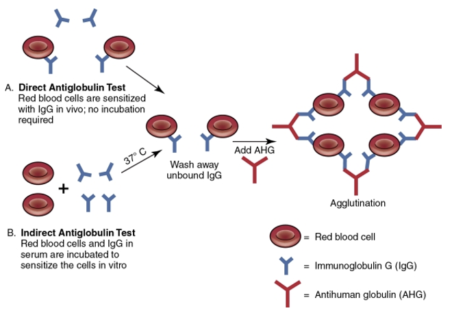
84
What are the CBC findings of Immune HA?
85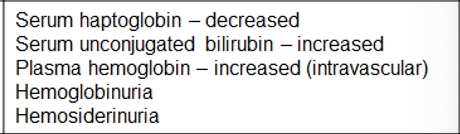
What are the Biochemistry findings of Immune HA?
86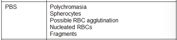
What are the PBS findings of Immune HA?
87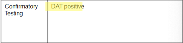
What are the confirmatory testing?
88
What causes premature RBC destruction and anemia in autoimmune hemolytic anemia (AIHA - Extrinsic defects)?
Premature RBC destruction and anemia caused by autoantibodies that bind to the RBC surface with or without complement activation.
89
How are autoimmune hemolytic anemias divided?
They are divided into 4 categories:
1. Warm autoimmune hemolytic anemia (WAIHA)
2. Cold agglutinin disease (CAD)
3. Paroxysmal cold hemoglobinura (PCH)
4. Mixed AIHA
1. Warm autoimmune hemolytic anemia (WAIHA)
2. Cold agglutinin disease (CAD)
3. Paroxysmal cold hemoglobinura (PCH)
4. Mixed AIHA
90
What is the most common type of autoimmune hemolytic anemias?
Warm Autoimmune Hemolytic Anemia (WAIHA)
91
What immunoglobulin is involved in warm autoimmune hemolytic anemia?
IgG
92
What is the optimum reactivity temperature of the autoantibody?
37°C
93
How is sensitization detected by DAT?
IgG or IgG + complement
94
What is the complement activation in warm AIHA?
variable
95
What type of hemolysis is seen in warm AIHA?
Extravascular primarily
96
What is the autoantibody specificity in warm AIHA?
Panreactive
97
What are the other laboratory findings in warm AIHA?
Polychromasia, spherocytes
98
What happens when IgM autoantibodies bind RBCs in cold temperatures?
IgM autoantibodies bind RBCs when patient exposed to cold temperatures.
99
What happens after IgM autoantibodies bind to RBCs?
Bound IgM antibodies activate complement.
100
What occurs when RBCs return to warmer areas of the body?
When RBCs return to central, warmer areas of body, IgM dissociates but complement stays attached.
101
How does hemolysis occur in cold agglutinin disease?
Hemolysis occurs predominately by hepatic macrophages but some intravascular hemolysis is possible if full complement occurs.
102
What immunoglobulin is involved in cold agglutinin disease?
IgM
103
What is the optimum reactivity temperature of the autoantibody?
4°C
104
How is sensitization detected by DAT?
Complement
105
Does complement activation occur in cold agglutinin disease?
Yes
106
What type of hemolysis is seen in cold agglutinin disease?
Extravascular; some intravascular
107
What is the autoantibody specificity in cold agglutinin disease?
Autoantibody specificity: I (most), i (some), Pr (rare)
108
What are the other laboratory findings in cold agglutinin disease?
RBC agglutination may be present; hemoglobinuria
109
What autoantibody usually causes PCH (CAIHA: Paroxysmal Cold Hemoglobinuria)?
PCH (extrinsic defect) is usually caused by Anti-P.
110
How does the autoantibody in PCH bind and activate complement?
Autoantibody binds the P antigen on RBCs in cold temps and partially activates complement BUT fully activates complement at warm temps thus exhibiting intravascular hemolysis.
111
In which population is PCH most commonly seen?
PCH is most commonly seen in young children after viral respiratory infection.
112
What immunoglobulin is involved in paroxysmal cold hemoglobinuria (PCH)?
IgG
113
What is the optimum reactivity temperature of the autoantibody in PCH?
4°C
114
How is sensitization detected by DAT in PCH?
Complement
115
Does complement activation occur in PCH?
Yes
116
What type of hemolysis occurs in PCH?
Intravascular
117
What is the autoantibody specificity in PCH?
Autoantibody specificity: P
118
What are the other laboratory findings in PCH?
Polychromasia, spherocytes, schistocytes, NRBCs, anisocytes, poikilocytosis; hemoglobinuria; Anti-P positive
119
What are the two types of alloimmune hemolytic anemias (extrinsic defect)?
There are usually 2 types of alloimmune hemolytic anemias:
1. Hemolytic Transfusion Reactions (HTR)
2. Hemolytic Disease of the Fetus and Newborn (HDFN)
1. Hemolytic Transfusion Reactions (HTR)
2. Hemolytic Disease of the Fetus and Newborn (HDFN)
120
What causes immune-mediated destruction of donor cells in hemolytic transfusion reactions (extrinsic defect)?
Immune–mediated destruction of donor cells by an antibody in the recipient
121
What are the two types of hemolytic transfusion reactions?
acute and delayed onset
122
When do acute hemolytic transfusion reaction (AHTR) occur?
AHTR occur within minutes to hours of the initiation of transfusion
123
What is the most common cause of acute hemolytic transfusion reaction?
Most common cause is accidental transfusion of ABO-incompatible donor RBCs into a recipient
124
What mediates hemolysis in AHTR?
IgM antibody, complement mediated intravascular hemolysis & activation of coagulation
125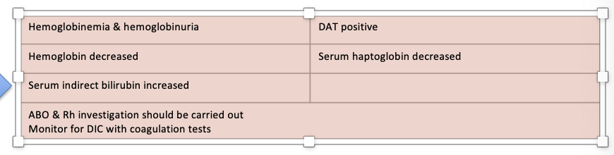
What are the lab findings and tasks of AHTR
126
When can delayed hemolytic transfusion reaction (DHTR) occur?
May occur days or weeks after the transfusion
127
What causes DHTR (extrinsic defect) after a subsequent transfusion?
The patient has been exposed to the antigen previously and the antibody generated from this initial exposure was below detectable levels. Then after subsequent exposure to the same antigen, the alloantibody has increased to levels where they are able to bind to transfused RBCs = extravascular hemolysis.
128
What is one sign that DHTR has occurred regarding hemoglobin levels?
Inadequate posttransfusion increase in hemoglobin
129
What is a DAT finding in DHTR?
Positive DAT IgG/complement
130
What morphologic evidence of hemolysis may be seen in DHTR?
polychromasia, spherocytes etc.
131
What biochemical evidence supports DHTR?
Increase in serum unconjugated bilirubin
132
How does hemolytic disease of the fetus and newborn (HDFN) occur?
IgG alloantibody produced by mom crosses the placenta into fetal circulation and bind to fetal cells.
133
What happens to sensitized fetal cells in HDFN?
Sensitized fetal cells are cleared by macrophages in fetal spleen and anemia develops.
134
How does the fetus compensate for anemia in HDFN?
To compensate, there is erythroid hyperplasia in fetal bone marrow and extramedullary erythropoiesis in fetal organs.
135
What can severe anemia in utero lead to?
If anemia is severe in utero, it can lead to generalized edema and death.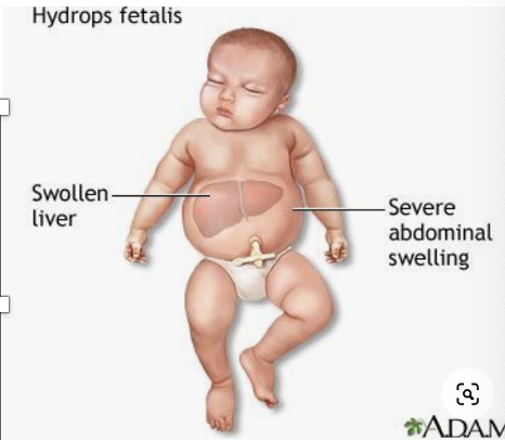
136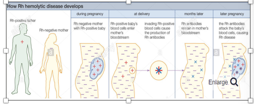
How does Rh hemolytic disease of the Fetus & Newborn (Rh HDFN) develops?
137
What are the Lab findings for Rh HDFN?
138
Why is ABO hemolytic disease of the fetus and newborn (ABO HDFN - extrinsic defect) usually milder than Rh HDFN?
Milder than Rh HDFN because A and B antigens are poorly developed on the newborn RBCs.
139
What reduces the maternal antibody effect in ABO HDFN?
Other cells and tissue express A and B antigens, which reduces the amount of maternal antibody directed toward fetal RBCs.
140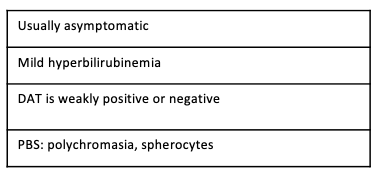
What are the laboratory findings of ABO HDFN? 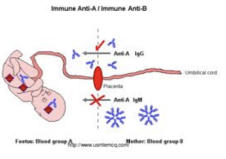
141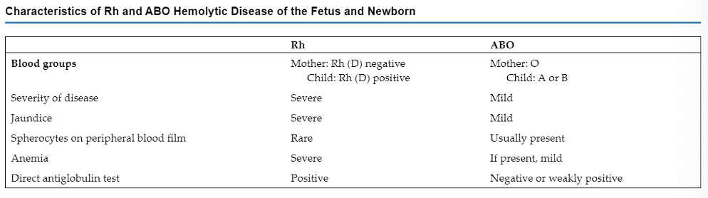
What are the characterisistics of Rh and ABO Hemolytic Disease of the Fetus and Newborn?
142
A group A Rh-negative mother gave birth to a group O Rh-positive baby. The baby is at risk for HDFN if:
A. This was the mother’s first pregnancy
B. The mother has IgG ABO antibodies
C. The mother was previously immunized to the D antigen
D. The mother received Rh immune globulin before delivery
A. This was the mother’s first pregnancy
B. The mother has IgG ABO antibodies
C. The mother was previously immunized to the D antigen
D. The mother received Rh immune globulin before delivery
C. The mother was previously immunized to the D antigen
143
What are Non-Immune Hemolytic Anemias (extrinsic defects)?
Conditions that cause physical or mechanical injury to the RBCs.
144
What are some causes of RBC injury leading to hemolysis?
This injury can be caused by abnormalities in the microvasculature or the heart and large blood vessels, infectious agents, chemicals, drugs, venoms or extensive burns.
145
What are MAHAs (Microangiopathic Hemolytic Anemias) characterized by?
MAHAs - a non-immune hemolytic anemia - are characterized by RBC fragmentation and thrombocytopenia.
146
How does RBC fragmentation occur in MAHAs?
The fragmentation occurs intravascularly by mechanical shearing of RBC membranes as they pass partially blocked microthrombi. The spleen clears the fragments.
147
What are the classical laboratory evidences of hemolytic anemia?
Classical evidence of hemolytic anemia:
↓ hemoglobin level
↑ reticulocyte count
↑ serum unconjugated bilirubin
↓ serum haptoglobin
↑ urine urobilinogen
↓ hemoglobin level
↑ reticulocyte count
↑ serum unconjugated bilirubin
↓ serum haptoglobin
↑ urine urobilinogen
148
What are the 3 main MAHAs?
1. TTP
2. HUS
3. DIC
2. HUS
3. DIC
149
What are the PBS findings in MAHAs?
PBS:
- Fragments
- Possible helmet cells & microspherocytes
- Polychromasia
- nRBCs
- Thrombocytopenia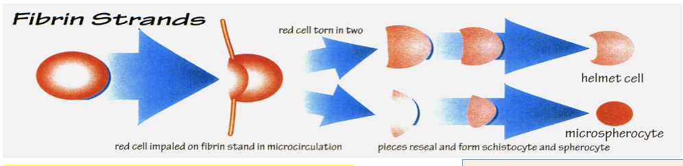
- Fragments
- Possible helmet cells & microspherocytes
- Polychromasia
- nRBCs
- Thrombocytopenia
150
What can severe fragmentation in MAHAs result in?
Hemoglobinemia, hemoglobinuria and marked decreases in serum haptoglobin.
151
What causes thrombotic thrombocytopenic purpura (TTP) - a non-immune hemolytic anemia?
TTP is caused by a deficiency of ADAMTS13 which results in ultralong VWF multimers that bind & activate platelets.
152
What does ADAMTS13 deficiency in TTP result in?
- Severe thrombocytopenia
- Ischemia particularly in the brain
- HA due to RBC rupture as they pass through microthrombi.
- Ischemia particularly in the brain
- HA due to RBC rupture as they pass through microthrombi.
153
What is the laboratory picture in Non-Immune Hemolytic Anemia?
- Severe thrombocytopenia
- ↑ WBC (immature granulocytes)
- ↓ hemoglobin
- Schistocytes
- Polychromasia/nRBCsHemoglobinuria
- ↑↑↑ serum LD
- ↑ serum unconjugated bilirubin
- ↓ serum haptoglobin
- Normal PT/PTT
- ↓↓↓ ADAMTS13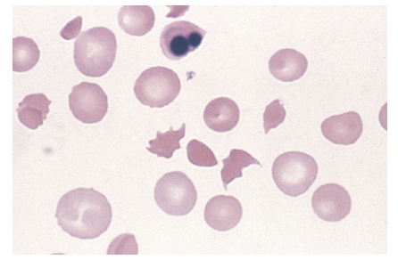
- ↑ WBC (immature granulocytes)
- ↓ hemoglobin
- Schistocytes
- Polychromasia/nRBCsHemoglobinuria
- ↑↑↑ serum LD
- ↑ serum unconjugated bilirubin
- ↓ serum haptoglobin
- Normal PT/PTT
- ↓↓↓ ADAMTS13
154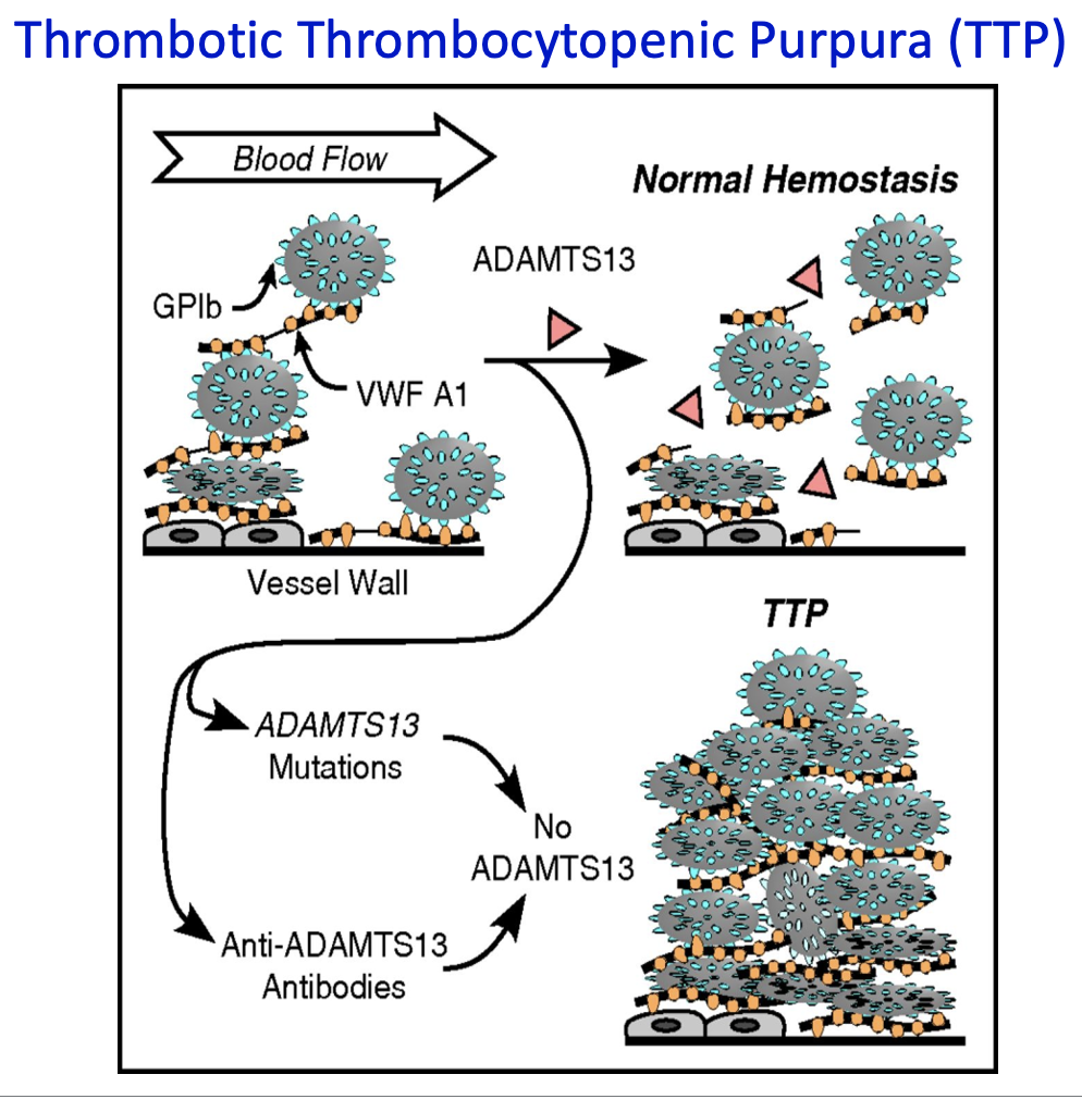
Normal Hemostasis:
1. Initial Platelet Adhesion
- When blood vessels are injured, VWF A1 domain binds to exposed subendothelial tissue.
- Platelets attach to VWF via the GPIb receptor.
2. Role of ADAMTS13
- ADAMTS13 is a protease that cleaves ultra-large VWF multimers.
- This prevents excessive platelet adhesion and aggregation.
3. Result
- A controlled clot forms at the injury site.
- Normal hemostasis is maintained without blocking blood flow.
Pathologic State:
1. ADAMTS13 Deficiency
- Can result from:
- ADAMTS13 gene mutations → congenital TTP
- Anti-ADAMTS13 autoantibodies → acquired TTP
2. No ADAMTS13 Activity
- Ultra-large VWF multimers remain uncleaved.
- Excessive platelet adhesion occurs.
3. Result
- Platelets form widespread microthrombi in small vessels.
- This leads to thrombocytopenia, hemolytic anemia, and organ damage, the hallmark of TTP.
1. Initial Platelet Adhesion
- When blood vessels are injured, VWF A1 domain binds to exposed subendothelial tissue.
- Platelets attach to VWF via the GPIb receptor.
2. Role of ADAMTS13
- ADAMTS13 is a protease that cleaves ultra-large VWF multimers.
- This prevents excessive platelet adhesion and aggregation.
3. Result
- A controlled clot forms at the injury site.
- Normal hemostasis is maintained without blocking blood flow.
Pathologic State:
1. ADAMTS13 Deficiency
- Can result from:
- ADAMTS13 gene mutations → congenital TTP
- Anti-ADAMTS13 autoantibodies → acquired TTP
2. No ADAMTS13 Activity
- Ultra-large VWF multimers remain uncleaved.
- Excessive platelet adhesion occurs.
3. Result
- Platelets form widespread microthrombi in small vessels.
- This leads to thrombocytopenia, hemolytic anemia, and organ damage, the hallmark of TTP.
155
What causes hemolytic uremia syndrome (HUS) - a non-immune hemolytic anemia?
HUS is caused by bacteria that produce Shiga toxin.
156
What is the most common cause of HUS?
The most common cause of disease is ingestion of improperly cooked meat especially in children.
157
What is the toxin’s affinity in HUS and what does it lead to?
The toxin has an affinity for endothelial cells particularly in the glomerulus leading to eventual apoptosis.
158
What happens due to endothelial cell damage in HUS?
The endothelial cell damage: activation of platelets, formation of platelet-fibrin thombi, blockages in the microvasculature of the glomeruli, acute renal failure.
159
What is the treatment for HUS?
There is no specific treatment for HUS — patients are provided supportive care as needed.
160
What are the hematologic findings of HUS?
- Decreased hemoglobin
- Decreased platelets
- Increased reticulocyte count
- Decreased platelets
- Increased reticulocyte count
161
What are the PBS findings of HUS?
- Schistocytes
- Polychromasia
- Nucleated red blood cells (severe cases)
- Polychromasia
- Nucleated red blood cells (severe cases)
162
What are the biochemical findings of HUS?
- Markedly increased lactate dehydrogenase activity
- Increased serum total and indirect bilirubin
- Decreased serum haptoglobin level
- Hemoglobinemia
- Hemoglobinuria
- Proteinuria, hematuria, casts
- Increased serum total and indirect bilirubin
- Decreased serum haptoglobin level
- Hemoglobinemia
- Hemoglobinuria
- Proteinuria, hematuria, casts
163
What is the laboratory picture of MAHA?
- ↑↑↑ serum LD
- ↑↑ serum unconjugated bilirubin
- ↓ serum haptoglobin
- Hemoglobinemia
- Hemoglobinuria
- ↓ hemoglobin
- Fragments
- Polychromasia
- Mild to moderate thrombocytopenia
- ↑ serum creatinine
- Proteinuria
- Hematuria
- Casts (hyaline, granular, RBC)
- Normal PT/PTT
- ↑↑ serum unconjugated bilirubin
- ↓ serum haptoglobin
- Hemoglobinemia
- Hemoglobinuria
- ↓ hemoglobin
- Fragments
- Polychromasia
- Mild to moderate thrombocytopenia
- ↑ serum creatinine
- Proteinuria
- Hematuria
- Casts (hyaline, granular, RBC)
- Normal PT/PTT
164
A 36-year-old woman was brought to the emergency department by her husband because she had experienced a seizure. He reported that she had been well until that morning, when she complained of a sudden headache and malaise. She was not taking any medications and had no history of previous surgery or pregnancy. Laboratory studies showed a WBC count of 15 × 109/L, a hemoglobin level of 7.8 g/dL, a platelet count of 18 × 109/L, and schistocytes and helmet cells on the peripheral blood film. Chemistry test results included markedly elevated serum lactate dehydrogenase activity and a slight increase in the level of total and indirect serum bilirubin. The urinalysis results were positive for protein and blood, but there were no RBCs in the urine sediment. Prothrombin time and partial thromboplastin time were within the reference interval. When the entire clinical and laboratory picture is considered, which of the following is the most likely diagnosis?
A. HUS
B. HELLP syndrome
C. TTP
D. Exercise-induced hemoglobinuria
A. HUS
B. HELLP syndrome
C. TTP
D. Exercise-induced hemoglobinuria
C. TTP
165
What mechanical factor can cause hemolysis?
Prosthetic cardiac valves (Traumatic Cardiac Hemolytic Anemia)
166
How severe is the hemolysis caused by Traumatic Cardiac Hemolytic Anemia?
This hemolysis is usually mild and compensated by the bone marrow however, it is possible to see more severe cases.
167
What is the lab picture of Traumatic Cardiac Hemolytic Anemia?
Fragments are characteristic feature found on the PBS due to mechanical fragmentation of RBCs
- ↑ reticulocytes
- Normal platelet count
- ↑ serum unconjugated bilirubin
- ↑ plasma hemoglobin
- ↓ serum haptoglobin
- Hemoglobinuria
- Hemosiderinuria & ↓ serum ferritin (with chronic hemoglobinuria)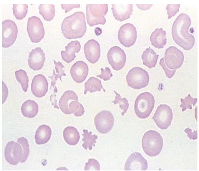
- ↑ reticulocytes
- Normal platelet count
- ↑ serum unconjugated bilirubin
- ↑ plasma hemoglobin
- ↓ serum haptoglobin
- Hemoglobinuria
- Hemosiderinuria & ↓ serum ferritin (with chronic hemoglobinuria)
168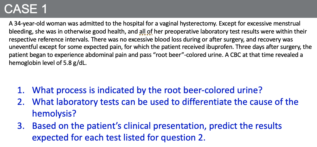
1. What process is indicated by the root beer–colored urine?
- Hemoglobinuria due to acute intravascular hemolysis.
- Dark, “cola” or “root beer” urine = free hemoglobin (not intact RBCs) being excreted.
2. What laboratory tests can be used to differentiate the cause of the hemolysis?
Key tests to distinguish intravascular vs extravascular hemolysis:
- Serum haptoglobin
- LDH
- Indirect bilirubin
- Urine hemoglobin
- Urine hemosiderin (Prussian blue stain of sediment)
- Peripheral blood smear (schistocytes vs spherocytes)
3. Based on the patient’s clinical presentation, expected results: This case is acute intravascular hemolysis (likely drug-induced hemolysis from ibuprofen or another oxidant stress):
- Serum haptoglobin: ↓ markedly (binds free Hgb, gets depleted)
- LDH: ↑↑ (released from lysed RBCs)
- Indirect bilirubin: ↑ (from heme breakdown)
- Urine hemoglobin: Positive
- Urine hemosiderin: May be positive later (after tubular uptake and storage)
- Peripheral smear: Schistocytes (fragmented RBCs)
- Hemoglobinuria due to acute intravascular hemolysis.
- Dark, “cola” or “root beer” urine = free hemoglobin (not intact RBCs) being excreted.
2. What laboratory tests can be used to differentiate the cause of the hemolysis?
Key tests to distinguish intravascular vs extravascular hemolysis:
- Serum haptoglobin
- LDH
- Indirect bilirubin
- Urine hemoglobin
- Urine hemosiderin (Prussian blue stain of sediment)
- Peripheral blood smear (schistocytes vs spherocytes)
3. Based on the patient’s clinical presentation, expected results: This case is acute intravascular hemolysis (likely drug-induced hemolysis from ibuprofen or another oxidant stress):
- Serum haptoglobin: ↓ markedly (binds free Hgb, gets depleted)
- LDH: ↑↑ (released from lysed RBCs)
- Indirect bilirubin: ↑ (from heme breakdown)
- Urine hemoglobin: Positive
- Urine hemosiderin: May be positive later (after tubular uptake and storage)
- Peripheral smear: Schistocytes (fragmented RBCs)
169
170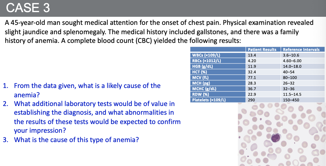
Case Summary
- 45-year-old man with chest pain, jaundice, and splenomegaly
History: gallstones, family history of anemia
CBC:
- WBC: 13.4 (high)
- RBC: 4.20 (low)
- Hgb 11.9 g/dL (low)
- Hct 32.4% (low)
- MCV 77.1 fL (low/microcytic)
- 36.7 g/dL (elevated)
- RDW 22.9% (very high)
- platelets: 290 (normal)
1. From the data given, what is a likely cause of the anemia?
Key clues:
- Family history → hereditary condition
- Jaundice + gallstones → chronic hemolysis (bilirubin stones)
- Splenomegaly → extravascular RBC destruction
- Elevated MCHC and high RDW → strongly suggest spherocytes
Likely cause = Hereditary Spherocytosis (HS)
2. What additional laboratory tests would be of value, and expected abnormalities?
To confirm HS, useful tests include:
- Peripheral smear → spherocytes present
- Reticulocyte count → elevated (marrow response)
- Indirect bilirubin & LDH → elevated (from RBC breakdown)
- Serum haptoglobin → decreased (consumed by binding free hemoglobin)
- Osmotic fragility test or EMA (Eosin-5-maleimide) binding test → increased fragility
- DAT (Coombs test) → negative (rules out autoimmune hemolytic anemia)
3. What is the cause of this type of anemia?
- Genetic defect in RBC membrane proteins → most commonly spectrin, ankyrin, band 3, or protein 4.2.
- Defective cytoskeleton → loss of RBC surface area → spherical RBCs→ reduced deformability → splenic sequestration and destruction (extravascular hemolysis).
- 45-year-old man with chest pain, jaundice, and splenomegaly
History: gallstones, family history of anemia
CBC:
- WBC: 13.4 (high)
- RBC: 4.20 (low)
- Hgb 11.9 g/dL (low)
- Hct 32.4% (low)
- MCV 77.1 fL (low/microcytic)
- 36.7 g/dL (elevated)
- RDW 22.9% (very high)
- platelets: 290 (normal)
1. From the data given, what is a likely cause of the anemia?
Key clues:
- Family history → hereditary condition
- Jaundice + gallstones → chronic hemolysis (bilirubin stones)
- Splenomegaly → extravascular RBC destruction
- Elevated MCHC and high RDW → strongly suggest spherocytes
Likely cause = Hereditary Spherocytosis (HS)
2. What additional laboratory tests would be of value, and expected abnormalities?
To confirm HS, useful tests include:
- Peripheral smear → spherocytes present
- Reticulocyte count → elevated (marrow response)
- Indirect bilirubin & LDH → elevated (from RBC breakdown)
- Serum haptoglobin → decreased (consumed by binding free hemoglobin)
- Osmotic fragility test or EMA (Eosin-5-maleimide) binding test → increased fragility
- DAT (Coombs test) → negative (rules out autoimmune hemolytic anemia)
3. What is the cause of this type of anemia?
- Genetic defect in RBC membrane proteins → most commonly spectrin, ankyrin, band 3, or protein 4.2.
- Defective cytoskeleton → loss of RBC surface area → spherical RBCs→ reduced deformability → splenic sequestration and destruction (extravascular hemolysis).