Obstetrics
Study these flashcardsFree · spaced repetition · iPhone, Android, Mac & web
All cards
1
LMP
last menstrual period
2
Gravidity *
number of times a woman has been pregnant, regardless of outcome
3
Parity *
number of times a woman has had pregnancies that have carried over the threshold of viability [-24 weeks]
4
Dysmenorrhoea
painful periods
5
Dyspareunia
pain during sex, can be superficial or deep
6
blood supply of the uterus / ovaries
lateral = ovarian arteries from the abdominal aorta
medial = ascendary branch of uterine artery from the internal iliac artery
medial = ascendary branch of uterine artery from the internal iliac artery
7
vulva
global term of all the structures that make up the external female genitalia
8
female sex hormones system
hypothalamus releases GnRH to the anterior pituitary gland which releases LH and FSH. These two stimulate the development of the folicles in the ovaries, the theca granulosa. This releases oestrogen which inhibits GnRH release.
9
Oestrogen role
promotes female secondary sexual characteristics:
breast tissue development, growth / development of female sex organs at puberty, blood vessel development in the uterus, development of the endometrium
breast tissue development, growth / development of female sex organs at puberty, blood vessel development in the uterus, development of the endometrium
10
Progesterone role
provided by the corpus luteum after ovulation. It thickens / maintains endometrium, thickens cervical mucus and increases body temperature
11
Trimesters
12 weeks or less = 1st trimester
13 weeks to 27 weeks = 2nd trimester
28 weeks onwards = 3rd trimester
13 weeks to 27 weeks = 2nd trimester
28 weeks onwards = 3rd trimester
12
Antenatal care routine
booking usually around less than 11 weeks. First scan is usally in first trimester, anomaly scan is offered routinely in 2nd trimester. Usually around 10 antenatal visits for 1st pregnancy and 7 for subsequent pregnanies
13
booking visit
usually identifying risk factors, general information like diet, alcohol, smoking, folic acid, BP, urine dipstick, BMI
Also can do bloods for FBC, group save, rhesus status, haemoglobinopathies, hepatitis, HIV test offered.
Downs syndrome screening can also be around this time
Also can do bloods for FBC, group save, rhesus status, haemoglobinopathies, hepatitis, HIV test offered.
Downs syndrome screening can also be around this time
14
Downs syndrome scan: what it entails
offered but no compulsory. it is an invasive procedure so carries the risk of miscarriage
Combined test is the standard which involves an US [nuchal translucency meansurement] and maternal blood tests
offer tests include the triple test [b-hCG, alpha fetoprotein, serum oestriol] and the quadruple test which involves inhibin-A added on
if the score of the foetus having Downs is more than 1 in 150, then aminocentesis or chorionic villous sampling is offered.
Non-invasive prenatal testing is being offered more and more now
Combined test is the standard which involves an US [nuchal translucency meansurement] and maternal blood tests
offer tests include the triple test [b-hCG, alpha fetoprotein, serum oestriol] and the quadruple test which involves inhibin-A added on
if the score of the foetus having Downs is more than 1 in 150, then aminocentesis or chorionic villous sampling is offered.
Non-invasive prenatal testing is being offered more and more now
15
foetal alcohol syndrome: what is it, signs, complications
baby shows signs of alcohol withdrawal at birth [hypotonic, irritation, tremors].
Can lead to learning difficulties, microcephaly, growth retardation, cardiac malformations, short palebral fissures, epicanthic folds
Can lead to learning difficulties, microcephaly, growth retardation, cardiac malformations, short palebral fissures, epicanthic folds
16
Vaccines offered to all pregnant women
whooping cough from 16 weeks gestation, influenza when available
live vaccines like MMR are avoided
live vaccines like MMR are avoided
17
Thyroid changes in pregnancy: management
increase in thyroxine binding globulin levels which leads to increase in total thyroxine.
Hypothyroidism untreated can lead to miscarriage, anaemia, pre-eclampsia, SGA. Levothyroxine is given
Hyperthyroidism untreated can lead to premature labour, foetal loss, maternal HF. Treat by propylthiouracil in 1st trimester, carbimazole in 2-3rd trimester. Aim is for free T4 levels to be kept in upper third of normal ranges
Hypothyroidism untreated can lead to miscarriage, anaemia, pre-eclampsia, SGA. Levothyroxine is given
Hyperthyroidism untreated can lead to premature labour, foetal loss, maternal HF. Treat by propylthiouracil in 1st trimester, carbimazole in 2-3rd trimester. Aim is for free T4 levels to be kept in upper third of normal ranges
18
Epilepsy and pregnancy
ideally, epilepsy should be controlled with a single anti-epileptic before becoming pregnant. Pregnancy can make seizure control worse. Avoid Sodium valproate [NTD risk] and phenytoin. 5mg folic acid daily to avoid NTD. Safer drugs are levetiracetam, lametrigine, carbamezepine
19
RA and pregnancy
ideally, arthritis is well controlled for more than 3 months before pregnancy. usually sx of RA will improve during pregnancy and flare up after delivery.
hydroxychloroquine and sulfasalzine are safe. corticosteroids given for flare ups. Avoid methotrexate and advise to stop 6 months before conception.
hydroxychloroquine and sulfasalzine are safe. corticosteroids given for flare ups. Avoid methotrexate and advise to stop 6 months before conception.
20
NSAIDs and pregnancy
NSAIDs block prostaglandins which are needed to maintain ductus arteriosus and stimulate uterine contractions. Uusally avoided unless very necessary and especially avoided in 3rd trimester as they can cause premature closure of ductus arteriosus and delay labour
21
beta blockers and pregnancy
used for HPTN, cardiac problems, migraine. Labetalol is the ideal one used to treat pre-eclampsia
can case FGR, hypoglycaemia / bradycardia in neonates
can case FGR, hypoglycaemia / bradycardia in neonates
22
ACEi / ARB and pregnancy
these can cross placenta and affect foetus kidneys, leads to less urine production. This can lead to miscarriage / foetal death, oligohydramnios, renal failure / hypotension in neonates, hypocalvaria [incomplete small bone formation]
23
opiates and pregnancy
can cause neonatal abstinence syndrome which leads to withdrawal sx in neonates after birth. Presents in 3-72hrs after birth with irritability, tachypnoea, high temperature and poor feeding
24
warfarin and pregnancy
crosses placenta and is teratogenic in pregnancy. can cause foetal loss, congenital malformations and leading during pregnancy
25
lithium and pregnancy
mood stabiliser for bipolar px, mania or recurrent depression. avoided in pregnant women unless all other options have failed, and needs close monitoring of lithium levels every 4 weeks until 36 weeks where it is weekly. Avoided in breastfeeding and 1st trimester due to congential cardiac problems risk
26
SSRIs and pregnancy
these can cross the placenta. need to balance risks vs benefits. congenital heart defects if used in 1st trimester and risk of persistent pulmonary HPTN in neonates if used in 3rd trimester
27
Rhesus and pregnancy
if mother is rhesus D +ve = no Tx
if mother is rhesus D -ve, consider baby is +ve which can lead to antibodies formation. The 1st pregnancy will be fine but subsequent pregnancies will lead to haemolytic disease of the newborn
Needs early prophylaxis via anti-D injections usually at 28 weeks Gest and at birth if baby is +ve.
if mother is rhesus D -ve, consider baby is +ve which can lead to antibodies formation. The 1st pregnancy will be fine but subsequent pregnancies will lead to haemolytic disease of the newborn
Needs early prophylaxis via anti-D injections usually at 28 weeks Gest and at birth if baby is +ve.
28
Kleihauer test
checks how much foetal blood has passed into mothers during a sensitisation event [antepartum haemorrhage, amniocentesis procedure, abdo trauma]. Assesses if further doses of anti-D are needed.
29
Small gestational age *
defined as a foetus below 10th centile for their gestational age. Severe is below 3rd centile
30
SGA measurement
based on US using estimated foetal weight and foetal abdominal circumference. Customised growth charts are used to assess the size of the foetus which are based on the mother’s height, weight, parity, ethnicity
31
SGA causes
can be constitutionally small due to mother OR be due to FGR
32
foetal growth restriction causes
Placenta mediated growth restriction: idiopathic, pre-eclampsia, maternal smoking / alcohol, malnutrition, infection
Non-placenta mediated growth restriction: genetic, structural, errors of metabolism
Non-placenta mediated growth restriction: genetic, structural, errors of metabolism
33
other signs of FGR
oligohydramnios, abnormal doppler, reduced foetal movements, abnormal CTGs
34
FGR complications: short and long term
Short term: foetal death / stillbirth, birth asphyxia, neonatal hypothermia / hypoglycaemia
Longer term: CVS disease, T2DM, obesity, mood / behaviour problems
Longer term: CVS disease, T2DM, obesity, mood / behaviour problems
35
RFs for SGA
previous SGA, obesity, smoking, diabetes, HPTN, older than 35
multiple pregnancies, low PAPPA, antepartum haemorrhage, antiphospholipid syndrome
multiple pregnancies, low PAPPA, antepartum haemorrhage, antiphospholipid syndrome
36
monitoring SGA
if below 10th centile, woman is booked for serial growth scans with umbilical artery doppler scan. They are also booked for this if they have 3 or more minor RFs, 1 or more major RFs OR issues measuring symphysis fundal height [obese / large fibroids]
37
large gestational age definition
more than 4.8kg at birth or higher than 90th centil during pregnancy
38
LGA risks to mother
shoulder dystocia, failure to progress, perineal tears, Caesarean or instrumental delivery, PPH, uterine rupture
39
LGA risks to baby
birth injury [foetal distress, hypoxia], neonatal hypoglycaemia, obesity in childhood, T2DM in adulthood
40
Monozygotic vs dizygotic
mono means identical twins, dizygotic means non-identical twins due to two different ovas being fertilised at the same time
41
mono vs diamniotic
just how many amniotic sacs there are
42
mono vs dichorionic
if they share one placenta or two separate placentas
43
lamda sign
this is seen in dichorionic diamniotic twins. The inter-twin membrane will be between the twins in a lamda shape 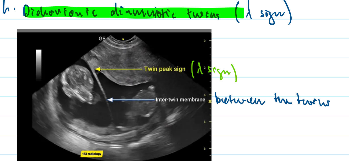
44
T sign
seen in monochorionic diamniotic twins. will see an abrupt membrane across the shared placenta 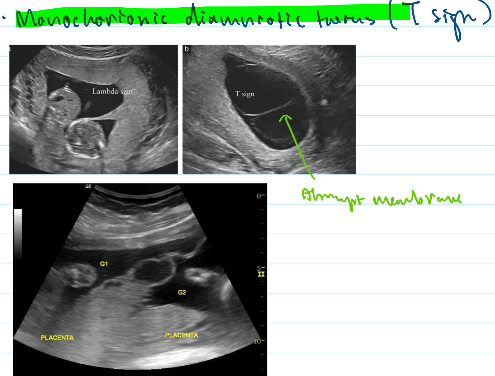
45
twin-twin transfusion syndrome
occurs when foetuses share a placenta. The connection of blood supples between the two foetuses may lead to one foetus receiving the majority of blood from the placenta whilst the other one is starved. The recipient foetus can become fluid overloaded, leading to HF and polyhydramnios. Donor foetus can have FGR, anaemia, olgohydramnios.
You will see a difference in the size of foetuses and px need to referred to tertiary specialist foetal medicine centre
You will see a difference in the size of foetuses and px need to referred to tertiary specialist foetal medicine centre
46
twin anaemia polycythaemia sequence
essentially twin-twin transfusion syndrome but less acute. one foetus will be anaemic, the other is polycythaemic
47
Antenatal care for multiple pregnancies
additional monitoring for anaemia with FBC at booking clinic, 20 and 28 weeks.
Additional US needed as well. 2 weekly scans from 16 weeks onwards for monochorionic. 4 weekly scans from 20 weeks for dichorionic
Additional US needed as well. 2 weekly scans from 16 weeks onwards for monochorionic. 4 weekly scans from 20 weeks for dichorionic
48
multiple pregnancies planned delivery
offered between:
32-33+6 for uncomplicated monochorionic monoamniotic
36-36+6 for uncomplicated monochorionic diamniotic twins
37-37+6 for uncomplicated dichorionic diamniotic twins
Before 35+6 for triplets
waiting beyond these dates is associated with increased foetal death risk
32-33+6 for uncomplicated monochorionic monoamniotic
36-36+6 for uncomplicated monochorionic diamniotic twins
37-37+6 for uncomplicated dichorionic diamniotic twins
Before 35+6 for triplets
waiting beyond these dates is associated with increased foetal death risk
49
multiple pregnancies and delivery
monoamniotic = elective C section at 32-33+6
Diamniotic can try vaginal delivery IF first baby is cephalic. C section otherwise
Diamniotic can try vaginal delivery IF first baby is cephalic. C section otherwise
50
UTI and pregnancy: causes and management
usually caused by e.coli, others include klebsiella, enterococcus, pseudomonas aeruginosa
Management: 7 DAYS ABX
1st line: cefalexin / amoxicillin
2nd line: nitrofurantoin [avoid near term]
3rd line: Fosfomycin [used if allergic to 1st line or resistant]
4th line: trimethoprim [avoided in 1st trimester]
5th line: co-amoxiclav [can cause necrotising enterocolitis]
Management: 7 DAYS ABX
1st line: cefalexin / amoxicillin
2nd line: nitrofurantoin [avoid near term]
3rd line: Fosfomycin [used if allergic to 1st line or resistant]
4th line: trimethoprim [avoided in 1st trimester]
5th line: co-amoxiclav [can cause necrotising enterocolitis]
51
Rubella and pregnancy: cause, risk, features, investigations, management
viral infection caused by togavirus. risk is highest in first 8-10 weeks. features in neonate include congenital deafness, cataracts, heart disease and learning disabilities. IgM antibodies will be raised in women recently exposed to virus. MMR vaccination is offered UNLESS during pregnancy where it is offered post-natally
52
chicken pox and pregnancy: cause, features, investigations, management
caused by VZV. it is more dangerous in pregnancy because it can lead to fetal varicella syndrome and more severe cases in mother like varicella encephalitis. neonatal affects include FGR, microcephaly, learning disabilities, skin changes in specific dermatomes. Test for IgG in mother.
VZIG can be given as soon as possible if before 20 weeks gestation and not immune. If after 20 weeks, offer oral aciclovir days 7-14 after explosure
VZIG can be given as soon as possible if before 20 weeks gestation and not immune. If after 20 weeks, offer oral aciclovir days 7-14 after explosure
53
Listerosis and pregnancy: cause, features
transmitted via unpasteurised dairy products, processed meats. Advise women to avoid eating these. Can lead to increased risk of miscarriage, foetal death, severe neonatal infection
54
Congenital cytomegalovirus and pregnancy: cause features
spreads via infected urine, occurs due to CMV infection in mother during pregnancy. Can lead to FGR, microcephaly, learning disability, seizures, hearing / vision loss
55
Congenital toxoplasmosis and pregnancy: cause, spread, features
cause is toxoplasma gondii parasite. spread via faeces of cats. Features include intracranial calcification, hydrocephalus, chorioretinitis
56
parovirus B19 and pregnancy: features, complications, investigations, management
usually affects children and is self-limiting. Features include viral sx, “slapped cheeks” rash, reticular erythematous rash affecting trunk and limbs. Risk of miscarriage, severe fetal anaemia, Hydrops fetalis [fetal HF]. Investigate via IgM [acute] or IgG [chronic], supportive management
57
Hydrops fetalis
foetal anaemia caused by parovirus infection of erythroid progenitor cells in foetal bone marrow. Causes faulty RBCs to be formed which results in heart failure.
Will need intrauterine blood transufions and can lead to maternal pre-eclampsia-like syndrome [triad of hydrops fetalis, placental oedema and oedema in mother]
Will need intrauterine blood transufions and can lead to maternal pre-eclampsia-like syndrome [triad of hydrops fetalis, placental oedema and oedema in mother]
58
Anaemia & pregnancy: screening when
at booking clinic and 28 weeks gestation
59
Why are pregnany women more prone to anaemia?
Plasma volume increases during pregnancy which results in a reduction in haemoglobin concentration. Important to optimise the Tx so that the woman has reasonable reserves in case there is significant blood loss during delivery.
60
what investigations are offered for anaemia in pregnancy?
Haemoglobin concentration, MCV looked at, haemoglobinopathy screening offered as well for thalassaemia and sickle cell disease
61
Management for anaemia in pregnant women
Cut off is below 110 Hb. Give Ferrous sulphate 200mg 3 times a day. Intramuscular hydroxycobalamin injection for low B12 levels
62
VTE and pregnancy RFs [too many]
smoking, parity of 3 or above, age over 35, BMI over 30, reduced mobility, multiple pregnancy, pre-eclampsia, immobility, FHx, thrombophilia, IVF pregnancy
63
Why are pregnant women at higher risk of VTE?
increase in factors 7,8,10, fibrinogen
decrease in protein S
Uterus presses on IVC leading to venous stasis
decrease in protein S
Uterus presses on IVC leading to venous stasis
64
When to start VTE prophylaxis in pregnant women?
28 weeks if there are 3 RFs
1st trimester if there are 4 or more RFs
there are many other situations as well like if they have had a previous VTE, hosital admission and surgical procedures
1st trimester if there are 4 or more RFs
there are many other situations as well like if they have had a previous VTE, hosital admission and surgical procedures
65
What VTE prophylaxis is given for pregnant women? *
Depends on policy and protocol but usually a LMWH unless contraindicated and continued until 6 weeks postnatally. Prophylaxis is temporarily stopped when women goes into labour and is started immediately after delivery [except in PPH, spina anaesthesia and epidurals].
If CI to LMWH = use mechanical prophylaxis like intermittent pneumatic compressions or anti-embolic compression stockings
If CI to LMWH = use mechanical prophylaxis like intermittent pneumatic compressions or anti-embolic compression stockings
66
Gestational diabetes definition and complications
diabetes triggered by pregnancy due to a decrease in insulin sensitivity during pregnancy, resolves after birth.
Usually leads to macrosomia which increases risk of shoulder dystocia OR T2DM longer term
Usually leads to macrosomia which increases risk of shoulder dystocia OR T2DM longer term
67
RFs of gestational diabetes
previous gestational diabetes, BMI over 30, ethnicity, FHx of diabetes, previous macrosomic baby
68
Gestational diabetes investigations and diagnostic criteria
OGTT for all pregnant women with RFs at 24-28 weeks. Diagnosed as higher than 5.6 fasting glucose and 7.8 2hr post glucose levels
69
Management of gestational diabetes
Education about condition, how to monitor and track blood sugar levels.
If fasting glucose is below 7, diet and exercise escalating to metformin and insulin
If fasting is above 7, insulin straight away
Glibenclamide is used if patients refuse insulin or are unable to tolerate metformin
If fasting glucose is below 7, diet and exercise escalating to metformin and insulin
If fasting is above 7, insulin straight away
Glibenclamide is used if patients refuse insulin or are unable to tolerate metformin
70
Obstetric cholestasis what is it
the reduction in outflow of bile acids from the liver with the condition resolving after the delivery of the baby. Usually develops in the 3rd trimester
71
Obstetric cholestasis clinical features
pruritus of the hands / soles of feet, fatigue, dark urine, pale / greasy stools, jaundice. NO RASH INVOLVED
72
Managment for obstetric cholestasis
monitor LFTs during and after pregnancy. Ursodeoxycholic acid is the primary Tx. Emollients can be used for itching and water soluble vitamin K for prothrombin time
73
Acute fatty liver of pregnancy pathophysiology
usually occurs in 3rd trimester.
Long-chain-3-hydroxyacyl-CoA dehydrogenase [LCHAD] deficiency [autosomal recessive] is the most common cause. LCHAD is a important enzyme to break down fatty acids for fuel. A deficiency leads to a build up of fatty acids within the liver leading to acute hepatitis
Long-chain-3-hydroxyacyl-CoA dehydrogenase [LCHAD] deficiency [autosomal recessive] is the most common cause. LCHAD is a important enzyme to break down fatty acids for fuel. A deficiency leads to a build up of fatty acids within the liver leading to acute hepatitis
74
Acute fatty liver of pregnancy management
obstetric emergency so it needs prompt admission and delivery of baby. most px will recover after delivery. There is a chance of acute liver failure which can lead to liver transplant consideration
75
Placenta praevia what is it
when the placenta is attached in the lower portion of the uterus, lower than the presenting part of the uterus which can lead to blocking the internal cervical OS. One of the causes of antepartum haemorrhage
76
Placenta praevia RFs
previous C-sections [embryo more likely to embed into scar], previous placenta praevia, older maternal age, maternal smoking, structural uterine abnormalities, assissted reproduction
77
Placenta praevia clinical features
usually ASx, may present with painless vaginal bleeding in pregnancy, no pain, uterus is not tender, foetal heart normal
78
placenta praevia investigations
20 week anomaly scan used to assess the position of the placenta and diagnose placenta praevia.
Do not do a digital vaginal exam before a transvaginal US as it may provoke a severe haemorrhage
Do not do a digital vaginal exam before a transvaginal US as it may provoke a severe haemorrhage
79
Placenta praevia management
if diagnosed before 20 weeks, repeat scan at 32 weeks. If present again, repeat scan at 36 weeks.
Plan delivery at 36-37 weeks. C-section is required with different incisions maybe being done.
Plan delivery at 36-37 weeks. C-section is required with different incisions maybe being done.
80
Vasa praevia: pathophysiology
When the foetal blood vessels are placed over the internal cervical OS leading to them being prone to bleeding when membranes are ruptured. This can be caused by Velamentous umbilical cord [where the umbilical cord inserts into the chorioamniotic membranes, leaving the foetal vessels unprotected] OR an Accessory lobe of the placenta
81
Vasa praevia RFs
low lying placenta, IVF pregnancy, multiple pregnancy
82
Vasa Praevia clinical features
diagnosed via US but this isnt reliable. Uusally presents with antepartum haemorrhage around 2nd / 3rd trimester with fetal bradycardia being seen.
Vaginal examination during labour may show pulsating fetal vessels through dilated cervix.
Vaginal examination during labour may show pulsating fetal vessels through dilated cervix.
83
Vasa praevia management
for ASx women, elective C-section planned for 34-36 weeks with corticosteroids from 32 weeks to mature the fetal lungs
antepartum haemorrhage = emergency c sections
antepartum haemorrhage = emergency c sections
84
Placental abruption what is it
when the placenta separates from the wall of the uterus during pregnancy. Bleeding can occur in this space which leads to antepartum haemorrhage
85
Placental abruption RFs
ABRUPTION: abruption previously, blood pressure, rupture membranes [premature / prolonged], uterine injury, polyhydramnios, twins, infection, older age, narcotic use [cocaine]
86
Placental abruption presentation
sudden onset severe abdominal pain that is continuous. vaginal bleeding and shock sx will be seen. Fetal distress seen on CTG.
Woody abdomen on palaption
Woody abdomen on palaption
87
Placental abruption management
obstetric emergency. ABCDE management, if fetus is alive and in distress = emergency c section.
If there is no distress, when monitor closely and if past 36 weeks, consider a vaginal birth
If there is no distress, when monitor closely and if past 36 weeks, consider a vaginal birth
88
Placenta accreta what is it
when the placenta implants deeper than the endometrium, making it difficult to separate the placenta after delivery of the baby.
layers: endo then myometrium and then perimetrium
layers: endo then myometrium and then perimetrium
89
placenta increta *
attaches deeply into myometrium
90
placenta percreta *
invades past perimetrium, potentially into other organs likebladder
91
placenta accreta RFs *
previous incidence, previous endometrial curettage procedures, previous c section, multigravida, increased maternal age, placenta praevia
92
placenta accreta clinical features *
very ambiguous sx, diagnosed on antenatal US scans particularly in women with previous incidence or C-section. It is a cause of PPH and MRI scans can help aid assessment of depth / width of invasion
93
placenta accreta management
specialist MDT approach. options include hysterectomy with placenta still in uterus [recommended]. Uterus preserving surgery with resection is also an option or expectant management [leaving placenta to be resorbed over time] which has the risk of infection
94
Pre-eclampsia definition
new onset of BP of 140 / 90 or higher after 20 weeks gestation AND 1 or more of:
proteinuria,
other organ involvement [renal insufficiency, neurological, haematological]
proteinuria,
other organ involvement [renal insufficiency, neurological, haematological]
95
Chronic HPTN definition
High BP that exists before 20 weeks gestation and is long-standing. Therefore not caused by dysfunction in placenta and so is no classed as pre-eclampsia
96
Pregnancy induced HPTN definition *
HPTN occuring after 20 weeks gestation without proteinuria
97
Eclampsia definition *
pre-eclampsia but with seizures
98
pre-eclampsia pathophysiology *
Lacunae [pools of blood] are formed from spinal arteries when they break down which reduces the vascular resistance of the maternal blood flow from the uterine arteries to the veins. If the formation of lacunae is inadequate, pre-eclampsia is formed due to the high vascular resistance in the spiral arteries and poor perfusion of the placenta.
99
Pre-eclampsia clinical features *
headache
visual disturbance / papilloedema
HPTN + proteinuria
RUQ / epigastric pain
hyperreflexia
low platelet / abnormal LFTs
HELLP syndrome
visual disturbance / papilloedema
HPTN + proteinuria
RUQ / epigastric pain
hyperreflexia
low platelet / abnormal LFTs
HELLP syndrome
100
Complications of pre-eclampsia *
can lead to eclampsia, foetal complications, haemorrhage, cardiac failure
101
Management of pre-eclampsia *
Aspirin 75-150mg daily from 12 weeks until birth if px has RFs. Close monitoring and scoring systems used to see if escalation is needed.
Labetalol is first line, Nifedipine is 2nd line, methyldopa is 3rd line.
IV hydralazine and magnesium sulphate can be given in severe cases or when seizures occur
After birth, Enalapril is first line, Nifedipine is 1st line Black African
Labetalol is first line, Nifedipine is 2nd line, methyldopa is 3rd line.
IV hydralazine and magnesium sulphate can be given in severe cases or when seizures occur
After birth, Enalapril is first line, Nifedipine is 1st line Black African
102
Pre-eclampsia diagnosis *
BP of 140 / 90 or higher + proteinuria or organ dysfunction or placental dysfunction
Placental growth factor can also be tested around 20-35 weeks where low levels are seen in pre-eclampsia
Placental growth factor can also be tested around 20-35 weeks where low levels are seen in pre-eclampsia
103
Magnesium sulphate: usage, doses, monitoring, reversing
used for seizures associated to eclampsia. Given as an IV bolus of 4g over 5-10 mins and continue treatment for up to 24hrs after last seizure / delivery
Need to monitor urine output, reflexes, RR and oxygen sats as overdose can lead to respiratory depression.
Reversed by calcium gluconate
Need to monitor urine output, reflexes, RR and oxygen sats as overdose can lead to respiratory depression.
Reversed by calcium gluconate
104
HELLP syndrome *
follows on from severe pre-eclampsia
Haemolysis + Elevated Liver enzymes + Low Platelets
will see n / v, RUQ pain, lethargy. Resolves with delivery of baby
Haemolysis + Elevated Liver enzymes + Low Platelets
will see n / v, RUQ pain, lethargy. Resolves with delivery of baby
105
Breech presentation classifications *
when the presenting part of the fetus is the legs / bottom
complete breech: legs fully flexed
Frank breech: legs extended
footling breech: foot is the presenting part
complete breech: legs fully flexed
Frank breech: legs extended
footling breech: foot is the presenting part
106
Breech RFs
uterine malformations / fibroids, placenta praevia, poly / oligohydramnios, fetal abnormalities, prematurity
107
Breech management *
if before 36 weeks usually resolves spontaneously.
External cephalic version: used at 36 weeks for nulliparous or after 37 weeks for multiparous, to manually move fetus from breech to cephalic [50% success]. Usaully given tocolysis like terbutaline to relax uterus
If ECV fails, woman can choose between vaginal delivery [safer for mother but needs experienced nurses and doctors] or C-section [safer for baby]
External cephalic version: used at 36 weeks for nulliparous or after 37 weeks for multiparous, to manually move fetus from breech to cephalic [50% success]. Usaully given tocolysis like terbutaline to relax uterus
If ECV fails, woman can choose between vaginal delivery [safer for mother but needs experienced nurses and doctors] or C-section [safer for baby]
108
Absolute CIs to external cephalic version *
when caesarean is required, antepartum haemorrhage in last 7 days, abnormal CTG, major uterine abnormalitiy, rupture of membrane, multiple pregnancy
109
Stillbirth definition *
birth of dead fetus after 24 weeks gestation
110
Stillbirth causes [many] *
unexplained [50%], pre-eclampsia, placental abruption, vasa praevia, cord prolapse, obstetric cholestasis, diabetes, thyroid disease, infection, congenital / genetic abnormalities
111
Stillbirth RFs *
FGR, smoking, alcohol, increased maternal age, maternal obesity, twins, sleeping on back
112
Women should always report if they experience: *
reduced fetal movements, abdominal pain, vaginal bleeding
113
Aortacaval compression & management*
After 20 weeks gestation, the uterus can compress the IVC and aorta when the woman lies down. This leads to reduced cardiac output / hypotension which can lead to cardiac arrest.
Manage via notifying women to lie on their left side to position the uterus away from the IVC
Manage via notifying women to lie on their left side to position the uterus away from the IVC
114
Differences in ALS for pregnant woman *
15 degree tilt to LHS for CPR to avoid compression of IVC.
Early intubation to protect airway as well as early oxygen
Aggressive fluid resuscitation
Delivery of the baby after 4 mins and within 5 mins of starting CPR via C-section, with it being performed at the site of arrest.
Early intubation to protect airway as well as early oxygen
Aggressive fluid resuscitation
Delivery of the baby after 4 mins and within 5 mins of starting CPR via C-section, with it being performed at the site of arrest.
115
Reduced fetal movements is a sign of
foetal distress, as a method of fetal compensation to chronic hypoxia
116
Quickening in pregnancy *
the first onset of fetal movements at around 18-20 weeks. Fetal movements should be established by 24 weeks gestation with multiparous women usually experiencing them sooner
117
Reduced fetal movements investigations *
before 24 weeks = hand held doppler, if not felt then refer to maternal fetal medicine unit
24-28weeks: hand held doppler
after 28 weeks: hand held doppler but if no fetal heartbeat, then immediate US offered
24-28weeks: hand held doppler
after 28 weeks: hand held doppler but if no fetal heartbeat, then immediate US offered
118
Miscarriage definition *
the spontaneous loss of pregnancy before 24 weeks gestation. Around 30% of pregnancies.
119
Complete miscarriage *
usually before 12 weeks, placenta expelled with fetus
120
incomplete miscarriage and management
around 12-24 weeks, parts of placenta remain in uterus therefore requires vaginal misoprostol OR surgical management
121
threatened miscarriage
vaginal bleeding with closed cervix and fetus is alive
122
missed miscarriage
no sx, but fetus is dead
123
Miscarriage clinical features *
vaginal bleeding that ranges from spotted to heavy
lower abdominal cramping pain
vaginal fluid / tissue discharge
loss of pregnancy sx [no nausea, breast tenderness]
LBP
lower abdominal cramping pain
vaginal fluid / tissue discharge
loss of pregnancy sx [no nausea, breast tenderness]
LBP
124
Referral for miscarriages *
any haemodynamic instability: emergency admission
if just bleeding w/ no pain and less than 6 weeks: repeat pregnancy test after a week, if -ve = miscarriage
otherwise: refer to early pregnancy assessment unit
if just bleeding w/ no pain and less than 6 weeks: repeat pregnancy test after a week, if -ve = miscarriage
otherwise: refer to early pregnancy assessment unit
125
Miscarriage investigations *
usually clinical diagnosis and transvaginal US [looking for mean gestational sac diameter, foetal pole and heartbeat], alongside a beta-hCG test for pregnancy status
126
Miscarriage management *
Expectant: waiting for spontaneous miscarriage, escalates to medical / surgical management if there is any sign of haemorrhage or infection
Medical: Oral mifeprisone to induce uterine contractions. 48hrs later, misoprostol. Pregnancy test 3 weeks later
Surgical: vacuum aspiration in theatre under LA / GA
Medical: Oral mifeprisone to induce uterine contractions. 48hrs later, misoprostol. Pregnancy test 3 weeks later
Surgical: vacuum aspiration in theatre under LA / GA
127
Recurrent miscarriage definition and possible causes *
3 or more, warrants investigations
causes include: antiphospholipid syndrome, endocrine disorders, uterine abnormalities, smoking, parental chromosomal abnormalities
causes include: antiphospholipid syndrome, endocrine disorders, uterine abnormalities, smoking, parental chromosomal abnormalities
128
Abortion gestational criteria *
24 weeks along with specific criteria AND 2 registered medical practitioners that agree. Must be carried out by a registered medical practitioner in an NHS hospital
Can be performed after 24 weeks IF:
Continuing the pregnancy is a risk to life of the woman, termination will prevent “grave permanenent injury” to the physical / mental health of the woman or if there is “substantial risk” that the child will suffer physical / abnormalities.
Can be performed after 24 weeks IF:
Continuing the pregnancy is a risk to life of the woman, termination will prevent “grave permanenent injury” to the physical / mental health of the woman or if there is “substantial risk” that the child will suffer physical / abnormalities.
129
Medical abortion
Mifepristone and Misoprostol [given 48hrs later]. Mimics a miscarriage. Can be done at home and a pregnancy test 2 weeks later is needed for confirmation
130
Surgical abortion
Vacuum aspiration, dilation / evacuation.
131
Hyperemesis gravidarum what is it
severe form of n / v that is a common sx of pregnancy.
132
Hyperemesis gravidarum RFs
multiple pregnancies, molar pregnancies, nulliparity, obesity, FHx of n / v
Smoking REDUCES the incidence of hyperemesis
Smoking REDUCES the incidence of hyperemesis
133
Hyperemesis gravidarum diagnostic criteria *
Protracted n / v AND:
more than 5% weight loss compared to before pregnancy AND dehydration AND electrolyte imbalance
Quantified by pregnancy-unique quantification of emesis [PUQE]. more than 12 is severe out of 15. less than 7 is mild
more than 5% weight loss compared to before pregnancy AND dehydration AND electrolyte imbalance
Quantified by pregnancy-unique quantification of emesis [PUQE]. more than 12 is severe out of 15. less than 7 is mild
134
Hyperemesis gravidarum management
conservative: avoid triggers, rest, eat plain food, wrist acupressure
First line: antihistamines [oral cyclisine], phenothiazines [prochlorperazine]
Second line: ondansetron [cleft lip risk if used in 1st trimester], domperidone / metaclopramide [extrapyramidal SEs, so dont use for longer than 5 days ]
First line: antihistamines [oral cyclisine], phenothiazines [prochlorperazine]
Second line: ondansetron [cleft lip risk if used in 1st trimester], domperidone / metaclopramide [extrapyramidal SEs, so dont use for longer than 5 days ]
135
molar pregnancies what is it and types
Hydatidiform mole which is a tumour that grows like a pregnancy in the uterus. Can be complete [where two sperm cells fertilise an empty ovum which leads to sperms cells to combine and divide, no fetal material] or partial [two sperms fertilise a normal ovum, new cells has 3 sets of chromosomes, can contain fetal material]
136
Molar pregnancies vs normal pregnancy *
can experience: worse morning sickness, vaginal bleeding, uterus size is greater than expected gestational age, abnormally high hCG, thyrotoxicosis
Snow storm appearance on pelvic US
Snow storm appearance on pelvic US
137
Chorioamnitis
a life threatning medical emergency usually a result of an ascending bacterial infection of the amniotic fluid / placenta. RF is preterm ROM
Needs prompt delivery of fetus and administration of IV ABx
Needs prompt delivery of fetus and administration of IV ABx
138
Prevention of neural tube defects
400mcg of folic acid until 12th week of pregnancy
Higher risk px need 5mg folic acid before conception until 12th week [BMI over 30]
Higher risk px need 5mg folic acid before conception until 12th week [BMI over 30]
139
High risk for neural tube defects
MORE: Metabolic disease, Obesity, Relative / FHx of NTDs, Epilepsy
+ Sickle cell / thalassaemia
+ Sickle cell / thalassaemia
140
Oligohydramnios definition
presence of lower than normal volume of amniotic fluid in the uterus
141
Oligohydramnios aetx
Uteroplacental insufficiency, fetal urinary system abnormalities, premature ROM, post term gestation, chromosomal abnormalities, maternal use of ACEi and prostaglandin inhibitors
142
Oligohydramnios clinical features and investigations
usually will see reduced fetal growth and development
can lead to Potter’s syndrome: clubbed feet, facial deformity, congenital hip dysplasia
US will show reduced amniotic fluid index of single deepest pocket
can lead to Potter’s syndrome: clubbed feet, facial deformity, congenital hip dysplasia
US will show reduced amniotic fluid index of single deepest pocket
143
Oligohydramnios management
depends on cause and GA
can rehydrate mother, amnio infusion or escalate to delivery via IOL or c section
can rehydrate mother, amnio infusion or escalate to delivery via IOL or c section
144
Polyhydramnios definition and causes *
too much fluid in the uterus
excess production: maternal DM, fetal renal disorders / anaemia, twin to twin transfusion syndrome
Insufficient removal: esophageal / duodenal atresia, diaphragmatic hernia, anencephaly, chromosomal disorders
excess production: maternal DM, fetal renal disorders / anaemia, twin to twin transfusion syndrome
Insufficient removal: esophageal / duodenal atresia, diaphragmatic hernia, anencephaly, chromosomal disorders
145
polyhydramnios clinical features
will see a tense / large uterus that is unusual for GA
difficulty feeling fetal parts on palpation
difficulty feeling fetal parts on palpation
146
Labour definition
the onset of REGULAR and PAINFUL contractions associated with cervical dilation and descent of the descending part
147
Signs of labour
regular and painful uterine contractions
show / shedding of mucous plug
ROM
dilation of cervix
show / shedding of mucous plug
ROM
dilation of cervix
148
Braxton Hicks contractions
occasional IRREGULAR contractions of the uterus, usually in 2nd or 3rd trimester. there also may be irregular cramping of the abdomen
149
Stage 1 of labour [phases]
latent phase: 0-3 dilation
active phase: 3-7cm
transition: 7-10cm
cervical dilation AND effacement
active phase: 3-7cm
transition: 7-10cm
cervical dilation AND effacement
150
Stage 2 of labour
from 10cm dilation to delivery of the baby.
3Ps: POWER, PASSENGER, PASSAGE
3Ps: POWER, PASSENGER, PASSAGE
151
Stage 3 of labour
delivery of the placenta afterwards: can be physiological or active [midwife / doctor assists taking the placenta out]. If there is haemorrhage or 60 mins delay, proceed with active management via giving IM oxytocin
152
Monitoring in labour
FHR monitored every 15 mins
contractions every 30 mins
maternal pulse rate every 60 mins
maternal BP / temp and vaginal examination and maternal urine every 4 hrs
contractions every 30 mins
maternal pulse rate every 60 mins
maternal BP / temp and vaginal examination and maternal urine every 4 hrs
153
Induction of labour beneficial instances to start labour early
prelabour ROM, FGR, pre-eclampsia, obstetric cholestasis, existing diabetes, stillbirths
154
Bishop score & its factors
determines whether we should induce labour yet. Score up to 13. Score of 7 or more means there is a high chance of spontaenous labour. If 5 or less, not ready to do IOL
assessment based on: cervical position [anterior is 2], cervical consistency [soft is 2], cervical effacement [60% is 2, 80% is 3], cervical dilation [3-4cm is 2, more than 5cm is 3], fetal station [-1,0 is 2 points, +1 and +2 is 3 points]
If score is 6 or less, vaginal prostaglandins or oral misoprostol
If bishop score is above 6, amniotomy and an IV oxytocin
assessment based on: cervical position [anterior is 2], cervical consistency [soft is 2], cervical effacement [60% is 2, 80% is 3], cervical dilation [3-4cm is 2, more than 5cm is 3], fetal station [-1,0 is 2 points, +1 and +2 is 3 points]
If score is 6 or less, vaginal prostaglandins or oral misoprostol
If bishop score is above 6, amniotomy and an IV oxytocin
155
Membrane sweep
Assissting IOL method. Used from 40 weeks gestation to attempt to initiate labour via inserting a finger into the cervix to separate the chorionic membrane from the decidua to initiate labour
156
Vaginal prostaglandins
E2 [dinoprostone] can be inserted as a gel, tablet, pessary [propess] into vagina, to stimulate the cervix and uterine contractions
157
Cervical ripening balloon
silicone balloon inserted into cervix and gently inflated to dilate the cervix,
Usually used in cases of women where previously prostaglandins have failed
Usually used in cases of women where previously prostaglandins have failed
158
Artificial ROM
Oxytocin infusion to IOL. Only used if there are reasons not to use vaginal prostaglandins
159
Uterine hyperstimulation: what is it, criteria and management
When contractions of uterus are prolonged, causing fetal distress, in response to vaginal prostaglandins.
Classified as individual contractions being longer than 2 mins and more than 5 contractions every 5 mins
Manage via removing vaginal prostalandin and possible giving tocolysis like terbutaline
Classified as individual contractions being longer than 2 mins and more than 5 contractions every 5 mins
Manage via removing vaginal prostalandin and possible giving tocolysis like terbutaline
160
Ergometrine use, SEs and CI
stimulates smooth muscle contraction via alpha adrenergic, dopaminergic, serotonergic receptors. Used in 3rd stage of labour or to prevent PPH
SEs: HPTN, diarrhoea, vomiting, angina, coronary artery spasm
CI: eclampsia px, caution in px with HPTN
SEs: HPTN, diarrhoea, vomiting, angina, coronary artery spasm
CI: eclampsia px, caution in px with HPTN
161
Simple analgesia for pregnancy
paracetamol is used often. Codeine may be added for extra, avoid NSAIDs
162
Entonox
Gas mixture of 50% nitrous oxide and 50% oxygen. User controlled pain control
SEs: lightheadedness, nausea, sleepiness
SEs: lightheadedness, nausea, sleepiness
163
IM pethidine / diamorphine
Opiod medication to help with anxiety and distress. Can cause drowsiness / nausea in mother AND respiratory depression in neonates
164
PCA [IV remifentanil] [Patient controlled analgesia]
Needs anaesthetic input and safety management in case of adverse effects
Naloxone for respiratory depression, atropine for bradycardia SEs
Naloxone for respiratory depression, atropine for bradycardia SEs
165
Epidural and SEs
catheter into epidural space to insert levobupivacaine
SEs: headache after insertion, hypotension, motor weakness in legs [URGENT review as catheter may be incorrectly sited into subarachnoid space], nerve damage, prolonged 2nd stage, increased probability of instrumental delivery
SEs: headache after insertion, hypotension, motor weakness in legs [URGENT review as catheter may be incorrectly sited into subarachnoid space], nerve damage, prolonged 2nd stage, increased probability of instrumental delivery
166
Failure to progress definition
When labour is not developing at a satisfactory rate, more likely in nulliparous women.
There is less than 2cm cervical dilation in 4 hours
leads to increased risk to foetus / mother
There is less than 2cm cervical dilation in 4 hours
leads to increased risk to foetus / mother
167
Partogram
is used to monitor women in 1st stage using all kinds of data like cervical dilation, contractions, fetal HR, maternal pulse / BP / temp / urine output
Has two lines of alert and action, if these cross each other then amniotomy is considered
Has two lines of alert and action, if these cross each other then amniotomy is considered
168
Delay in 2nd stage requirement
when active pushing lasts over 2hrs in nulliparous and 1hr in multiparous
169
Delay in 3rd stage is defined as
more than 30mins with active management, more than 60 mins with physiological management
170
instrumental delivery definition
refers to vaginal delivery assissted by either a ventouse suction cup or forceps
co-amoxiclav is given after to reduce risk of maternal infection
co-amoxiclav is given after to reduce risk of maternal infection
171
instrumental delivery indications
failure to progress, foetal distress, maternal exhaustion, control of head in various fetal positions
172
instrumental delivery risks to mother
PPH, episiotomy, perineal tears, anal sphincter injury, incontinence of the bladder / bowel, nerve injury [obturator / femoral n]
173
instrumental delivery risks to the baby
cephalohaematoma [collection of blood between the skull and periosteum] with ventouse
facial nerve palsy with forceps
facial nerve palsy with forceps
174
Ventouse
suction cup on a cord, goes on the babies head and traction is applied to pull baby out of vagina
175
Forceps
2 curved metal pieces around babies head to apply traction to delivery baby. Bruises can form which mostly resolve spontaneously, fat necrosis is a very rare complication
176
umbilical cord prolapse definition
umbilical cord descends below presenting part of the foetus and through the cervix, after ROM
significant danger of presenting part compressing the cord, leading to fetal hypoxia
significant danger of presenting part compressing the cord, leading to fetal hypoxia
177
Umbilical cord prolapse RFs
when fetus is in abnormal lie after 37 weeks AND artificial ROM
178
Umbilical cord prolapse clinical features
fetal distress signs on CTG and vaginal examination to see prolapsed umbilical cord
179
Umbilical cord prolapse management
Emergency C section. Cord should be kept warm and wet + minimal handling [risk of vasospasm] whilst waiting for delivery
180
shoulder dystocia definition
obstetric emergency where the anterior shoulder of the baby becomes stuck behind the pubic symphysis of the pelvis after the head has been delivered. Often caused by macrosomia secondary to gestational diabetes
181
Shoulder dystocia presentation
Difficulty delivering face / head + obstruction in delivering shoulders
Failure of restitution where the head remains face downwards and does not turn sideways as expected
Turtle neck sign [retraction of head back into vagina]
Failure of restitution where the head remains face downwards and does not turn sideways as expected
Turtle neck sign [retraction of head back into vagina]
182
Shoulder dystocia RFs
fetal macrosomia, high maternal BMI, DM, prolonged labour
183
Shoulder dystocia management
Emergency: needs senior involvement ASAP
needs additional manoeuvres like McRoberts or episiotomy or apply pressure to the anterior shoulder through the abdomen to encourage it under the pubic symphysis
needs additional manoeuvres like McRoberts or episiotomy or apply pressure to the anterior shoulder through the abdomen to encourage it under the pubic symphysis
184
McRoberts manoeuvre
flexion + abduction of maternal hips, bringing thighs to abdomen to increase the relative anterior-posterior angle of the pelvis
185
Rubins and Woods screw manoevre
Rubins involves reaching into vagina to put pressure on posterior aspect of anterior shoulder. Woods screw is then to push the top shoulder forwards through the abdomen to try and delivery the baby
186
Perineal tears what is it
occurs when external vaginal OS is too narrow to accommodate the baby, leading to the tearing of skin / tissues as babys head passes
187
Perineal tears RFs
nulliparity, macrosomia, shoulder dystocia, Asian, occipito-posterior position, instrumental delivery
188
Perineal tears classification *
1st degree: limited to frenulum of labia minora and superficial skin, no repair needed
2nd degree: involves perineal muscles but not anal sphincter, needs suturing on ward
3rd degree: involves anal sphincter BUT NOT rectal mucosa, needs surgery
2nd degree: involves perineal muscles but not anal sphincter, needs suturing on ward
3rd degree: involves anal sphincter BUT NOT rectal mucosa, needs surgery
189
Perineal tears complication *
Urinary incontinence, Anal incontinence, fistula between vagina / bowel, sexual dysfunction / dyspareunia, psychological / mental health consequence
190
Episiotomy & angle of cut
used in anticipation of needing additional room for delivery of the baby, used with LA
45 degree cut, down and laterally to avoid anal sphincter
45 degree cut, down and laterally to avoid anal sphincter
191
Perineal massage
reduces risk of perineal tears
Involves massaging skin / tissue between vagina and anus. Done from 34 weeks onwards to stretch / prepare tissues for delivery
Involves massaging skin / tissue between vagina and anus. Done from 34 weeks onwards to stretch / prepare tissues for delivery
192
Elective C section indications
previous c section, placenta praevia, vasa praevia, breech, multiple pregnancy, cervical cancer, uncontrolled HIV infection
193
C section incisions
Pfannenstiel incision [2 fingers above public symphysis]
Joel Cohen incision [recommended]
vertical incision is in only very rare cases
Joel Cohen incision [recommended]
vertical incision is in only very rare cases
194
Vaginal birth after C section CI
previous uterine rupture, vertical incision C section scar, other usual CI to vaginal delivery like placenta praevia
195
Amniotic fluid embolism what is it
amnitoic fluid passes into mothers blood, usually around labour / delivery. Amniotic fluid contains fetal tissue which can trigger the mothers immune system causing a systemic anaphylactic reaction [mortality 20%]
196
Amniotic fluid embolism RFs
increased maternal age, IOL, C-section, multiple pregnancy
197
Uterine rupture what is it
when myometrium ruptures either completely [uterine contents released into peritoneal cavity] OR incomplete [uterine intact, perimetrium surrounds uterus].
Leads to lots of bleeding
Leads to lots of bleeding
198
Uterine rupture RFs
previous C section scar is a source of weakness, VBAC, previous uterine surgery, high BMI, high parity, older age
199
Uterine rupture clinical features
Acutely unwell mother and abnormal CTG
Abdo pain, vaginal bleeding, ceasing of uterine contractions, hypotension, tachycardia, collapse
Abdo pain, vaginal bleeding, ceasing of uterine contractions, hypotension, tachycardia, collapse
200
Uterine rupture management
obstetric emergency, A2E with emergency C-section, stop bleeding and repair / remove uterus
201
Uterine inversion what is it
when the fundus of the uterus drops down through the uterine cavity and cervix, flipping the uterus inside out
May be a result of pulling too hard on umbilical cord during active management of 3rd stage of labour
May be a result of pulling too hard on umbilical cord during active management of 3rd stage of labour
202
Uterine inversion presentation *
usually with PPH so shock and collapse. Incomplete inversion can be felt with manual vaginal examination. Complete inversion, the uterus may be seen at introitus
203
Uterine inversion management *
Johnson manoeuvre: manual push fundus back into correct posn and hold it there for several minutes + oxytocin to stimulate tension
Hydrostatic methods via filling the vagina with fluid to inflate the uterus back [requires a tight seal, may be difficult to do practically]
Laparotomy of abdomen to reutrn uterus to normal position
Hydrostatic methods via filling the vagina with fluid to inflate the uterus back [requires a tight seal, may be difficult to do practically]
Laparotomy of abdomen to reutrn uterus to normal position
204
Prematurity classifications
before 37 weeks is premature. before 28 weeks is extreme, 29-32 is very preterm, 32-37 is moderate preterm
babies considered non-viable before 23 weeks so no resuscitation is considered
babies considered non-viable before 23 weeks so no resuscitation is considered
205
Prelabour ROM
amniotic sac ruptured before labour
206
Preterm prelabour ROM [P-PROM]
rupture before 37 weeks gestation and labour
207
Prolonged rupture of membranes
amniotic sac ruptures more than 18hrs before delivery
208
Preterm labour prophylaxis *
Vaginal progesterone to maintain pregnancy and prevent labour
Cervical cerclage is to stitch the cervix and keep it closed.
Both given around 16-24 weeks gestation and cervical length less than 25mm
Cervical cerclage is to stitch the cervix and keep it closed.
Both given around 16-24 weeks gestation and cervical length less than 25mm
209
Preterm prelabour ROM investigations
usually speculum will show pooling of amniotic fluid in posterior vaginal wall.
Can test IGF binding protein 1 levels in vaginal fluid which is usually high in amniotic fluid OR PAMG-1 [placental alpha microglobulin-1] is an alternative
Can test IGF binding protein 1 levels in vaginal fluid which is usually high in amniotic fluid OR PAMG-1 [placental alpha microglobulin-1] is an alternative
210
CTG normal levels
Contractions: 4-5 in labour is normal
Baseline Rate: 110-160
Variability: 5-25 is normal
Accelerations of more than 15bpm for more than 15 seconds is healthy
Decelerations of more than 15 bpm for more than 15 seconds are always PATHOLOGICAL
Baseline Rate: 110-160
Variability: 5-25 is normal
Accelerations of more than 15bpm for more than 15 seconds is healthy
Decelerations of more than 15 bpm for more than 15 seconds are always PATHOLOGICAL
211
Fetal bradycardia rule of 3
3 mins: call for help
6 mins: prepare theatre
9mins: prepare for delivery
12 mins: delivery by 15 mins
6 mins: prepare theatre
9mins: prepare for delivery
12 mins: delivery by 15 mins
212
Lactational amenorrhoea
occurs in women who are breastfeeding, may not return to regular menstrual periods for 6 months or longer.
98% effective as contraception
98% effective as contraception
213
Contraception postnatally
not considered fertile until 21 days after giving birth. After this, they need contraception. POP or implant are considered safe
214
COCP and pregnancy postnatally *
UKMEC4: if breastfeeding for less than 6 weeks post partum OR within first 21 days due to increased risk of VTE
UKMEC2: 6weeks to 6 months breast feeding post partum
UKMEC2: 6weeks to 6 months breast feeding post partum
215
IUD / IUS and pregnancy postnatally
within 48hrs of birth or more than 4 weeks after birth is UKMEC 1
between this time period is UKMEC3
between this time period is UKMEC3
216
Interpregnancy interval of less than 12 months is associated with:
increased risk of preterm birth, low birthweight, SGA babies
217
Postpartum endometritis what is it
inflammation of the endometrium, usually caused by infection travelling from vagina upwards. Occurs most commonaly after C-section
If endometritis occurs unrelated to pregnancy and delivery, then its pelvic inflammatory disease
If endometritis occurs unrelated to pregnancy and delivery, then its pelvic inflammatory disease
218
Postpartum endometritis clinical features *
presents shortly after birth with:
Foul smelling discharge / lochia, bleeding heavier or does not improve over time, lower abdominal pain, fever, sepsis
Foul smelling discharge / lochia, bleeding heavier or does not improve over time, lower abdominal pain, fever, sepsis
219
Postpartum endometritis diagnosis *
vaginal swabs, urine culture and sensitivities, US considered to rule out retained products of conception
220
Postpartum endometritis management
SEPSIS 6 and clindamycin / gentamicin
if milder, use community ABx like co-amoxiclav
if milder, use community ABx like co-amoxiclav
221
Retained products of conception what is it
pregnancy related tissue remain in uterus after delivery. Can also occur after miscarriage / abortions.
Diagnosis is made by US
Diagnosis is made by US
222
Retained products of conception presentation *
vaginal bleeding that gets heavier and does not improve over time, abnormal vaginal discharge, lower abdominal pain, fever
223
Retained products of conception management
Surgery to evacuate the retained products of conception under GA, via vacuum aspiration and curettage
224
When is iron infusion considered in pregnany women? CI?
px who have poor adherence to oral iron Tx, fail to respond / cannot tolerate oral iron or cannot absorb iron
CI in people with active infecctions as pathogen feeds on iron, worsening infections
CI in people with active infecctions as pathogen feeds on iron, worsening infections
225
Edinburgh postnatal depression scale
used to assess how mother has felt over past week as a screening tool for postnatal depression. Score of 30, more than 10 indicates postnatal depression
226
Baby blues sx *
mood swings, low mood, anxiety, irritability, tearfulness
227
baby blues is a result of: *
hormonal changes, recovery after birth, fatigue / sleep deprivation, responsibility of caring for neonate, established feeding, life events
228
Baby blues management *
usually mild sx that resolve within 2 weeks of delivery. Only needs reassurance and support
229
Post natal depression triad *
low mood, anhedonia, low energy
must last for longer than 2 weeks for diagnosis
must last for longer than 2 weeks for diagnosis
230
Postnatal depression managment *
mild cases: support, self help, follow up with GP
moderate: SSRIs and CBT
severe: specialist psychiatry services
moderate: SSRIs and CBT
severe: specialist psychiatry services
231
Puerperal psychosis sx *
onset of 2-3 weeks after delivery with:
delusions, hallucinations, depression, mania, confusion, thought disorder
delusions, hallucinations, depression, mania, confusion, thought disorder
232
Puerperal psychosis management *
urgent assessment and input from specialist mental health services. Admit to mother / baby unit, CBT, Meds, Electroconvulsive therapy
233
Mastitis what is it and aetx *
inflammation of breast tissue, sometimes associated with infection
caused by obstruction in ducts and accumulation of milk OR infection entering from nipple into ducts
caused by obstruction in ducts and accumulation of milk OR infection entering from nipple into ducts
234
Mastitis presentation
breast pain / tenderness, erythema, local warmth / tenderness, nipple discharge, fever
235
Mastitis management *
Duct blockage: conservative with regular milk feeds, breast massage, simple analgesia
Infections: first line is flucloxacillin, if allergic then erythromycin. Send samples for culture and sensitivites
Infections: first line is flucloxacillin, if allergic then erythromycin. Send samples for culture and sensitivites
236
Candidal infection of the nipple: what is it and cause *
usually occurs after a course of ABx, leading to recurrent mastitis as the cracked skin on nipple is an entrance for infection.
237
Candidal nipple presentation *
sore nipples after feeding, nipple tenderness / pruritus, cracked / flaky areola, sx in baby [oral thrush in baby]
238
Candidal nipple management *
topical miconazole 2% after every breastfeed
give baby miconazole gel / nystatin
give baby miconazole gel / nystatin
239
Sheehans syndrome what is it *
rare PPH complications where the drop in circulating blood volume leads to avascular necrosis of anterior pituitary gland. This is due to the ant pit gland only having the hypothalamo-hypophyseal portal system for blood supply
240
Sheehans presentation *
decreased lactation [prolactin loss], amenorrhoea, adrenal crisis, hypothyroidism
241
Sheehans management
specialist endocrinologist needed, hormone replacement therapy for all of the hormones affected
242
Engorgement of breast
usually occurs in first few days after delivery with pain being worse before a feed. The milk doesnt flow well leading to the infant having difficulties suckling. Hand expression of milk may help
243
Raynauds disease of nipple
intermittent pain, during and after feeding. Blanching of the nipple seen followed by cyanosis and erythema.
Advise px to have minimal exposure to cold, use heat packs, avoid caffeine, can use nifedipine
Advise px to have minimal exposure to cold, use heat packs, avoid caffeine, can use nifedipine
244
Group B streptococcus RFs
prematurity, prolonged ROM, previous sibling GBS infection, maternal pyrexia
245
GBS management
offer intrapartum ABx prophylaxis which is benzylpenicillin
246
Fitz-Hugh Curtis syndrome *
Complication of pelvic inflammatory disease where perihepatitis occurs in 10% of cases. Characterised by RUQ pain and can be confused with cholecystitis
247
what antibiotic is given in preterm prelabour ROM?
10 days oral erythromycin
248
Poor infant weight gain threshold and management
10% threshold in the first week of life. NICE recommends an “expert” review of feeding if this occurs via midwife-led breastfeeding clinics AND monitoring of weight until weight gain is satisfactory
249
High risk factors of pre-eclampsia
Hypertensive disease in a previous pregnancy
CKD
Autoimmune disease
T1 / 2DM
Chronic HPTN
CKD
Autoimmune disease
T1 / 2DM
Chronic HPTN
250
Moderate risk factos of pre-eclampsia
First pregnancy
40yrs or over
pregnancy interval of more than 10yrs
BMI > 35
FHx of pre-eclampsia
Multiple pregnancy
40yrs or over
pregnancy interval of more than 10yrs
BMI > 35
FHx of pre-eclampsia
Multiple pregnancy