Dermatology
Study these flashcardsFree · spaced repetition · iPhone, Android, Mac & web
All cards
1
Dermatology history specifics
Any atophy in PMH: astham, eczema, hayfever
Childhood skin problems
SH: ask about sun exposure, occupation, regular burn, going abroad
DH: any new medications started, new creams / cosmetics
Childhood skin problems
SH: ask about sun exposure, occupation, regular burn, going abroad
DH: any new medications started, new creams / cosmetics
2
Acne vulgaris what is it
chronic inflammatory dermatosis notable for open / closed comedones [black and whiteheads], and papules / pusutles / nodules
3
Acne vulgaris aetiology
+ve FHx [genetic]
Androgens [testosterone and DHEAS] stimulates sebaceous gland activity and sebum production
Adolescence age
External factors like diet, stress, pollutant exposure
Androgens [testosterone and DHEAS] stimulates sebaceous gland activity and sebum production
Adolescence age
External factors like diet, stress, pollutant exposure
4
Acne vulgaris pathophysiology
Increased sebum production from increased androgens. This provides a lipid rich environment which favours the proliferation of skin microorganisms and leads to follicular occlusion
Hyperkeratinisation leads to keratinous plugs / microcomedones
Inflammation with interleukins and TNF-alpha
Hyperkeratinisation leads to keratinous plugs / microcomedones
Inflammation with interleukins and TNF-alpha
5
Acne vulgaris clincal features
comedones like whiteheads [closed] and blackheads [open]
Papules and pustules formation leading to nodules and cysts = leads to scarring
Papules and pustules formation leading to nodules and cysts = leads to scarring
6
Acne fulminans
a very severe acne form associated with systemic upset 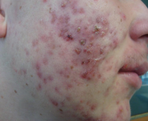
7
Acne vulgaris classification
mild: white / blackheads +/- sparse inflammatory lesions
moderate: widespread non-inflammatory lesions + papules and pustules
severe: extensive inflammatory lesions including nodules, pitting and scarring
moderate: widespread non-inflammatory lesions + papules and pustules
severe: extensive inflammatory lesions including nodules, pitting and scarring
8
Acne vulgaris management
mild-moderate: 12 weeks course of topical combination therapy [topic adapalene + benzoyl peroxide OR topical tretinoin + clindamycin]
moderate to severe: 12 weeks cause of same as above + / - oral doxycycline
COCp can be used as an alternative to oral ABx in women
moderate to severe: 12 weeks cause of same as above + / - oral doxycycline
COCp can be used as an alternative to oral ABx in women
9
Isoretinoin contraception advice
Use either 1 highly effective methods like an IUD or 2 complementary user dependent like barrier + oral contraceptives.
this is because Isoretinoin is a potent teratogen and strict adherence to the pregnancy prevention programme (PPP) is needed.
this is because Isoretinoin is a potent teratogen and strict adherence to the pregnancy prevention programme (PPP) is needed.
10
When to refer to dermatologist about acne
px with acne conglobata [v severe form]
px with nodulo-cystic acne
consider referall: if not responding to 2 completed courses, OR if not responding to previous Tx that has an oral ABx, OR acne with scarring OR acne causing significant psychological distress
px with nodulo-cystic acne
consider referall: if not responding to 2 completed courses, OR if not responding to previous Tx that has an oral ABx, OR acne with scarring OR acne causing significant psychological distress
11
Cellulitis what is it, causes and pathophysiology
bacterial infection of the dermis and deeper subcutaenous tissue
causes: staph aureus, group A / C strep, MRSA
Usually via a breach in the skin barrier which provides an entry. Breach via trauma, eczema, fungal, ulcers
causes: staph aureus, group A / C strep, MRSA
Usually via a breach in the skin barrier which provides an entry. Breach via trauma, eczema, fungal, ulcers
12
Cellulitis clinical features
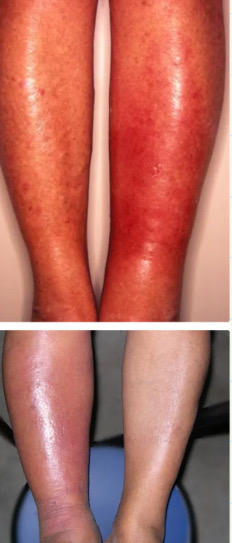
usually on shins unilaterally
Erythema is reasonably well defined. Blisters / bullae in more severe cases
Swelling and if golden yellow crust seen, indicates staph aureus
systemic: fever, malaise, nausea
Erythema is reasonably well defined. Blisters / bullae in more severe cases
Swelling and if golden yellow crust seen, indicates staph aureus
systemic: fever, malaise, nausea
13
cellulitis investigations / diagnosis
clinical, no further investigations needed
if px is admitted and speticaemia is suspected, do bloods and cultures
if px is admitted and speticaemia is suspected, do bloods and cultures
14
Cellulitis classification
Eron classification
class 1: no systemic signs and no uncontrolled co-morbidities
class 2: systemically unwell or well with co-morbidities like obesity or PAD which may complicated Tx
class 3: significant systemic upset or unstable co-morbidities
class 4: sepsis syndrome or severe life threatning infection like necrotising fasciitis
class 1: no systemic signs and no uncontrolled co-morbidities
class 2: systemically unwell or well with co-morbidities like obesity or PAD which may complicated Tx
class 3: significant systemic upset or unstable co-morbidities
class 4: sepsis syndrome or severe life threatning infection like necrotising fasciitis
15
When to admit for IV ABx for cellulitis:
Eron class 3 and 4
severe / rapidly deteriorating cellulitis
very young px < 1yr, or frail
immunocompromised
significant lymphoedema
facial / periorbital cellulitis
severe / rapidly deteriorating cellulitis
very young px < 1yr, or frail
immunocompromised
significant lymphoedema
facial / periorbital cellulitis
16
Management for Eron class 1
oral flucloxacillin first line
ALT: give clarithromycin / erthryomcin or doxycycline
ALT: give clarithromycin / erthryomcin or doxycycline
17
Eron class 2 management
admission may not be necessary, check local guidelines
18
Eron class 3-4 management
oral / IV co-amoxiclav or clindamycin
IV cefuroxime / cefriaxone
IV cefuroxime / cefriaxone
19
Erysipelas what is it
form of cellultis caused by group A strep pyogenes. Usually sharp demarcated borders from normal skin and affects the upper dermis and extends into the superficial cutaneous lymphatics
same management as cellulitis
same management as cellulitis
20
Plaque psoriasis presentation
the most common subtype. Well demarcated red, scaly patches affecting extensor surfaces, sacrum and scalp
21
Flexural psoriasis vs plaque psoriasis
will see smoother skin compared to plaque psoriasis
22
Guttate psoriasis trigger and appearance
transient psoriatic rash frequently triggered by strep infection. Multiple red / tear drop lesions on the body 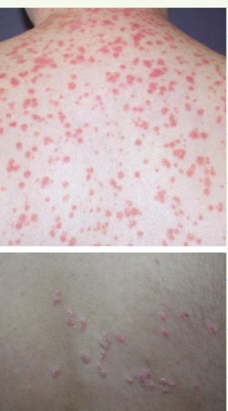
23
Guttate psoriasis management
usually resolves spontaneously within 2-3 months. Otherwise treat like psoriasis
Tonsillectomy may be necessary with recurrent episodes
Tonsillectomy may be necessary with recurrent episodes
24
Pustular psoriasis and management
commonly on palms / sores. Not infectious pus but the px are systemically unwell. Treated as a medical emergency and requires admission to hospital
25
Erythrodermic psoriasis
rare severe form of psoriasis with extensive erythematous inflamed areas covering most of the surface area of the skin. Will see large patches of exfoliation resulting in raw exposed areas.
Medical emergency and needs admission
Medical emergency and needs admission
26
Psoriasis pathophysiology
skin changes are caused by the rapid generation of new skin cells, resulting in an abnormal buildup and thickening of the skin.
Genetic associations: HLA B-13 / 17
worsened by skin trauma and stress. Triggered by strep infection
Genetic associations: HLA B-13 / 17
worsened by skin trauma and stress. Triggered by strep infection
27
Psoriasis clinical features
generally, dry / red / scaly patches on the skin, usually over the extensor surfaces of elbows / knees / scalp
think about psychosocial implications: low mood, low self-esteem, depression
think about psychosocial implications: low mood, low self-esteem, depression
28
specific psoriasis signs
Auspitz, Koebner
residual pigmentation of skin after lesions resolve
residual pigmentation of skin after lesions resolve
29
Auspitz sign
small bleeding where the plaques are scraped off
30
Koebner phenomenon
development of psoriatic lesions to areas of skin affected by trauma
31
Chronic plaque psoriasis management
1st line: potent topical steroid [beclamethasone] + vitamin D analogue [cholecalciferol], daily
2nd line: if no improvement after 8 weeks, double the vit D to BD
3rd line: no improvement after 12 weeks, potent corticosteroid BD for 3 weeks OR coal tar preparation applied OD / BD
2nd line: if no improvement after 8 weeks, double the vit D to BD
3rd line: no improvement after 12 weeks, potent corticosteroid BD for 3 weeks OR coal tar preparation applied OD / BD
32
Psoriasis secondary care management
phototherapy: narrow UVB light given
Photochemotherapy: psoralen + UVA light [PUVA]
Systemic therapy: methotrexate, ciclosporin, systemic retinoids, biologic agents
Photochemotherapy: psoralen + UVA light [PUVA]
Systemic therapy: methotrexate, ciclosporin, systemic retinoids, biologic agents
33
Simple psoriasis management
potent topical steroids OD / 4 weeks
if no improvement, use a different combination of topical agents
if no improvement, use a different combination of topical agents
34
face, flexural and genital psoriasis management
mild-moderate potency corticosteroid applied OD / BD daily for 2 weeks
35
Topical steroids notes
can cause skin atrophy / striae / depigmentation and rebound sx
should not be used for more than 1-2 weeks
recommend a 4 week break between topical steroids if starting a new regime
should not be used for more than 1-2 weeks
recommend a 4 week break between topical steroids if starting a new regime
36
Vitamin D analogues notes
causes less cell division leading to less epidermal proliferation. adverse effects are uncommon and these can be used long term.
Avoid in pregnancy, max dose is 100g for adults
Avoid in pregnancy, max dose is 100g for adults
37
Eczema what is it
chronic atopic condition caused by defects in the normal continuity of the skin barrier [irritants, allergens, microbes], leading to inflammation of the skin
38
Discoid eczema
coin shaped eczema patches that are itchy. Treat via moderately
potent corticosteroid + emollients like betamethasone
potent corticosteroid + emollients like betamethasone
39
Eczema clinical features
usually present in infancy with dry / red / itchy / sore patches of skin over the flexor surfaces and on the face / neck
40
Eczema and infections
eczema gives bacteria an opportunity to cause cellulitis, usually staph aureus
treat via flucloxacillin
treat via flucloxacillin
41
Eczema management conservative
avoid hot water baths, scratching / scrubbing skin, using soaps / body washes that remove the natural oils in the skin
reduce environmental triggers that can make sx worse like temperature changes, certain foods, washing powders, emotional event
reduce environmental triggers that can make sx worse like temperature changes, certain foods, washing powders, emotional event
42
Maintenance management of eczema
artificial barrier over the skin via an emollient, used after washing and before bed
ex) E45, Aveeno, Cetraben
ex) E45, Aveeno, Cetraben
43
Eczema flare management
thicker emollients + topical steroids + wet wraps
treat any bacterial / viral complications
treat any bacterial / viral complications
44
how are doses measured for topical steroids?
finger tip units. 1 FTU = 0.5g
45
eczema herpeticum
severe primary infection of the skin by HSV 1 / 2. more common in children. Will see monomorphic, punched out erosions, 1-3mm in diameter
admit and give IV aciclovir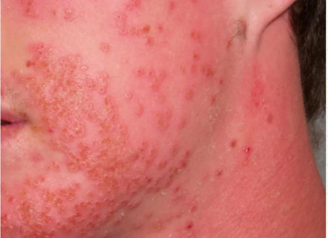
admit and give IV aciclovir
46
How to assess the extent of burns
Lund & Bowder Chart Or Wallace’s rule of 9
47
Wallace’s rule of 9s
head + neck = 9%
each arm, each anterior / post part of the leg = 9%
ant / post chest = each 9 %
ant / post abdomen = each 9 %
each arm, each anterior / post part of the leg = 9%
ant / post chest = each 9 %
ant / post abdomen = each 9 %
48
first degree burn
superifical epidermal: red and painful, no blisters
49
second degree burn [superficial]
partial thickness. pale pink painful, blistered. Slow CRT
50
second degree burn [deep]
partial thickness to the deep dermal. typically white but may have patches of non-blanching erythema. Reduced sensation , painful to pressure
51
third degree burn
full thickness. white / leathery brown / black in colour. no blisters, no pain [no sensation]
52
when to refer to secondary care for burns
all deep dermal and full thickness burns [2nd and 3rd degree]
any 1st degree over 3% of total body surface area [2% in children]
any second degree involving face / hands / feet / genitalia / limbs
Any inhalation injury
Any electrical / chemical burn injury
Suspicion of non-accidental injury
any 1st degree over 3% of total body surface area [2% in children]
any second degree involving face / hands / feet / genitalia / limbs
Any inhalation injury
Any electrical / chemical burn injury
Suspicion of non-accidental injury
53
immediate first aid for burns
ABC. remove appendage from source, irrigate heat burn with cool water and cover burn.
Electical burns: turn off the power supply
Chemical burns: brush off any powder and irrigate with water. Neutralising attempts are NOT recommended
Electical burns: turn off the power supply
Chemical burns: brush off any powder and irrigate with water. Neutralising attempts are NOT recommended
54
Burns pathophysiology and sequelae
inflammatory response to burn area. Will see systemic CVS effects due to fluid loss and sequestration of fluid into the third space.
Immunosuppression is common with large burns and sepsis is a common cause of death following major burns. [commonly bacterial translocation from the gut lumen]
Immunosuppression is common with large burns and sepsis is a common cause of death following major burns. [commonly bacterial translocation from the gut lumen]
55
ABCDE management of severe burns
early intubation for smoke inhalation or injury to face / neck
IV fluids via Parkland formula, give half of the amount in the first 8hrs
insert catheter, give analgesia and transfer to burns unit
Escharotomy may be needed to divide burnt tissue. Skin grafting is considered.
IV fluids via Parkland formula, give half of the amount in the first 8hrs
insert catheter, give analgesia and transfer to burns unit
Escharotomy may be needed to divide burnt tissue. Skin grafting is considered.
56
Parkland formula
Volume = TBSA of burn in % * kg * 4
57
Urticaria or hives: what is it
a local / generalised superficial swelling of the skin. Can be acute or chronic
58
acute urticaria aetx
usually allergies to food / medication / animals
contact with chemicals / latex / stinging nettles
insect bites
viral infections
dermatographism [rubbing of skin]
contact with chemicals / latex / stinging nettles
insect bites
viral infections
dermatographism [rubbing of skin]
59
chronic urticaria aetx
Idiopathic
Inducible by: sunlight, temp change, exercise, strong emotions, hot / cold weather, pressure
Autoimmune like SLE
Inducible by: sunlight, temp change, exercise, strong emotions, hot / cold weather, pressure
Autoimmune like SLE
60
Urticaria clinical features
small itchy lumps on skin ~ associated with patchy erythematous rash. Can be localised to a specific area or widespread. Angioedema and flushing of the skin may be seen 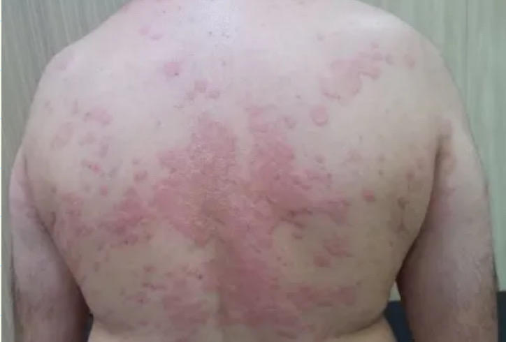
61
Urticaria management
non-sedating antihistamine [Fexofenadine]
for flare ups, oral steroids can be considered
refer to specialist for problematic cases with Tx like cyclosporin and LTRAs
for flare ups, oral steroids can be considered
refer to specialist for problematic cases with Tx like cyclosporin and LTRAs
62
Stevens Johnson syndrome what is it and RF
severe systemic reaction of skin / mucosa due to a drug reaction
RF: HIV
RF: HIV
63
Stevens Johnson syndrome causes
penicillins, sulphonamides [cotrimoxazole], anti-psychotics [phenytoin, carbamazepine, lamtrogine], allopurinol, NSAIDs, COCp
Non-drug causes: infection, vaccination
Non-drug causes: infection, vaccination
64
Stevens Johnson syndrome clinical features
maculopapular rash with large lesions, may later develop into vesicles / bullae
+ve Nikolsky sign
Mucosal involvement, systemic sx [fever, arthralgia]
scalp / palms / soles spared
+ve Nikolsky sign
Mucosal involvement, systemic sx [fever, arthralgia]
scalp / palms / soles spared
65
Nikolsky sign
+ve sign is when blisters / erosions appear when skin is rubbed gently
66
Stevens Johnson syndrome: investigations / diagnosis
clinical but skin biopsy can be done to confirm diagnosis
will see epithelial necrosis with few inflammatory cells
will see epithelial necrosis with few inflammatory cells
67
Stevens Johnson syndrome: management
admit for supportive tx, refer to dermatology / burns unit for tx
specialist guided steroids, immunoglobulins, immunosuppressants
specialist guided steroids, immunoglobulins, immunosuppressants
68
Toxic epidermal necrolysis: what is it
seen secondary to a drug reaction, will see a scalded appearance over an extensive area.
A more severe SJS
A more severe SJS
69
Toxic epidermal necrolysis: aetx
phenytoin, sulphonamides, allopurinol, penicillins, carbamazepine, NSAIDs
70
Toxic epidermal necrolysis: clinical features
pyrexia / tachycardia
+ve Nikolskys sign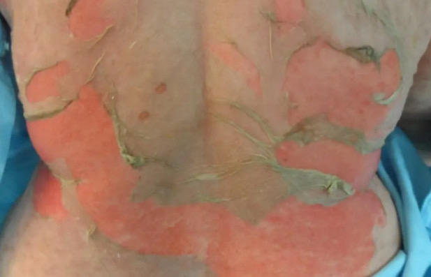
+ve Nikolskys sign
71
Toxic epidermal necrolysis: investigations
biopsy will show: full thickness epidermal necrosis with subepidermal detachment
72
Toxic epidermal necrolysis: management
stop precipitating factor. ICU + IV fluids
1st line: IV immunoglobulins
1st line: IV immunoglobulins
73
difference between SJS and TEN
SJS < 10% TBSA, TEN is > 30%
between the two, is an overlap
between the two, is an overlap
74
polymorphic eruption of pregnancy: what is it, features and management
pruritus seen in the last trimester
lesions are often on abdominal striae, sparing the periumbilical area
management depends on severity: emollients, mild topical / oral steroids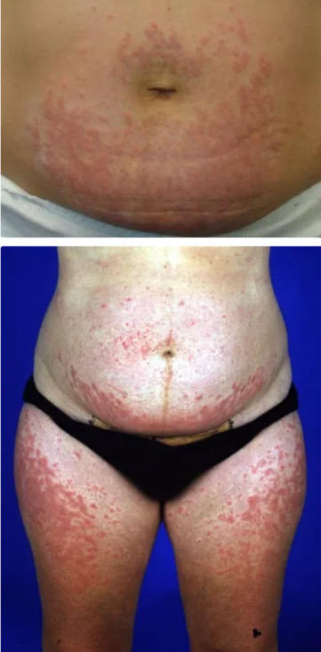
lesions are often on abdominal striae, sparing the periumbilical area
management depends on severity: emollients, mild topical / oral steroids
75
Pityriasis rosea: what is it and Tx
acute, self-limiting rash affecting young adults. Thought to be linked to herpes hominis virus 7
resolves after around 6 weeks
resolves after around 6 weeks
76
Pityriasis rosea clinical features
majority have no prodrome, might see a viral infection beforehand
Herald patch on trunk, followed by erythematous / oval / scaly patches which follow a characteristic distribution parallel to the line of Langer = fir tree appearance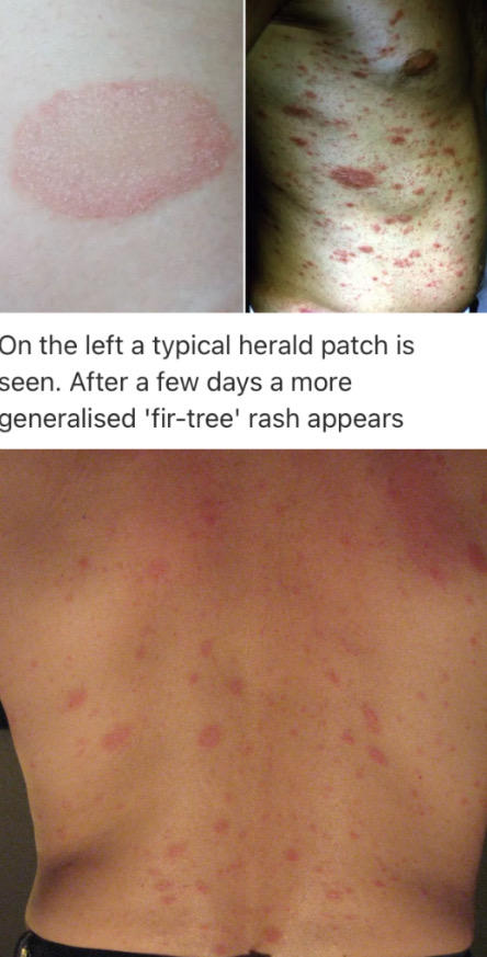
Herald patch on trunk, followed by erythematous / oval / scaly patches which follow a characteristic distribution parallel to the line of Langer = fir tree appearance
77
Erythroderma what is it
when >95% of the skin is involved in a rash of any kind
78
Erythroderma causes
eczema, psoriasis, drugs, lymphomas, leukaemias, idiopathic
79
Erythrodermic psorasis
Results from progression of chronic plaque psoriasis. Associated with mild systemic upset and can be triggered by many things such as withdrawal of systemic steroids. Need to be admitted for management 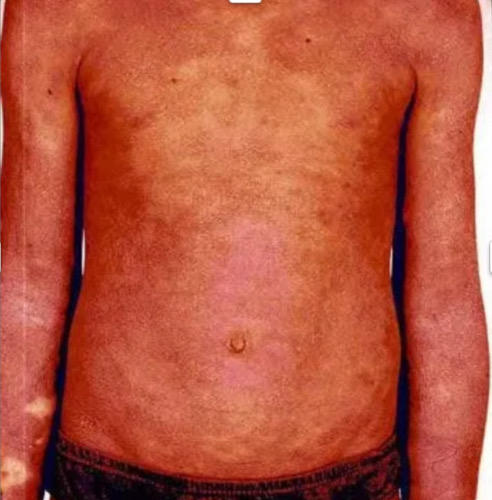
80
Erythema multiforme what is it
hypersensitivity reaction triggered by infection. divided into major and minor
81
erythema multiforme causes
HSV virus, idiopathic, streptococcus, drugs [penicillins, sulphonamides, allopurinol, NSAIDs, carbamezepine], CT disease like SLE sarcoidosis, malignancy
82
Erythema multiforme clinical features 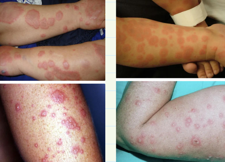
target lesions initially on dorsal hands / feet before spreading to torso. mostly on UL. + mild pruritus 
83
Erythema multiforme major
more severe form and is associated with mucosal involvement 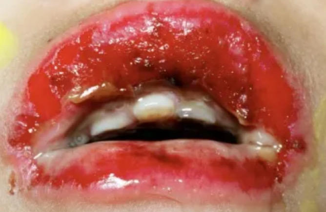
84
Erythema nodosum causes
infection [strep, TB], systemic disease [sarcoid, IBD, Behcets], Malignancy, drugs [penicillins, sulphonamides, COCp], pregnancy
85
Erythema nodosum clinical features
inflammation of subcutaneous fat usually causing tender, erythematous, nodular lesions. Usually on shins / forearms
resolves within 6 weeks , no scarring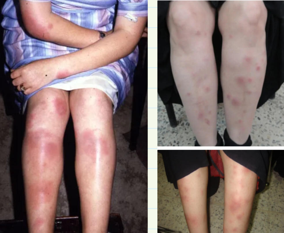
resolves within 6 weeks , no scarring
86
Bullous phemigoid: what is it
autoimmune condition causing sub-epidermal blistering of the skin. Secondary to development of antibodies against hepidesmosomal proteins BP 180 and 230
87
Bullous phemigoid clinical features
itchy, tense blisters around flexures. usually heals w / o scarring 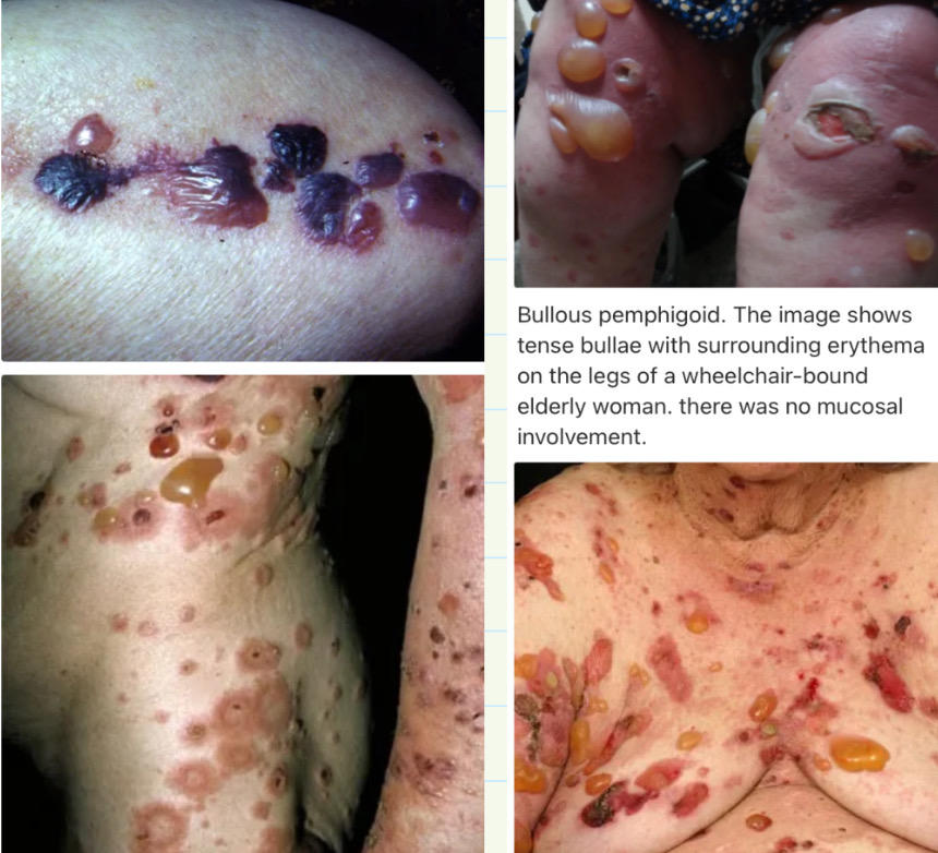
88
DRESS syndrome what is it
Type 4 hypersensitivity drug reaction with eosinophilia and systemic sx
89
DRESS syndrome causes
anti-epileptics, allopurinol, olanzapine, sulphonamide ABx
90
DRESS syndrome clinical features
high fever / tachycardia. Morbilliform [rash that looks likes measles] eruption. Haematological abnormalities, high WCC count, inflammation of internal organs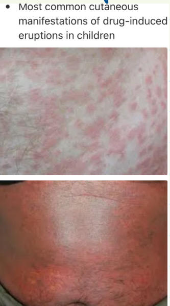
91
DRESS syndrome management
stop suspected causative medication. monitor + supportive care. consider systemic steroids
92
salmon patches: what are they
vascular birthmarks seen on half of newborn babies, often called stork marks / bites. Usually on forehead / eyelids / nape of neck
usually fade over a few months but can persist
usually fade over a few months but can persist
93
Pityriasis versicolor: what is it and RFs
superficial cutaneous fungal infection caused by Malassezia furfur
RFs: healthy individuals, immunosuppression, malnutrition, cushings
RFs: healthy individuals, immunosuppression, malnutrition, cushings
94
Pityriasis versicolor: clinical features
mostly trunk. Hypopigemented pink / brown patches. Mild pruritus and scale is common 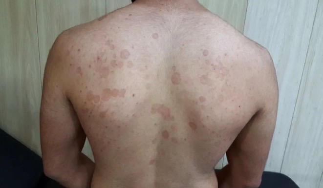
95
Pityriasis versicolor: management
topical antifungal: ketoconazole shampoo
if this fails, consider alternative diagnoses and give oral itraconazole
if this fails, consider alternative diagnoses and give oral itraconazole
96
Pyoderma gangrenosum: what is it
rare, non-infectious, inflammatory disorder. A very uncommon cause of very painful skin ulceration. Usually on LL
97
Pyoderma gangrenosum: aetx
idiopathic, IBD, rheumatological, haematological [myeloproliferative], granulomatosis with polyangiitis, primary biliary cirrhosis
98
Pyoderma gangrenosum: pathophysiology
neutrophilic dermatoses: dense infiltration in the affected tissue, seen on biopsy
99
Pyoderma gangrenosum: clinical features
on LL. initially, a sudden small pustule / red bump / blood blister. Later the skin breaks down and ulcerates. Borders of ulcer is purple. Systemic features 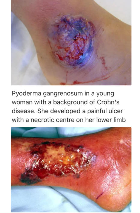
100
Pyoderma gangrenosum: investigations
clinical, histology only really useful to rule out other causes of an ulcer
101
Pyoderma gangrenosum: management
oral steroids first line.
Immunosuppressive therapy via ciclosporin and infliximab.
Surgery once disease process is controlled on immunosuppression
Immunosuppressive therapy via ciclosporin and infliximab.
Surgery once disease process is controlled on immunosuppression
102
Seborrhoeic dermatitis: what is it and associations
chronic dermatitis thought to be associated with fungus malassezia furfur. Associated with HIV and Parkinsons
103
Seborrhoeic dermatitis: clinical features
Eczematous lesions on the sebum rich areas like scalp / periorbital / auricular / nasolabial fold.
Salmon pink, scaly, ill defined plaques seen
otitis externa [common] and blepharitis may develop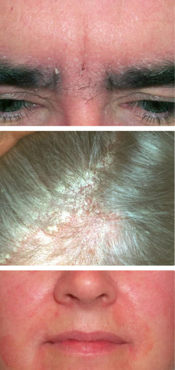
Salmon pink, scaly, ill defined plaques seen
otitis externa [common] and blepharitis may develop
104
Seborrhoeic dermatitis: management
for scalp: ketaconazole 2% shampoo
for face and body: topical ketoconazole, steroids
for face and body: topical ketoconazole, steroids
105
cradle cap
Seborrhoeic dermatitis seen in nappy area / scalp / face / limb flexures of babies. Will see erythematous rash with coarse yellow scales
reassure, topical emollient on scalp. Imidazole if persists. will resolve spontaenously by around 8 months of age
reassure, topical emollient on scalp. Imidazole if persists. will resolve spontaenously by around 8 months of age
106
Dermatitis [irritant]
non-allergic, due to weak acids or alkalis from detergents
will be seen on hands with erythema and crusting. more painful than itchy
will be seen on hands with erythema and crusting. more painful than itchy
107
Dermatitis [allergic]
type 4 hypersensitivity reaction. often seen after dyeing hair. will see intense pruritus.
give topical potent steroid and avoid offending agent
give topical potent steroid and avoid offending agent
108
Pemphigus vulgaris: what is it
autoimmne disease caused by antibodies directed against desmoglein 3, a cadherin type epithelial cell adhesion molecule. Common in Jewish population
109
Pemphigus vulgaris clinical features
will see mucosal ulceration. .skin blistering and easily rupturing vesicles and bullae. lesions are painful but no itchy. +ve Nikolskys sign
acantholysis on biopsy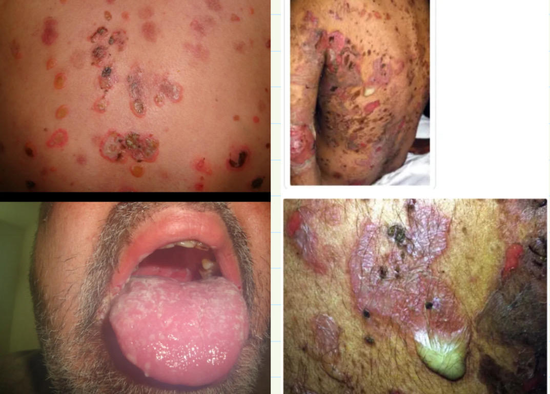
acantholysis on biopsy
110
Basal cell carcinoma: what is it
most common type of cancer in the West. Lesions are also known as “rodent ulcers”. Characterised by slow-growth and local invasion
111
BCC RFs
UV exposure
Hx of sunburn in childhood
Skin type 1 [always burns, no tanning]
increasing age
Male
Immunosuppression
Hx of sunburn in childhood
Skin type 1 [always burns, no tanning]
increasing age
Male
Immunosuppression
112
BCC clinical features
occurs mostly in sun-exposed sites like head / neck. Initially pearly, flesh-coloured papule with telangiectasia. Can later ulcerate, leaving a central crater 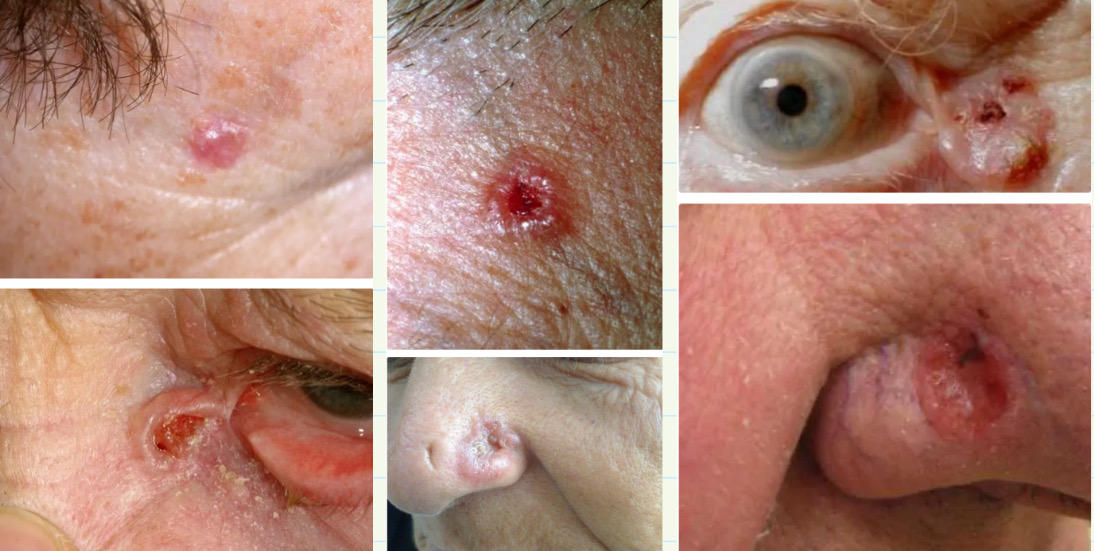
113
BCC: management
Routine derm referral. 2WW if concerned that delay will have significant impact
Medical: topical imiquimod / fluorouracil, cryotherapy, radiotherapy, curettage
Surgical removal is also an option
Medical: topical imiquimod / fluorouracil, cryotherapy, radiotherapy, curettage
Surgical removal is also an option
114
Squamous cell carcinoma: RFs
excessive sun exposure / PUVA therapy
Acitinic keratoses and Bowens disease
Immunosuppression
Smoking
Long-standing leg ulcers
Genetic condition [xeroderma pigmentosum]
Associated with HPV
Acitinic keratoses and Bowens disease
Immunosuppression
Smoking
Long-standing leg ulcers
Genetic condition [xeroderma pigmentosum]
Associated with HPV
115
SCC clinical features
typically on sun exposed sites such as head / neck OR dorsum of hands / feet
Rapidly expanding, painless, ulcerating nodules
Cauliflower like appearance, there may be areas of bleeding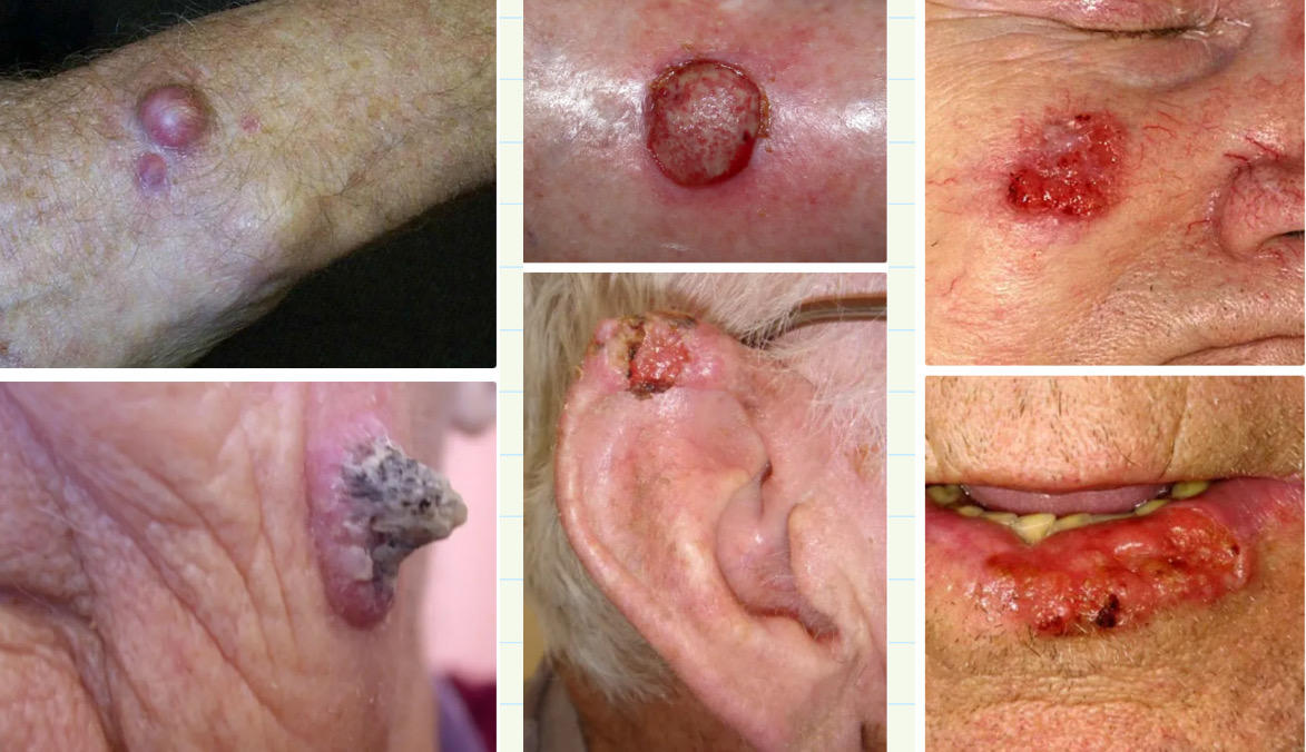
Rapidly expanding, painless, ulcerating nodules
Cauliflower like appearance, there may be areas of bleeding
116
SCC management
Surgical excision with 4mm margins IF lesion is less than 20mm in diameter
If larger than 20mm, margins increase to 6mm
Mohs micrographic surgery is used in cosmetically important sites or in high risk px
If larger than 20mm, margins increase to 6mm
Mohs micrographic surgery is used in cosmetically important sites or in high risk px
117
Melanoma RFs
sun exposure, fair skin, FHx, naevi count, immunosuppression
118
Lay people skin lesion identification ABCDE
Asymmetry, Border irregularity, colour variation, diameter of 7mm or more, evolving
119
Superficial spreading melanoma
70% of cases. typically in young people’s arms / legs / back and chest. Will see a growing mole with changes in size / shape / colour with perhaps oozing or bleeding
120
nodular melanoma
second most common. usually in middle aged people’s sun exposed areas.
will see a red / black lump that bleeds or oozes
will see a red / black lump that bleeds or oozes
121
Lentigo maligna melanoma
seen in older people, chronically sun exposed skin
will see a growing mole that is evolving and oozing / bleeding
will see a growing mole that is evolving and oozing / bleeding
122
Acral lentiginous melanoma
will see in nails / palms / soles of people with darker skin tone.
Subungual pigmentation [Hutchinsons sign] or on palms / feet
Subungual pigmentation [Hutchinsons sign] or on palms / feet
123
Weighted Glasgow Scale: major and minor criteria
Major criteria: change in size / shape / colour
Minor criteria: diameter of 7mm or more, inflammation, oozing / bleeding, altered sensation
Minor criteria: diameter of 7mm or more, inflammation, oozing / bleeding, altered sensation
124
Melanoma management
if suspicious: excision biopsy with lesion removed completely. Then review biopsy to see if further re-excision of margins is needed [usually 1-3cm].
Further tx include: sentinel lymph node mapping, isolated limb perfusion, block dissection of regional lymph node groups
Further tx include: sentinel lymph node mapping, isolated limb perfusion, block dissection of regional lymph node groups
125
Melanoma prognosis
measured using Breslow Thickness. if less than 0.75mm, survival is 95-100%. If more than 4mm, 5yr survival is 50%
126
Acitinic keratosis: what is it
a common premalignant skin lesion that develops as a consequence of chronic sun exposure
127
Acitinic keratosis: clinical features
small, crusty, scaly lesions. Will be pink / red / brown / flesh colour. On sun exposed areas. 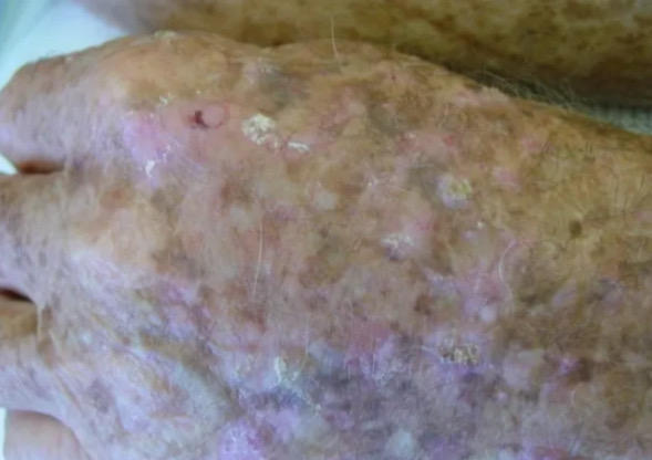
128
Acitinic keratosis: management
conservative: prevent further risk via sun avoidance / sun cream
Medical: 2-3 weeks of fluorouracil cream + topical hydrocortisone, topical diclofenac, topical imiquimod, cryotherapy, curettage and cautery
Medical: 2-3 weeks of fluorouracil cream + topical hydrocortisone, topical diclofenac, topical imiquimod, cryotherapy, curettage and cautery
129
Athelets foot / tinea pedis
caused by fungi in the genus Trichophyton. Will see scaling / flaking / itching between the toes.
Management is topical imidazole or terbinafine
Management is topical imidazole or terbinafine
130
Molluscum contagiosum: what is it and transmission
common skin condition caused by molluscum contagiosum virus. Mostly occurs in children with atopic eczema [1-4yr olds].
Transmission via direct close personal contact OR indirectly via fomites [contaminated surfaces] like shared towels / flannels
Transmission via direct close personal contact OR indirectly via fomites [contaminated surfaces] like shared towels / flannels
131
Molluscum contagiosum: clinical features
Typical small, flesh coloured papules that have a central dimple. Typically appears in “crops” of multiple lesions in a local area.
Commonly on trunk / flexures but anogenital lesions may also occur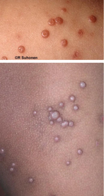
Commonly on trunk / flexures but anogenital lesions may also occur
132
Molluscum contagiosum: management
conservative: reassure px that it is self-limiting within 18months. Explain it is contagious so avoid sharing towels / clothing / baths. Encourage not to scratch the lesions. self isolation is not needed
medical: treatment is not recommended but cryotherapy can be done
medical: treatment is not recommended but cryotherapy can be done
133
when to consider referral in px w/ Molluscum contagiosum
people who are HIV +ve with extensive lesions = urgent HIV specialist referral
for px with eyelid / ocular lesions = refer to opthalmologist
adults with anogenital lesions = refer to GUM for STI screening
for px with eyelid / ocular lesions = refer to opthalmologist
adults with anogenital lesions = refer to GUM for STI screening
134
Keloid scars: what is it
tumour like lesions that arise from the connective tissue of a scar, and extend beyong dimensions of original wound
135
Keloid scars RFs
ethnicity [dark skin], young adults, commonly on sternum, shoulder, neck, faces
136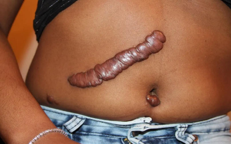
keloid scar image
137
keloid scar management
if early, intralesional steroids can be used [triamcinolone]
excision can be considered but can cause further scarring
excision can be considered but can cause further scarring
138
Impetigo: what is it
superficial bacterial skin infection caused by staph aureus OR strep pyogenes
Can be primary OR a secondary complication of an existing skin condition like eczema / scabies / insect bites
Can be primary OR a secondary complication of an existing skin condition like eczema / scabies / insect bites
139
Impetigo transmission
direct contact with discharge from the scabs of an infected person
Invasion via minor abrasions / scratching
Mainly via hands / indirectly via toys / clothing / equipment
Incubation period is 4-10 days
Invasion via minor abrasions / scratching
Mainly via hands / indirectly via toys / clothing / equipment
Incubation period is 4-10 days
140
Impetigo: clinical features
if staph infection, will see a golden crust. can be anywhere but usually on face, flexures, limbs
can see presence of bullous / vesicles with pus, which burst to form a golden crust. These will eventually heal without scarring but will be painful / itching
it is common to have systemic sx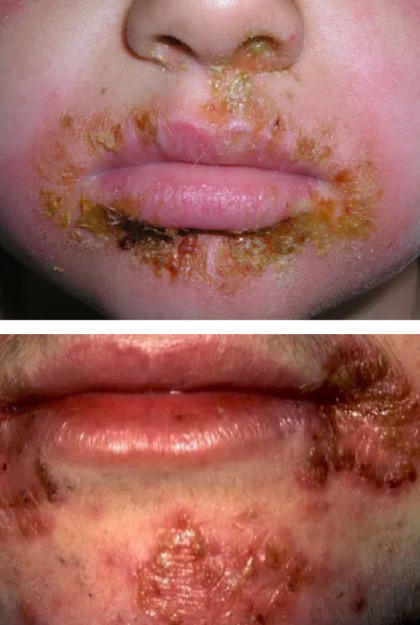
can see presence of bullous / vesicles with pus, which burst to form a golden crust. These will eventually heal without scarring but will be painful / itching
it is common to have systemic sx
141
Impetigo: management
exclude from school until lesions are crusted OR post 48hrs after ABx tx
Hydrogen peroxide is used if not systemically unwell OR not at high risk of complication. If ABx is needed, fusidic acid can be used.
if extensive, use oral flucloxacillin
Hydrogen peroxide is used if not systemically unwell OR not at high risk of complication. If ABx is needed, fusidic acid can be used.
if extensive, use oral flucloxacillin
142
Scabies: cause and pathophysiology
Mite Sarcoptes scabies is the cause and is spread by prolonged skin contact.
Scabies mite will burrow into skin, laying its eggs which causes a delayed type 4 hypersensitivity reaction which will cause intense pruritus.
It can take up to 8 weeks for any sx or rash to appear after the initial infestation
Scabies mite will burrow into skin, laying its eggs which causes a delayed type 4 hypersensitivity reaction which will cause intense pruritus.
It can take up to 8 weeks for any sx or rash to appear after the initial infestation
143
Scabies: clinical features
Intensely itchy small red spots with track marks where the mites have burrowed. Will see widespread pruritus but can be localised to finger webs
Will see excoriation from scratching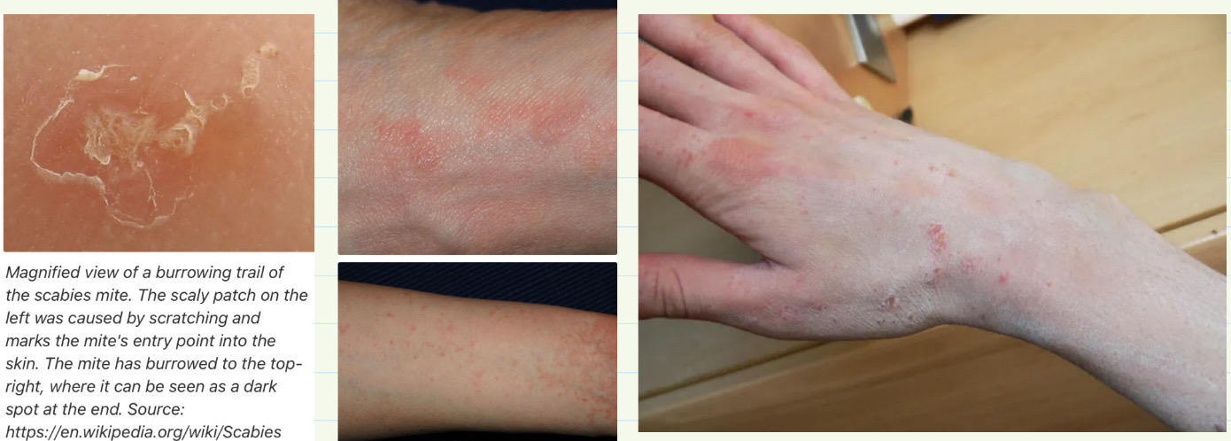
Will see excoriation from scratching
144
Scabies: management
avoid close physical contact. Need to hot wash all clothes / bedding / towels with scabies + hoovering of carpets and furniture. Need to treat all household / close physical contacts even if ASx
1st line med: permethrin 5% is applied to the whole body for 8-12 hours.
2nd line is malathion
Pruritus can persist for up to 4-6 weeks post-eradication
1st line med: permethrin 5% is applied to the whole body for 8-12 hours.
2nd line is malathion
Pruritus can persist for up to 4-6 weeks post-eradication
145
Crusted [Norweigan] Scabies
Will see crusting skin that will be teeming with hundred of thousands of organisms. will be extremely contagious and there will be scaly plaques just like psoriasis
admission may be needed for treatment with oral ivermectin and isolation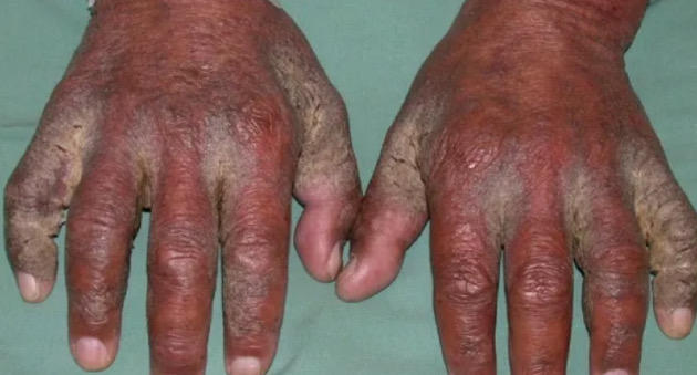
admission may be needed for treatment with oral ivermectin and isolation
146
immunocompromisation and scabies
immunocompromised px may not notice pruritus as they do not mount an immune response to the infestation
147
Tinea capitis: cause
trichopyton tonsurans OR microsporium canis from cats / dogs
148
tinea capitis clinical features
scarring alopecia in children. if untreated a raised / pustular / spongy mass called a kerion can form 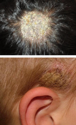
149
tinea capitis: investigations
scalp scrapings. lesions from microsporium canis will see a green fluorescence under Wood’s lamp
150
tinea capitis: management
Give topical ketoconazole shampoo for first 2 weeks to reduce transmission
oral antifungals like terbinafine for Trichophyton tonsurans OR griseofulvin for Microsporium infections
oral antifungals like terbinafine for Trichophyton tonsurans OR griseofulvin for Microsporium infections
151
Tinea corporis cause
caused by trichophyton rubrum and trichophyton vervucosum [contact with cattles]
152
tinea corporis clinical features
well defined annular erythematous lesions with pustules / papules 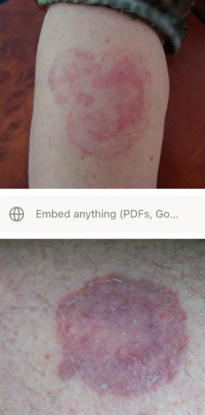
153
tinea corporis management
oral fluconazole
154
tinea incognito: what is it
arises from the use of steroids to treat an initial fungal infection. Usually caused by a misdiagnosis of ringworms for dermatitis, so a steroid is prescribed.
the steroid improves sx BUT accelerates the growth of the fungal infection by immune suppression.
the steroid improves sx BUT accelerates the growth of the fungal infection by immune suppression.
155
Hand foot and mouth disease: cause
caused by coxsackie A16 virus and enterovirus 71
156
Hand foot and mouth disease: clinical features
illness starts with typical viral URTI sx: tiredness, sore throat, dry cough, raised temp
after 1-2 days, small painful mouth ulcers will appear, followed by blistering red spots across the body [hands / feet / mouth].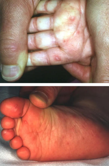
after 1-2 days, small painful mouth ulcers will appear, followed by blistering red spots across the body [hands / feet / mouth].
157
Hand foot and mouth disease: management
no medical management. give fluids / analgesia. will resolve after 10 days. contagious so should avoid sharing towels / bedding
158
Measles: what is it
viral spread via RNA paramyxovirus. done through aerosol transmission. rash starts 4 days after infection. rarely seen now due to immunisation programmes
159
measles: clinical features
prodromal phase: irittable, conjunctivitis, fever
Koplik spots: white spots on buccal mucosa
diarrhoea seen in 10% of px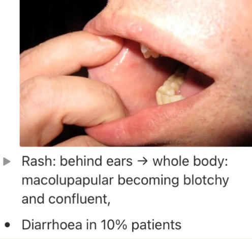
Koplik spots: white spots on buccal mucosa
diarrhoea seen in 10% of px
160
Measles investigations
IgM antibodies can be detected within a few days of rash onset
161
Measles management
supportive. admit if immunosuppressed or pregnant. inform public health as it is a notifiable disease
162
Measles complications
otitis media [most common], pneuomonia is the most common cause of death.
Encephalitis, diarrhoea, keratoconjunctivitis, myocarditis
Encephalitis, diarrhoea, keratoconjunctivitis, myocarditis
163
Staphylococcal scalded skin syndrome: what is it
usually affects children under 6yrs old
infection caused by staph, producing epidermolytic toxins, causing widespread painful blisters and desquamation
infection caused by staph, producing epidermolytic toxins, causing widespread painful blisters and desquamation
164
Staphylococcal scalded skin syndrome: clinical features
lesion is initially superficial and crusted. in 24hrs, the surrounding skin will become painful and scarlet. Skin will become tender / paper like = forms blisters and desquamotus [peels off].
looks like scalded skin.
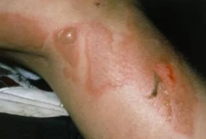
looks like scalded skin.
165
Staphylococcal scalded skin syndrome triad
erythema, bullae / blisters, desquamation
166
Staphylococcal scalded skin syndrome: management
admit for IV flucloxacillin and analgesia.
monitor and maintenance via fluid and electrolytes
monitor and maintenance via fluid and electrolytes
167
Acute paronychia: what is it
infection of the skin and soft tissue of the proximal and lateral nail fold, caused by staph aureus. usually seen in the tx of wound infection
168
Acute paronychia: clinical features
swelling, erythema, tenderness, pussy discharge
recent wound history
recent wound history
169
Acute paronychia: investigations
wound swab if discharge is present
170
Acute paronychia: management
flucloxacillin 500mg QDS, seek advice if MRSA is suspected.
if wound does not improve, refer to surgeons
if wound does not improve, refer to surgeons
171
viral wart: cause
HPV via direct contact
172
viral wart clinical features
papules with rough, papillomatous and hyperkeratotic surface, ranging from 1mm to 1cm.
will see plantar warts [localised to soles which are painful], plane warts on hands / face, and filiform warts [on a long stalk]
will see plantar warts [localised to soles which are painful], plane warts on hands / face, and filiform warts [on a long stalk]
173
viral warts: management
topical tx with salicyclic acid OD. most px will resolve with 12 weeks.
cryotherapy is also an option
cryotherapy is also an option
174
Alopecia areata what is it and features
presumed autoimmune condition, causing localised well demarcated patches of hair loss. There may be “exclamation mark” hairs at the borders.
No signs of inflammation / erythema / atrophy
No signs of inflammation / erythema / atrophy
175
Alopecia areata management
1st line: topical steroids OR intralesional corticosteroids
Minoxidil to stimulate hair growth
psychological support
Minoxidil to stimulate hair growth
psychological support
176
lichen planus what is it and causes
immune mediated skin disorder that has an unknown aetx but drug causes include: gold, quinine, thiazides
177
lichen planus clinical features
itchy, papular rash mostly on palms / soles / genitalia and flexor surfaces of arm
rash is often polygonal in shape with “white lines” pattern on surface [Wickhams striae]. Will see Koebner phenomenon.
Oral involvement of white lace pattern on the buccal mucosa in 50% of cases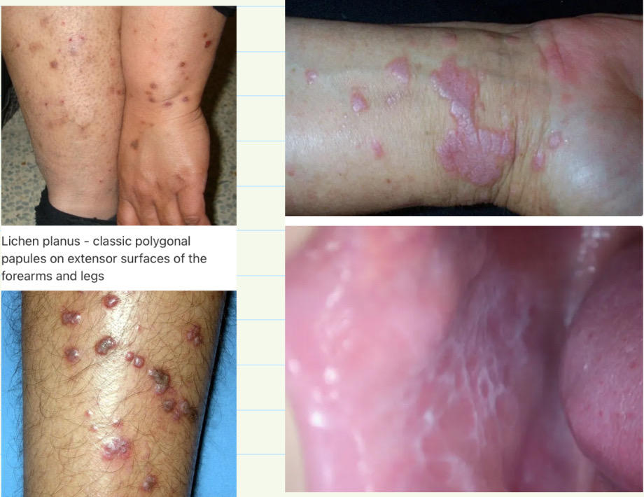
rash is often polygonal in shape with “white lines” pattern on surface [Wickhams striae]. Will see Koebner phenomenon.
Oral involvement of white lace pattern on the buccal mucosa in 50% of cases
178
lichen planus management
1st line: topical steroids
Benzydamine mouthwash for oral lichen planus
Benzydamine mouthwash for oral lichen planus
179
Dermatitis herpetiformis what is it
autoimmune skin disorder associated with coeliac disease. Caused by deposition of IgA in the dermis. 90% of px will have small bowel biopsy findings consistent with glutent sensitive enteropathy
180
dermatitis herpetiformis clinical features
Blistering, itchy, vesicular skin lesions on the extensor surfaces 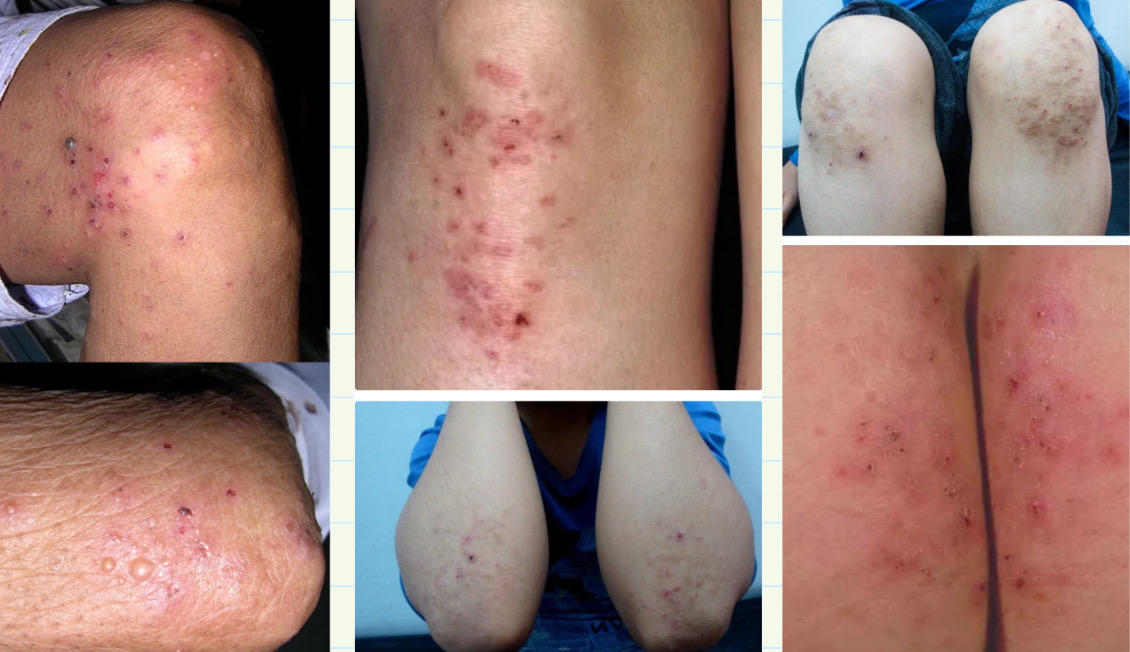
181
Dermatitis herpetiformis investigations
Skin biopsy of direct immunoflourescence will show deposition of IgA in a granular pattern in the upper dermis. Will also see the presence of anti-tTG antibodies consistent with coeliac disease
182
Dermatitis herpetiformis management
gluten free diet
Dapsone for severe pruritus [SE of haemolytic anaemia]
Dapsone for severe pruritus [SE of haemolytic anaemia]
183
Hereditary haemorrhagic telangiectasia / Osler Weber Rendu syndrome: what is it
autosomal dominant condition that presents with multiple telangiectasia over the skin and mucous membranes
forms telangiectasia and arteriovenous malformations [AVM]. This lead to significant bleeding, epistaxis and GI bleeding
forms telangiectasia and arteriovenous malformations [AVM]. This lead to significant bleeding, epistaxis and GI bleeding
184
Hereditary haemorrhagic telangiectasia: investigations
need 3 out of the 4:
epistaxis [spontaneous or recurrent]
Telangiectasia
FHx: 1st degree relative
Visceral lesions: GI bleeding or AVM seen on CT / CXR [pulmonary nodules]
epistaxis [spontaneous or recurrent]
Telangiectasia
FHx: 1st degree relative
Visceral lesions: GI bleeding or AVM seen on CT / CXR [pulmonary nodules]
185
Hereditary haemorrhagic telangiectasia: management
if blood loss is severe: blood transfusion
if IDA, give iron supplements
Embolisation of AVMs within organs can help manage the condition
if IDA, give iron supplements
Embolisation of AVMs within organs can help manage the condition
186
Hidradenitis suppurativa: what is it
chronic painful inflammatory skin disorder, characterised by the development of inflammatory nodules, pustules, sinus tracts and scars in intertriginous areas
should be suspected in pubertal / post pubertal px who have a diagnosis of recurrent furuncles and boils
should be suspected in pubertal / post pubertal px who have a diagnosis of recurrent furuncles and boils
187
intertriginous areas
areas where two skin surfaces rub each other, like the axilla or inner thighs
188
Hidradenitis suppurativa: RFs
FHx, smoking, obesity, diabetes, PCOS, mechanical stretching of skin
189
Hidradenitis suppurativa: clinical features
initial manifestation involves recurrent painful inflamed nodules usually on intertriginous skin.
Nodules can rupture, leading to purulent discharge, malodorous material [foul smelling]
nodules can result in plaques, sinus tracts, “rope like” scarring
Nodules can rupture, leading to purulent discharge, malodorous material [foul smelling]
nodules can result in plaques, sinus tracts, “rope like” scarring
190
Hidradenitis suppurativa: management
good hygiene, loose clothing, stop smoking, lose weight
give steroids / flucloxacillin for acute flares. Longer term, give topical clindamycin / oral rifampicin
surgical excision and drainage is also an option
give steroids / flucloxacillin for acute flares. Longer term, give topical clindamycin / oral rifampicin
surgical excision and drainage is also an option
191
vitiligo: what is it and associations
autoimmune condition which results in the loss of melanocytes = depigmentation of the skin
associated with T1DM, addisons, autoimmune thyoid disorders, pernicious anaemia, alopecia areata
associated with T1DM, addisons, autoimmune thyoid disorders, pernicious anaemia, alopecia areata
192
Vitiligo causes
genetics involving tyrosine enzyme
immune destruction of melanocytes
Associated with treatments like transdermal patches of methylphenidate for ADHD and immune checkpoint inhibitors like pembrolizumab
Triggered by stress
Correlates with metastatic melanoma, stem cell transplants
immune destruction of melanocytes
Associated with treatments like transdermal patches of methylphenidate for ADHD and immune checkpoint inhibitors like pembrolizumab
Triggered by stress
Correlates with metastatic melanoma, stem cell transplants
193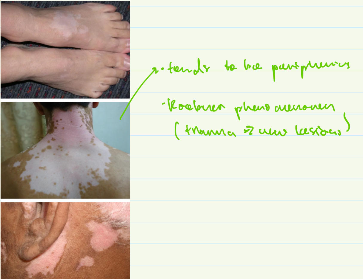
vitiligo clinical features
194
vitiligo management
sun block for affected areas of skin, camouflage make up
topical corticosteroids can reverse changes if early enough
Tacrolimus / phototherapy
topical corticosteroids can reverse changes if early enough
Tacrolimus / phototherapy
195
Rosacea clinical features
typically nose / cheeks / forehead
will see flushing as the first sx. Telangiectasia and development into persistent erythema with papules and pustules
Rhinophyma / blepharitis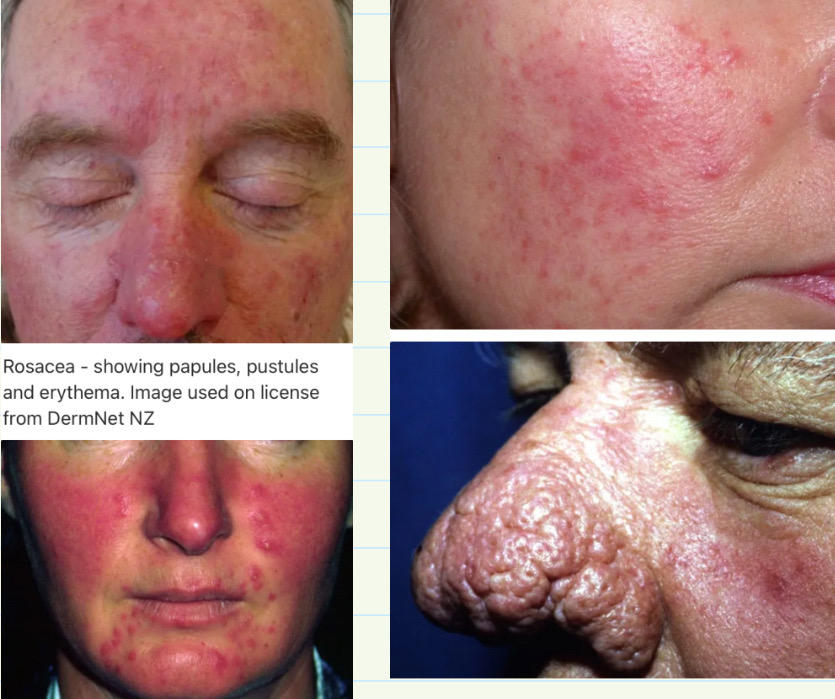
will see flushing as the first sx. Telangiectasia and development into persistent erythema with papules and pustules
Rhinophyma / blepharitis
196
Rhinophyma
associated with rosacea where the nose enlarges and becomes red / bumpy / bulbous
197
Rosacea conservative management
apply daily sunscreen, camouflage creams to hide redness
198
rosacea management for predominantly erythema / flushing
topical brimonidine gel [alpha adrenergic agonist]
199
rosacea management for mild-moderate papules + / - pustules
topical ivermectin 1st line
alt: topical metronidazole
alt: topical metronidazole
200
rosacea management for moderate to severe papules + / - pustules
topical ivermectin + oral doxycycline
201
when to consider referral in rosacea
when the sx are not improved with primary care = laser therapy
any px with rhinophyma
any px with rhinophyma
202
Acanthrosis nigricans: what is it
symmetrical, brown velvety plaques found on neck / axilla / groin
203
Acanthrosis nigricans: causes
T2DM, GI cancer / malignancy, obesity, PCOS, acromegaly, cushings disease, hypothyroidism, familial, prader-willi syndrome, COCp or nicotinic acid
204
Acanthrosis nigricans: pathophysiology
insulin resistance leads to hyperinsulinaemia. this stimulates keratinocytes and dermal fibroblast proliferation via interaction with IGFR1
205
Hirsutism what is it
androgen dependent hair growth in women
206
hirsutism causes
PCOS
cushings, CAH, androgen therapy, obesity, adrenal tumour, phenytoin / corticosteroids
cushings, CAH, androgen therapy, obesity, adrenal tumour, phenytoin / corticosteroids
207
Hirsutism assessment
Ferriman-Gallwey scoring system
9 body areas scored from 0-4. More than 15 total is moderate / severe
9 body areas scored from 0-4. More than 15 total is moderate / severe
208
Hirsutism management
weight loss, waxing / bleaching, COCp use
topical eflornithine for facial hirsutism [CI in pregnancy and breast feeding]
topical eflornithine for facial hirsutism [CI in pregnancy and breast feeding]
209
Hypertrichosis causes
androgen dependent hair growth
minoxidil, ciclosporin, diazoxide
Congenital hypertrichosis terminalis / lanuginosa
anorexia nervosa
minoxidil, ciclosporin, diazoxide
Congenital hypertrichosis terminalis / lanuginosa
anorexia nervosa
210
Koebner phenomenon
skin lesions that appear at the site of injury 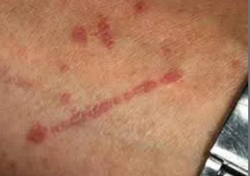
211
Livedo reticularis what is it
purplish, non-blanching, reticulated rash caused by obstruction of the capillaries, leading to swollen venules
212
Livedo reticularis causes
idiopathic, polyarteritis nodosa, SLE, antiphospholipid syndrome, Ehlers Danlos syndrome, homocystinuria
213
Shingles what is it
acute, unilateral painful blistering rash caused by reactivation of VZV. follows primary VZV infection which lies dormant in dorsal root or cranial n ganglia
214
Shingles RFs
increasing age, HIV [strongest RF], other immunosuppressive conditions [steroids, chemotherapy]
215
Shingles clinical features
Prodrome: burning pain over affected dermatome for 2-3 days. Pain is severe and can interfere with sleep. Systemic sx like fever, headache, lethargy
Rash itself is erythematous and macular. Well demaracated by the dermatome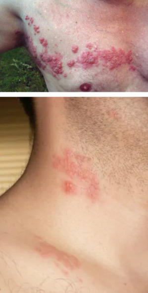
Rash itself is erythematous and macular. Well demaracated by the dermatome
216
Shingles management
Infectious so avoid pregnant / immunocompromised until vesicles have crusted over. Cover the lesions to reduce risk and one-off vaccine for shingles is offered in px over 70yrs old
NSAIDs / paracetamol for neuropathic pain. Corticosteroids given if immunocompetent and NSAIDs is not working
Antivirals [aciclovir] within 72hrs unless px is younger than 50 AND has a mild rash with no RFs.
if severe, admit to hospital for IV antivirals [or if suspecting opthalmic or meningitis]
NSAIDs / paracetamol for neuropathic pain. Corticosteroids given if immunocompetent and NSAIDs is not working
Antivirals [aciclovir] within 72hrs unless px is younger than 50 AND has a mild rash with no RFs.
if severe, admit to hospital for IV antivirals [or if suspecting opthalmic or meningitis]
217
Shingles sequelae
post herpetic neuralgia, herpes zoster ophthalmicus [Hutchinsons nose]
Ramsay Hunt syndrome
Ramsay Hunt syndrome
218
Head lice / nits: what is it
pediculus capitis is organism. feeds on human blood and lays eggs [grey / brown]. Nits are the empty egg shells [white] that have hatched
219
Head lice transmission
via direct HEAD-HEAD CONTACT
nits cannot jump / fly / swim
nits cannot jump / fly / swim
220
head lice management
fine-toothed combing of wet / dry hair
only give meds IF living lice is indicated like malathion, isopropyl myristate, dimeticone
household contacts DO NOT need to be treated unless also affected
only give meds IF living lice is indicated like malathion, isopropyl myristate, dimeticone
household contacts DO NOT need to be treated unless also affected
221
Purpura what is it
bleeding into skin from small blood vessels: leads to non-blanching rash
typically seen in low platelets BUT also in bleeding disorder like vWf
typically seen in low platelets BUT also in bleeding disorder like vWf
222
Children with new purpuric rash
ADMIT for meninogoccal septicaemia or Acute lymphoblastic leukaemia
give parenteral ABx prior to transfer
give parenteral ABx prior to transfer
223 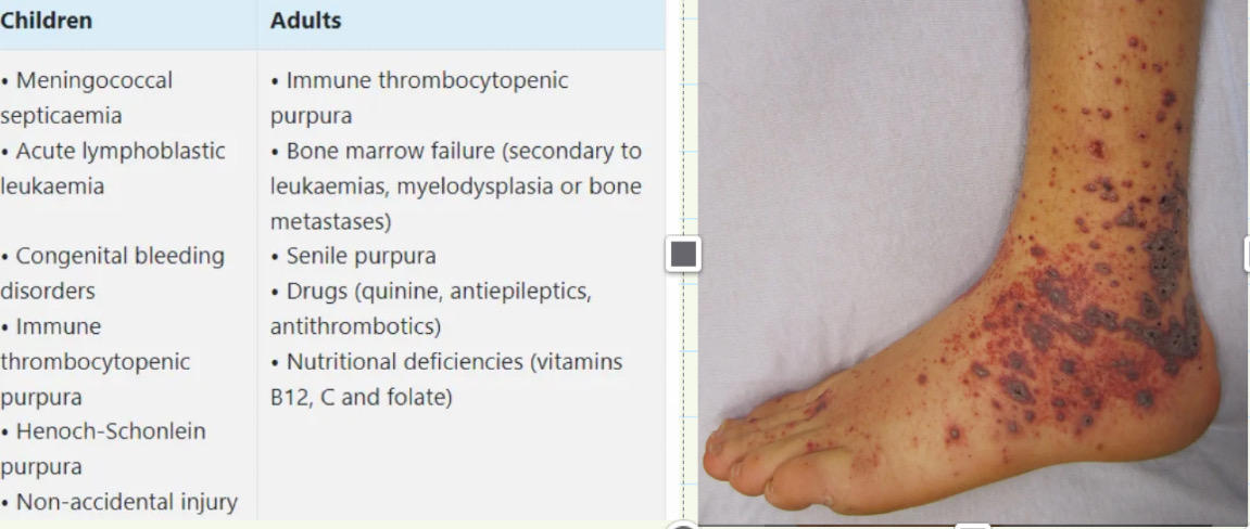
Purpura causes
224
Chicken pox vaccination
live attenuated vaccine [varivax]
indicated in healthcare workers and contacts of immunocompromised px
indicated in healthcare workers and contacts of immunocompromised px
225
Zostavox
shingles vaccine for all px aged over 70yrs. Live attenuated, SC route
SEs: injection site reactions, chickenpox development
SEs: injection site reactions, chickenpox development
226
Alopecia bloods investigations to do
FBC, Ferritin and TFT
227
Androgenetic alopecia
caused due to the change in androgen levels
reassure px
reassure px
228
scarring alopecia: causes and appearance
causes include infection, SLE, lichen planopilaris
will see atrophy / tufting of hairs and the loss of follucular opening
will see atrophy / tufting of hairs and the loss of follucular opening
229
scarring vs non-scarring alopecia
there will be destruction and loss of follicular opening in scarring. This is not seen in non-scarring
230
Seborrhoeic keratoses what is it
benign epidermal skin lesions seen in older people
231
Seborrhoeic keratoses features
large variation in colour from flesh to light borwn to black. Stuck on appearance and keratotic plugs may be seen on surface 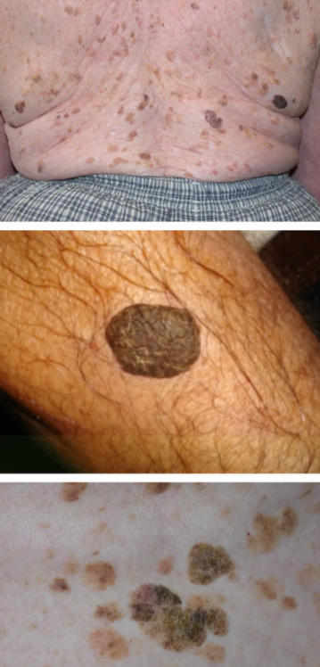
232
Seborrhoeic keratoses management
reassure that it is benign. Remove via curettage, cryotherapy
233
Dermatofibroma: what is it
common benign fibrous skin lesions, caused by the abnormal growth of dermal dendritis histiocyte cells, often preceded by an injury
mostly on legs and arms. Found in immune conditions like HIV / SLE
mostly on legs and arms. Found in immune conditions like HIV / SLE
234
Dermatofibroma: clinical features
solitary firm papule / nodule, 5-10mm in size
dimple / crusting depression when lesion is squeezed and picked. Slow growth over time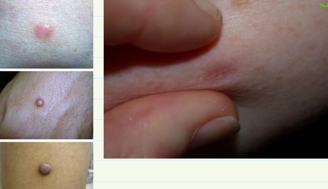
dimple / crusting depression when lesion is squeezed and picked. Slow growth over time
235
Dermatofibroma: management
if asx, no Tx unless bothersome or cosmetically undesirable
surgical excision, cryotherapy, laser therapy
surgical excision, cryotherapy, laser therapy
236
Lipoma: what is it
benign tumour of adipocytes, found in subcutaenous tissues
malignant transformation into liposcarcoma is very rare
malignant transformation into liposcarcoma is very rare
237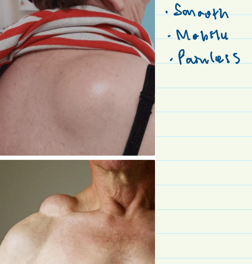
lipoma features
238
Lipoma management
observe, if diagnosis is uncertain OR compressing on surrounding structures = removal
239
liposarcoma suggestive features
size over 5cm
increasing size
pain
deep anatomical location
increasing size
pain
deep anatomical location
240
pyogenic granuloma: causes
trauma, pregnancy, young adult women
241
pyogenic granuloma: features
common on head, neck, upper trunk, hands
initial small red brown spots that develop into raised spherical red / brown lesions. can bleed and ulcerate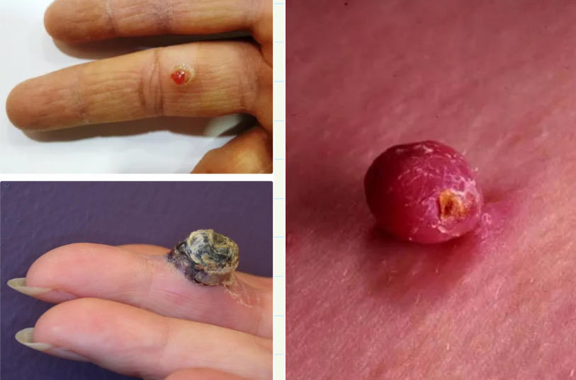
initial small red brown spots that develop into raised spherical red / brown lesions. can bleed and ulcerate
242
pyogenic granuloma: management
lesions in pregnancy usualy resolve spontaneously post partum
removal via curettage + cauterisation, cryotherapy, excision
removal via curettage + cauterisation, cryotherapy, excision
243
Sebaceous cysts
encompasses epidermal cysts and pilar cysts [outer root of the hair follicle]
excision of cyst wall is needed to prevent reoccurence
excision of cyst wall is needed to prevent reoccurence
244
spider naevi vs telagiectasia
spider naevia fill from the centre but telagiectasia fill from edges, when pressing on them and watching them fill
245
spider naevi associations
liver disease, pregnancy, COCp
246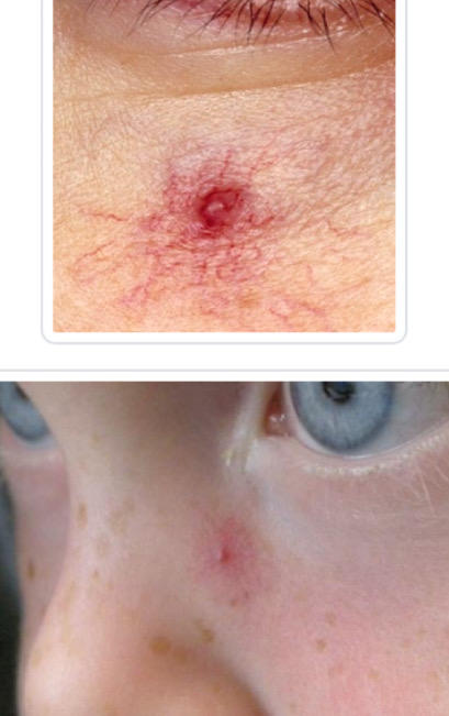
spider naevi
247
Erythrasma: cause, sx, investigations, Tx
ASx, flat, slightly scaly pink / brown rash found on groin / axilla. Caused by an overgrowth of the diphtheroid Corynebacterium Minutissum.
Will see coral-red fluorescence on Wood’s light.
Treat via oral erythromycin
Will see coral-red fluorescence on Wood’s light.
Treat via oral erythromycin
248
Curlings ulcers
Acute gastric ulcers tht develop in response to severe physiological stress such as burns. Involves an increase in gastric acid secretion and a decrease in mucosal blood flow folowing severe burns
249
Bowen’s disease
Precancerous dermatosis that is a precursor to SCC. More common in elderly px.
Will see red / scaly patches in sun-exposed areas
Manage via topical 5-fluorouracil BDS / 4 weeks
Will see red / scaly patches in sun-exposed areas
Manage via topical 5-fluorouracil BDS / 4 weeks
250
Telogen effluvium
loss and thinning of hair in response to severe stress
251
Zinc deficiency
Acrodermatitis which are red, crusted lesions around the perianal / periorificial region. Can also lead to hypogonadism, alopecia, cognitive impairment, short stature