exam 1 - nervous system
Study these flashcardsFree · spaced repetition · iPhone, Android, Mac & web
All cards
1
CNS vs PNS
CNS = brain and spinal cord
PNS = all the nerves that exit from brain (cranial nerves) or spinal cord (spinal nerves)
-- responsible for all sensory and motor signaling!
PNS = all the nerves that exit from brain (cranial nerves) or spinal cord (spinal nerves)
-- responsible for all sensory and motor signaling!
2
"tracks" vs "nerves"
tracks are axon bundles in the CNS
nerves are all the axon bundles in the PNS
nerves are all the axon bundles in the PNS
3
2 divisions of the sensory PNS
1) visceral (focuses on whats going on in ur internal organs; nausea, gas, pain, etc)
2) somatic (focuses on whats happening everywhere else!)
2) somatic (focuses on whats happening everywhere else!)
4
2 divisions of the motor PNS
1) visceral (output to smooth and cardiac muscle; all the involuntary responses!)
-- further divided into sympathetic and parasympathetic
2) somatic (output to skeletal muscle; so ur reflexes and ur voluntary motions)
-- further divided into sympathetic and parasympathetic
2) somatic (output to skeletal muscle; so ur reflexes and ur voluntary motions)
5
2 types of cells that make up nervous tissue
NEURONS: control activities, process info
- excitable
- high metabolic rate
- extreme longevity
- nonmitotic
GLIAL CELLS: support and protect the neurons
- nonexcitable
- mitotic asf
- mop up excess K+ to support extracellular enviro; aid in recovery from injuries
- excitable
- high metabolic rate
- extreme longevity
- nonmitotic
GLIAL CELLS: support and protect the neurons
- nonexcitable
- mitotic asf
- mop up excess K+ to support extracellular enviro; aid in recovery from injuries
6
true or false: since they never have to extend very far before delivering their action potential, neurons are generally short cells
FALSE! they can be pretty long, esp in the PNS when they have to travel through like half of ur body
7
what's going on in this diagram?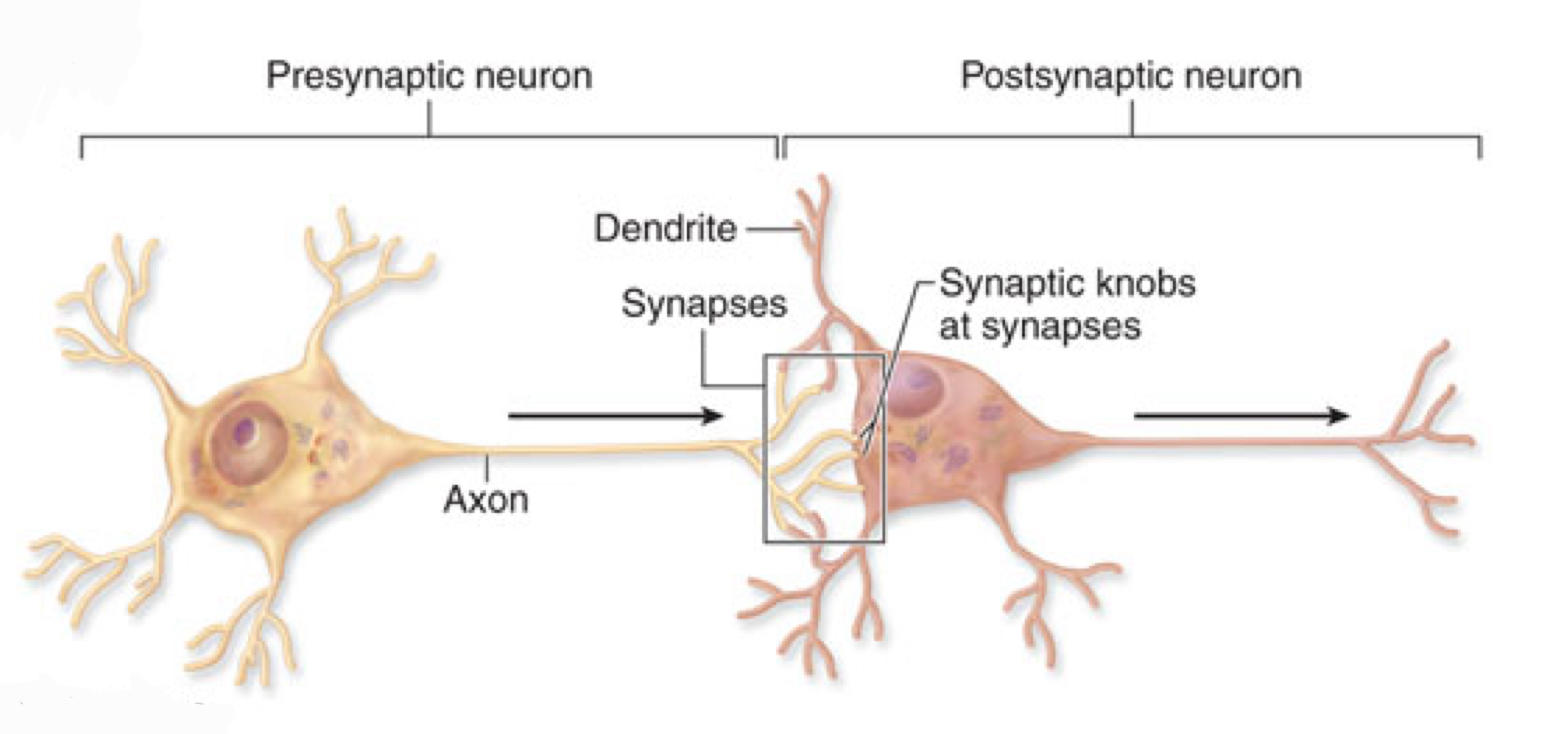
showing the movement of an action potential down a neuron's axon, and across its synapse to the second cell (shown as a neuron here, but in the body it can also just be a muscle cell)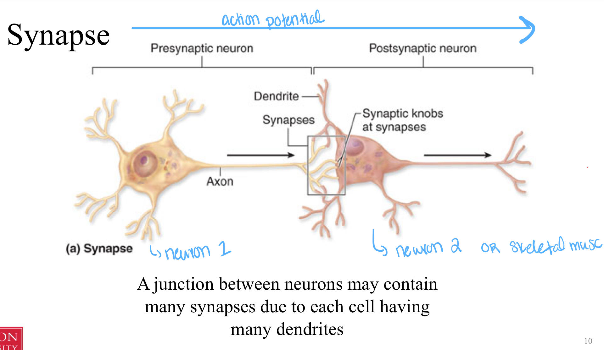
8
how is myelin produced in the PNS vs the CNS?
PNS: schwann cells that kinda live on the axon
CNS: oligodendracytes that live in between diffferent neurons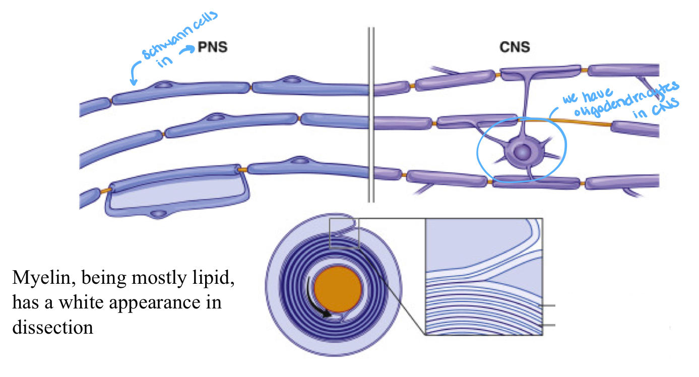
CNS: oligodendracytes that live in between diffferent neurons
9
identify each meningeal layer and describe them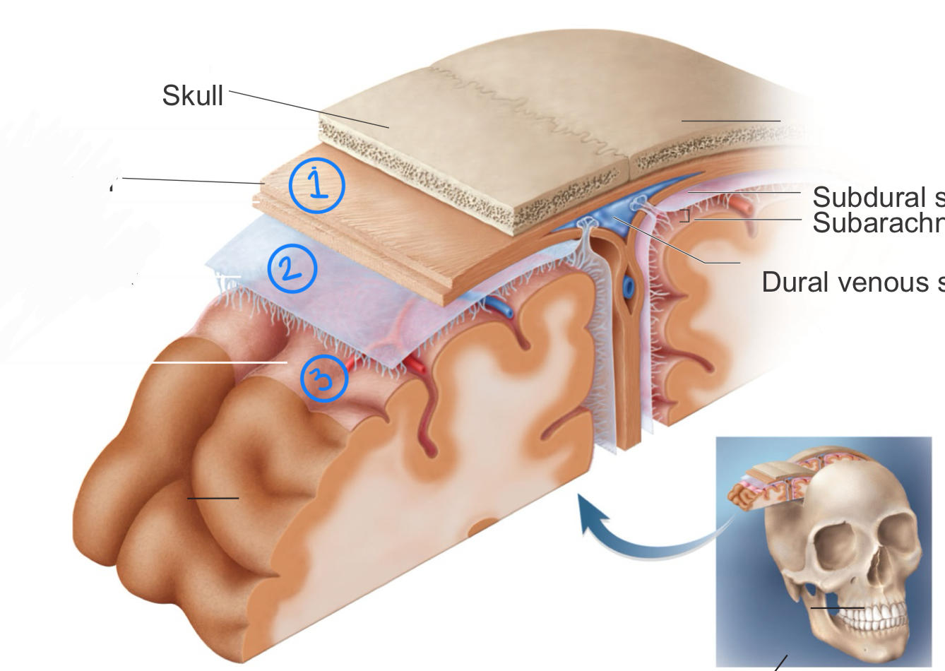
1) dura mater: super thick and fibrous layer! has 2 layers: periosteal and menigeal
2) arachnoid mater: loose-ish shrink wrap vibes! has little "legs" under it that create a space for cerebrospinal fluid
3) pia mater: tight shrink wrap! it's basically adhered to the structure beneath it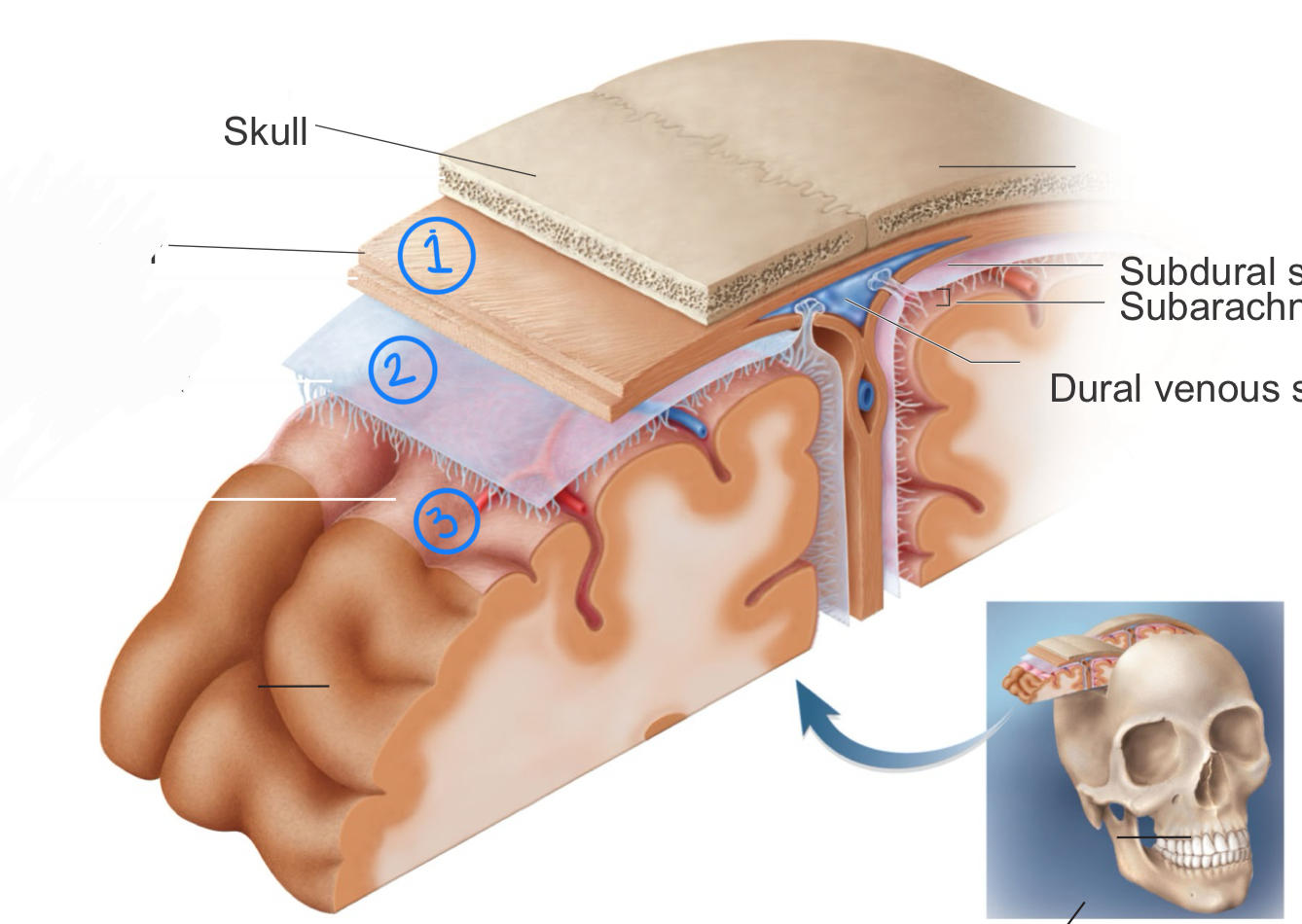
2) arachnoid mater: loose-ish shrink wrap vibes! has little "legs" under it that create a space for cerebrospinal fluid
3) pia mater: tight shrink wrap! it's basically adhered to the structure beneath it
10
why does the subarachnoid space exist?
CSF lives there!
11
t/f: the meningeal layers protect both the brain and spinal cord
true!!
12
identify each structure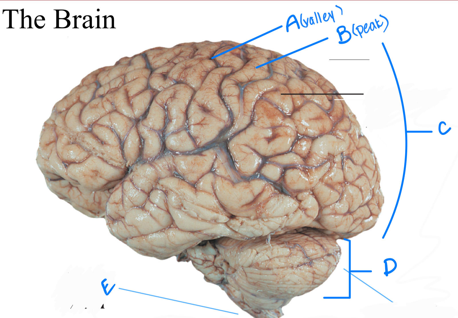
A- sulci
B - gyri
C- cerebrum
D- cerebellum
E - brain stem
B - gyri
C- cerebrum
D- cerebellum
E - brain stem
13
gray matter vs white matter
gray matter is made of the cell bodies of all the neurons in the area
white matter is all the myelin covered axons of the neurons
white matter is all the myelin covered axons of the neurons
14
generally speaking, in the brain, white matter is found ________ and gray matter is found ________
- more superficial; deeper
- deeper; more superficial
- honestly, it's kinda mixed up in there
15
in the spinal cord, white matter is found _________ and gray matter is found _______
- more superficial; deeper
- deeper; more superficial
- honestly, it's kinda mixed up in there
16
name the 5 cerebral lobes
frontal
parietal
occipital
temporal
insula
parietal
occipital
temporal
insula
17
what structure is this?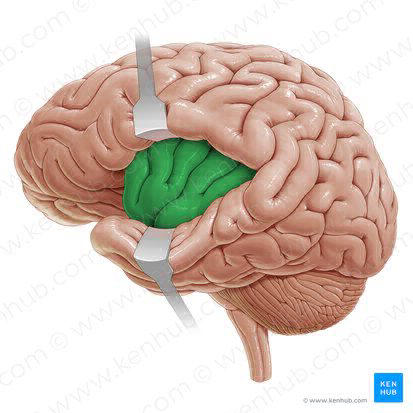
insula (deep lobe of brain)
18
"The brain is incredibly anatomocially specific." What do we mean by this?
like very specific parts of the brain do very specific things; like there's not rlly overlap
19
frontal lobe function
planning, coordinating and executing skeletal muscle movement;
controlling personality, cognition, decision making , planning, language functions
(this is the one that doesnt develop until ur 25 ...)
controlling personality, cognition, decision making , planning, language functions
(this is the one that doesnt develop until ur 25 ...)
20
primary motor cortex
- found in frontal lobe
- aka the precentral gyrus
- controls skeletal muscle activity
(highlighted red here)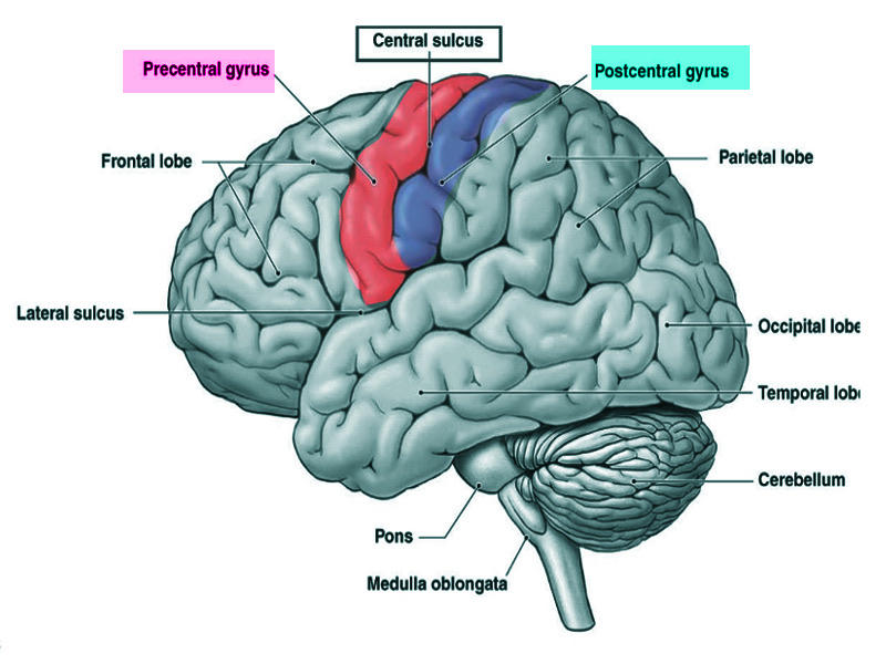
- aka the precentral gyrus
- controls skeletal muscle activity
(highlighted red here)
21
pre-motor cortex
- found in frontal lobe
- plans and coordinates skeletal muscle activity (hence the "pre-motor")
* pre-motor is found before the primary motor cortex duhhh
(highlighted blue here)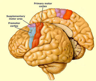
- plans and coordinates skeletal muscle activity (hence the "pre-motor")
* pre-motor is found before the primary motor cortex duhhh
(highlighted blue here)
22
parietal lobe function
interpretting sensory info (via the somatosensory cortex)
23
somatosensory cortex
- found in parietal lobe
- aka the postcentral gyrus
- interprets sensory info
(highlighted blue here)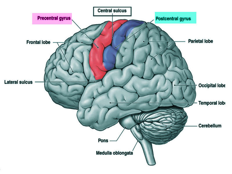
- aka the postcentral gyrus
- interprets sensory info
(highlighted blue here)
24
homunculus diagram
basically shows us how much the precentral/primary motor cortex and the postcentral/somatosensory cortex favor each part of ur body; the bigger the body part is, the more sensitive/controlled it is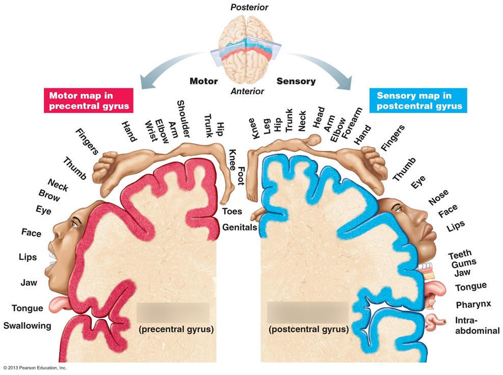
25
temporal lobe function
olfactory and auditor info
26
occipital lobe function
receives and interprets visual input; interprets associated memories with visual input
27
primary visual cortex
- found in occipital lobe
- receives visual input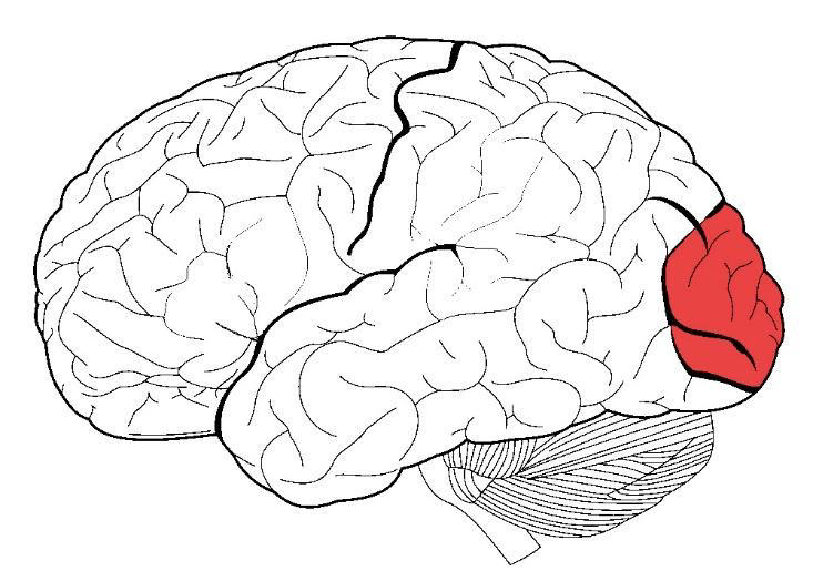
- receives visual input
28
insula lobe function
recieiving and interpretting taste
29
cerebellum
responsible for proprioception (where u are in space)
30
hypothalamus
- control over autonomic nervous system and endocrine system
- nuclei in the hypothalamus control:
--- reg of body temp
--- control of emotional behavior
--- control of food and water intake (so hunger and thirst innit)
* regulatory asf!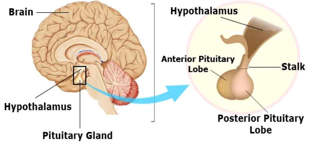
- nuclei in the hypothalamus control:
--- reg of body temp
--- control of emotional behavior
--- control of food and water intake (so hunger and thirst innit)
* regulatory asf!
31
limbic system
collection of deep brain structures that regulate emotions, memories and emotional responses to sensory stimuli
-- includes amygdala and hippocampus
-- connects to reticular formation to coordinate physical responses to fear, emotion and memory (like tensing up, high BP, nausea, etc)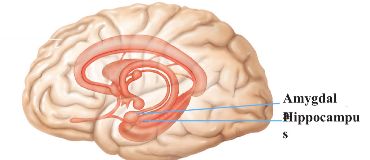
-- includes amygdala and hippocampus
-- connects to reticular formation to coordinate physical responses to fear, emotion and memory (like tensing up, high BP, nausea, etc)
32
identify the amygdala and describe its functions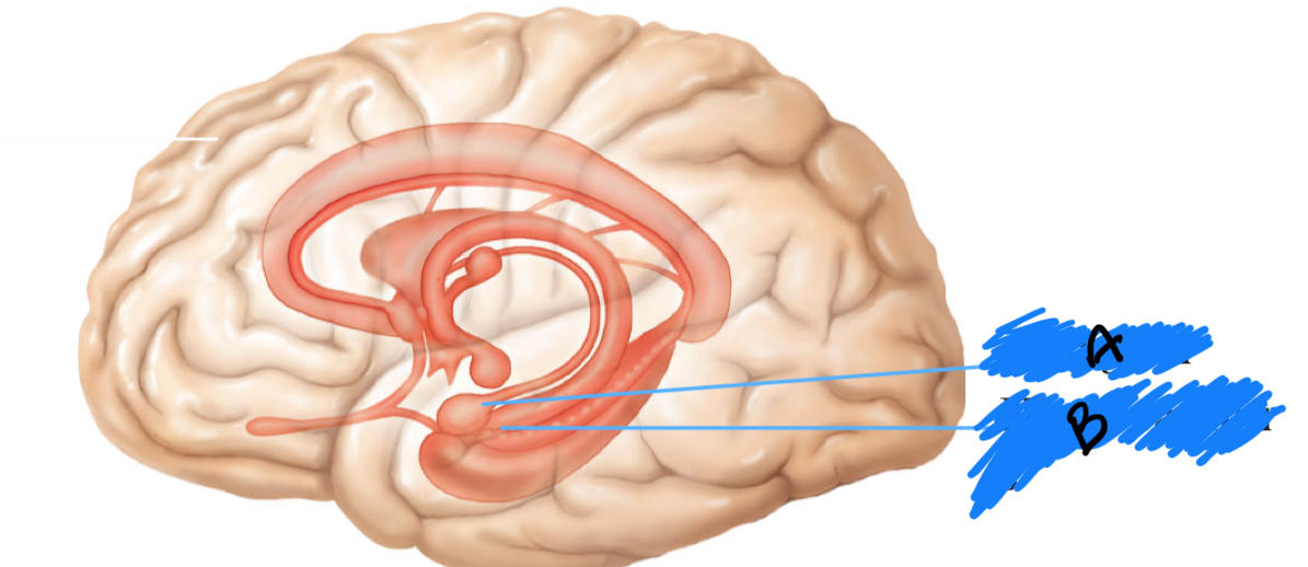
A)
- fear center!
- processes fear and coordinates appropriate nervous system response
- recognizes menacing facial expression
- fear center!
- processes fear and coordinates appropriate nervous system response
- recognizes menacing facial expression
33
identify the hippocampus and describe its functions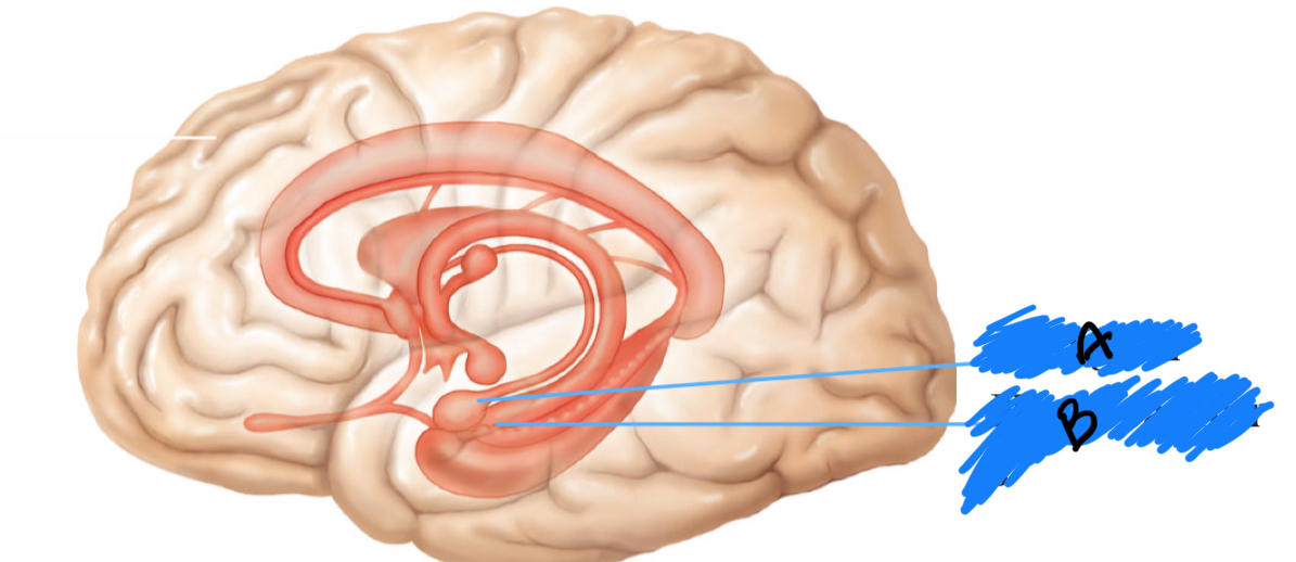
B)
- forms, consolidates and retrieves memories
- forms, consolidates and retrieves memories
34
thalamus
- acts as a filter for somatosensory information
- directs sensory info to correct cerebrum location; also sends info ab where sensation is coming from
- can edit, amplify or diminish info depending on what's important in the moment
--- like, after u put on your clothes, you're not super aware of the sensation of them on your skin all day; you start to focus on other details despite that continuous sensory info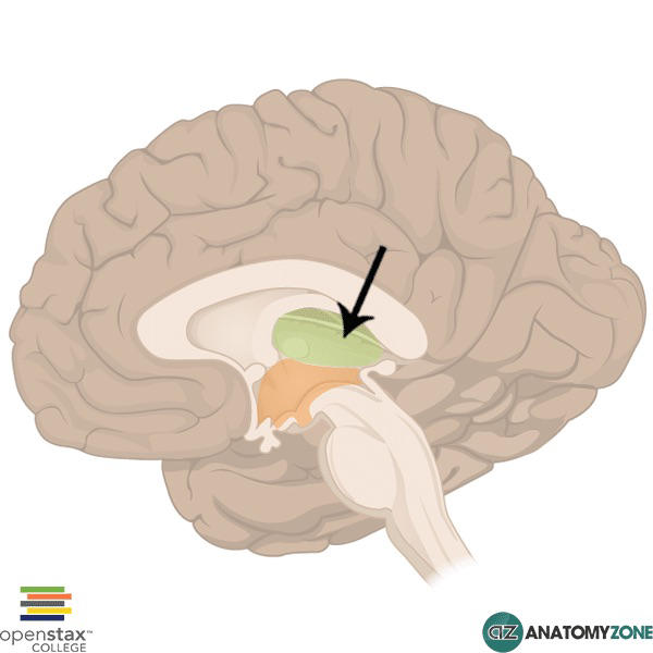
- directs sensory info to correct cerebrum location; also sends info ab where sensation is coming from
- can edit, amplify or diminish info depending on what's important in the moment
--- like, after u put on your clothes, you're not super aware of the sensation of them on your skin all day; you start to focus on other details despite that continuous sensory info
35
what is this structure?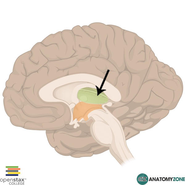
the thalamus
36
midbrain
- start of the brain stem
- made up of individual nuceli with discrete functions
- many centers of control for limb, eye motor movements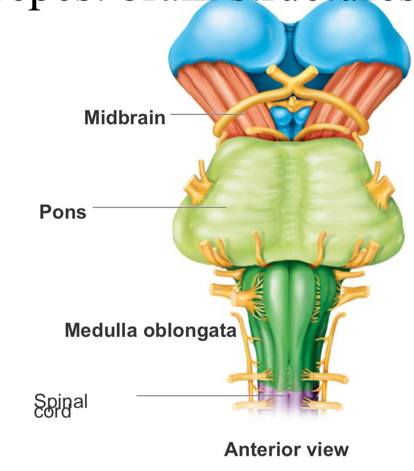
- made up of individual nuceli with discrete functions
- many centers of control for limb, eye motor movements
37
pons
- part of brain stem
- sensory and motor tracts that connect the brain to the spinal cord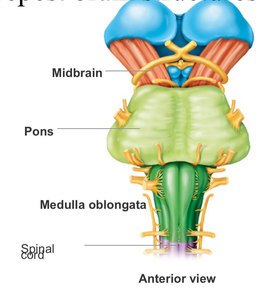
- sensory and motor tracts that connect the brain to the spinal cord
38
medulla oblongata
- last part of brain stem; directly continuous with spinal cord
- all comm btwn brain and spinal cord involves tracts through the medulla!
- important role in cardiac and respiratory control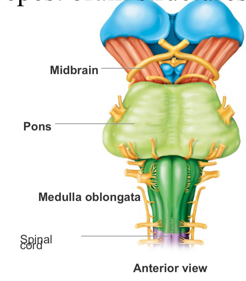
- all comm btwn brain and spinal cord involves tracts through the medulla!
- important role in cardiac and respiratory control
39
spinal cord
- major conduction pathway btwn brain and body
- continuous with brainstem
- extends from the foramen magnum of the skull to L1/L2
- ends in the cauda equina
- protected by bone, ligaments, meninges and CSF
- continuous with brainstem
- extends from the foramen magnum of the skull to L1/L2
- ends in the cauda equina
- protected by bone, ligaments, meninges and CSF
40
identify the following structures on the spinal cord:
- conus medullaris
- filum terminale
- cauda equina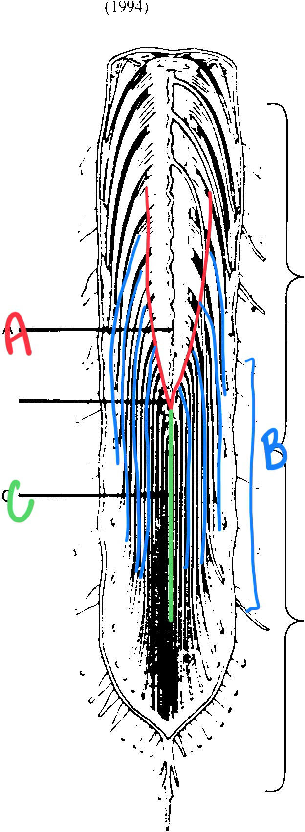
- conus medullaris
- filum terminale
- cauda equina
A - conus medullaris
B - cauda equina
C - filum terminale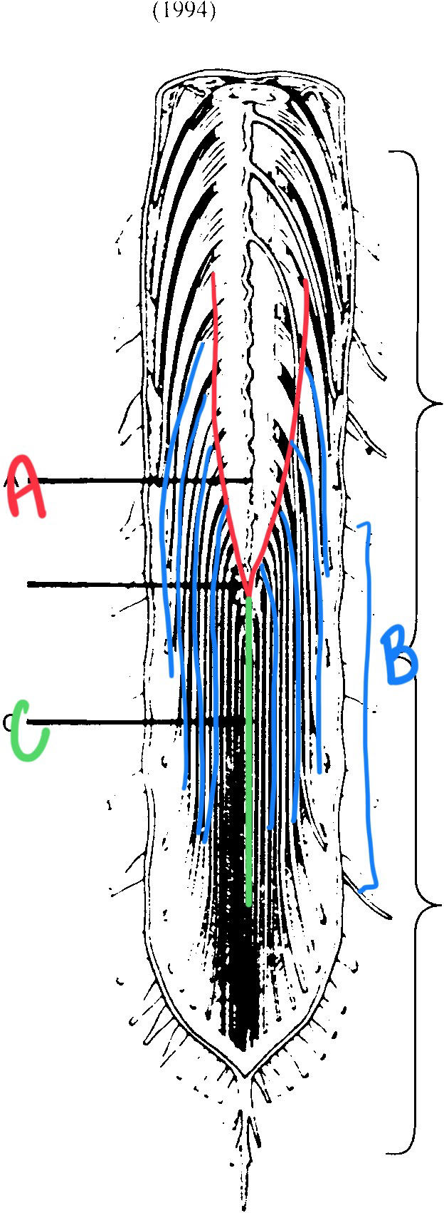
B - cauda equina
C - filum terminale
41
label each spinal cord/vertebral region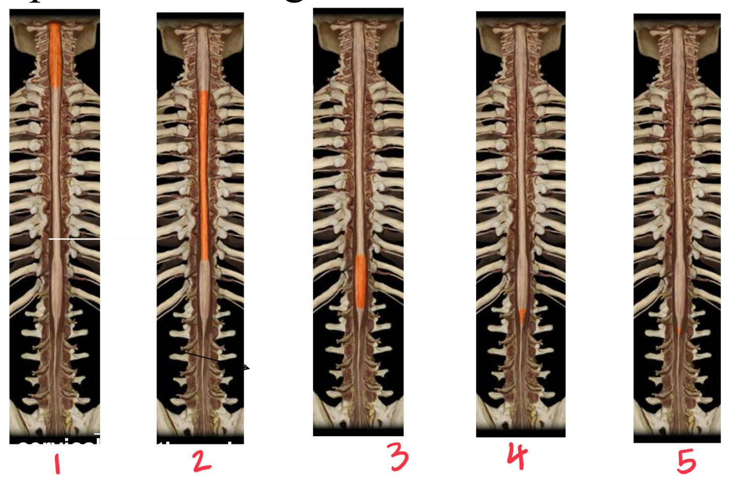
1 - cervical
2- thoracis
3 - lumbar
4- sacral
5- coccygeal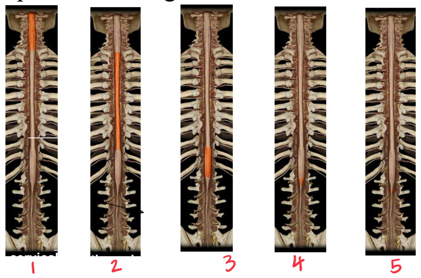
2- thoracis
3 - lumbar
4- sacral
5- coccygeal
42
identify the following spinal cord structures and decide whether they are sensory, motor, autonomic or mixed:
- anterior horn
- lateral horn
- posterior horn
- anterior root
- posterior root
- spinal nerve
- posterior ramus
- anterior ramus
- spinal ganglion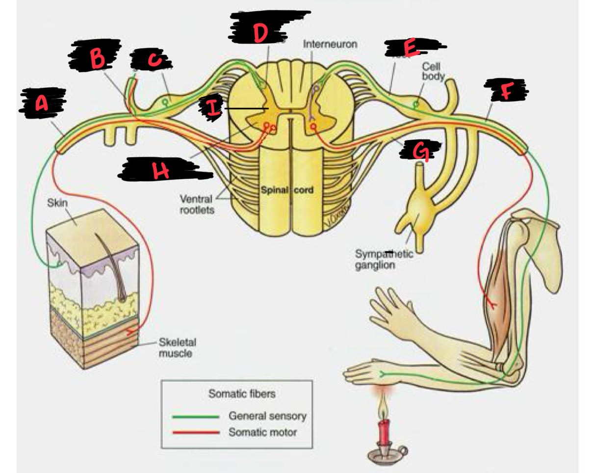
- anterior horn
- lateral horn
- posterior horn
- anterior root
- posterior root
- spinal nerve
- posterior ramus
- anterior ramus
- spinal ganglion
A - anterior ramus (mixed, to trunk and limbs)
B - posterior ramus (mixed, to back muscles and skin)
C - spinal ganglion (sensory, cell bods)
D - posterior horn (sensory)
E - posterior root (sensory)
F - spinal nerve (mixed)
G - anterior root (motor)
H - anterior horn (motor)
I - lateral horn (autonomic; reflexive interneurons)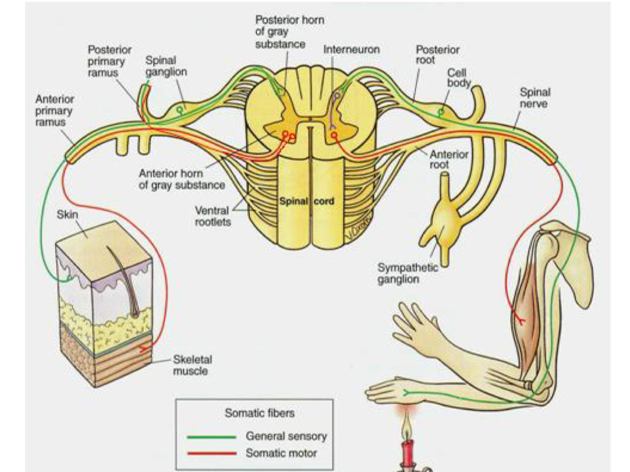
B - posterior ramus (mixed, to back muscles and skin)
C - spinal ganglion (sensory, cell bods)
D - posterior horn (sensory)
E - posterior root (sensory)
F - spinal nerve (mixed)
G - anterior root (motor)
H - anterior horn (motor)
I - lateral horn (autonomic; reflexive interneurons)
43
the anterior/ventral structures of the spinal cord (horn, root) are exclusively __________ pathways
- motor
- sensory
- mixed
- autonomic
44
the posterior/dorsal structures of the spinal cord (horn, root, spinal ganglion) are exclusively ___________ pathways
- motor
- sensory
- mixed
- autonomic
45
true or false: spinal nerves and rami are all mixed pathways, regardless of whether they are posterior or anterior.
true!!!!! once you pass the roots, the pathways converge into one mixed spinal nerve
* think ab it: all parts of your body need to send sensory info and then recieve a motor response duh
* think ab it: all parts of your body need to send sensory info and then recieve a motor response duh
46
what structure are you in if you are traveling towards the spinal cord with only signals that have sensory info?
dorsal/posterior root
47
which structure are you currently in if the signal is traveling from the front of your hand towards your spine?
ventral rami
* ventral bc it's from a limb, not ncessarily bc it's sensory info!
* ventral bc it's from a limb, not ncessarily bc it's sensory info!
48
what structure are in if you are motor info about to leave the spinal cord?
ventral horn
49
what structure are you in if you are conneting to a motor axon in the spinal cord?
interneuron
50
what is the spinal ganglion?
it's a little bulb btwn the spinal nerve and dorsal root that contains sensory cell bodies!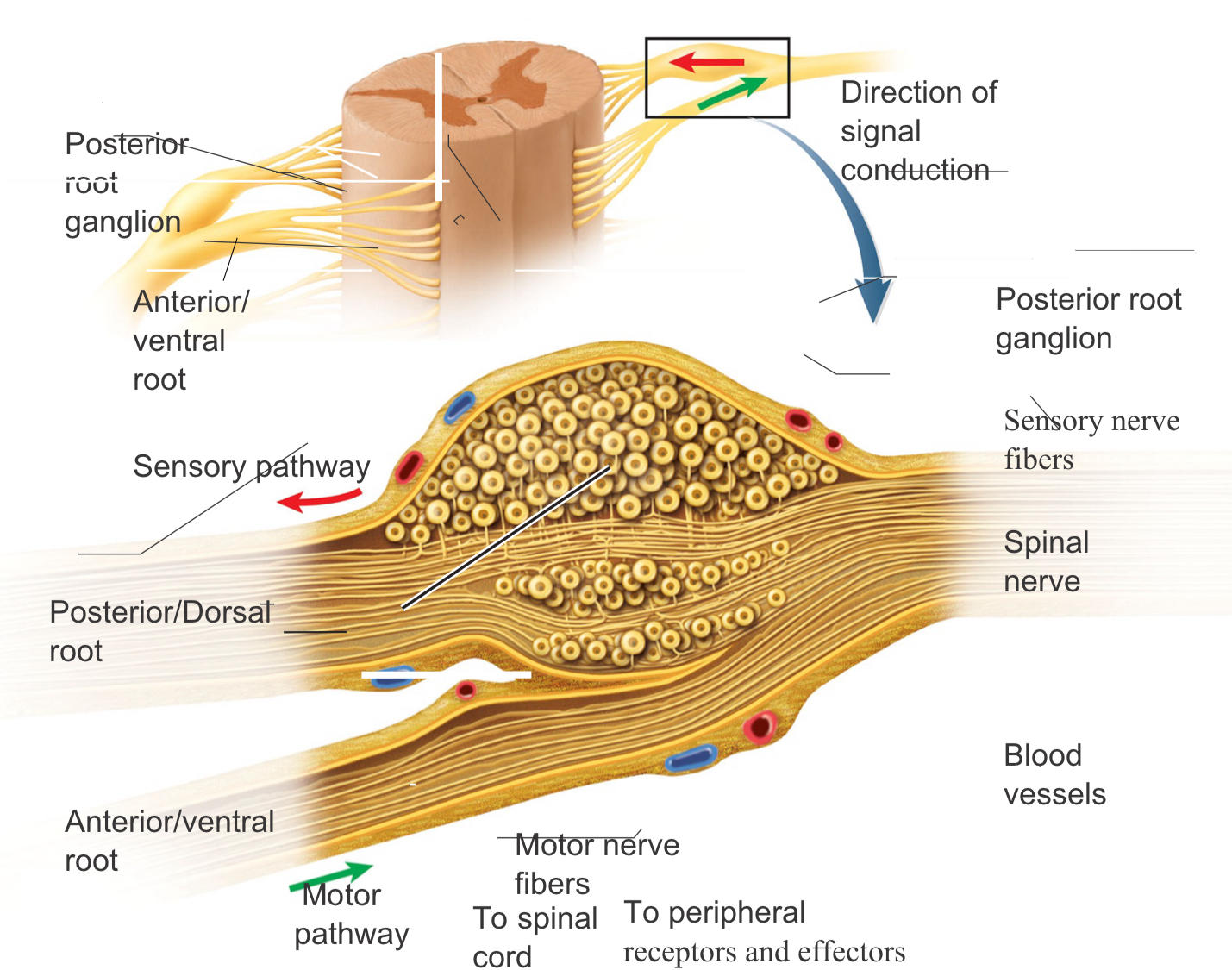
51
if all the sensory cell bodies ar ein the spinal gaglions, where are the motor and interneuron cell bodies located?
in the gray matter of the spinal cord (central ofc)
52
nerve plexus
- plexus literally means "braid"
- it's where branches of a spinal nerve will split, merge, split, merge and split again
- mostly done so a lot of nerves can get into a relatively tight space
- also it's like a little back up plan: if one nerve gets damaged at a more peripheral location, then it's still innervated by several diff sources more upstream
- big "named" nerves split off from the plexus to innervate muscles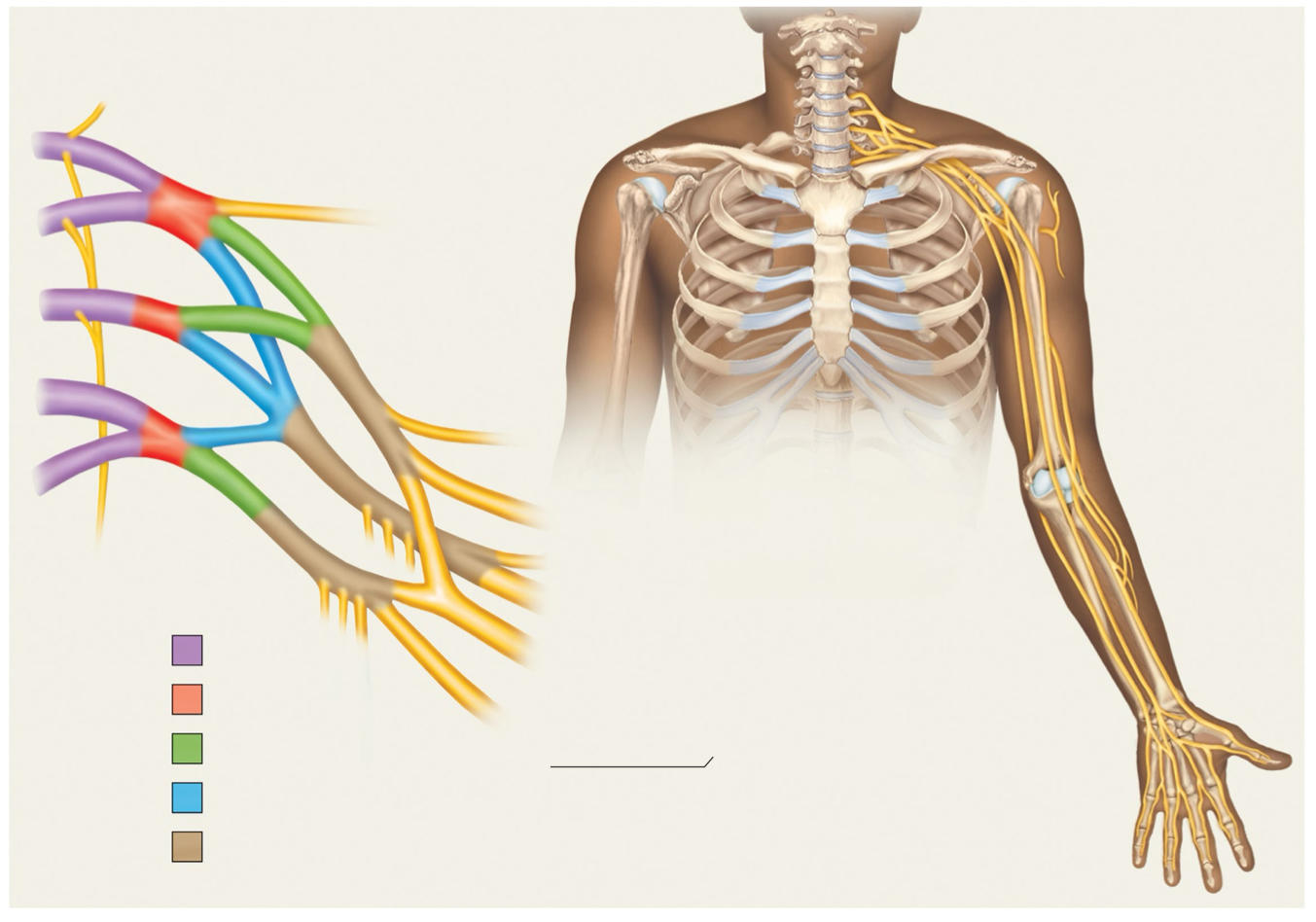
- it's where branches of a spinal nerve will split, merge, split, merge and split again
- mostly done so a lot of nerves can get into a relatively tight space
- also it's like a little back up plan: if one nerve gets damaged at a more peripheral location, then it's still innervated by several diff sources more upstream
- big "named" nerves split off from the plexus to innervate muscles
53
what do we mean by "a system of redundancy" when discussing nerve plexuses?
it's like a little back up plan: if one nerve gets damaged at a more peripheral location, then it's still innervated by several diff sources more upstream
* in the pic, the cervical plexus is a junction of spinal nerves C1,2,3,4 -- for every peripheral nerve, there's a little innervation from each -- if one happens to get damaged, ur still gonna be innervated by smthn!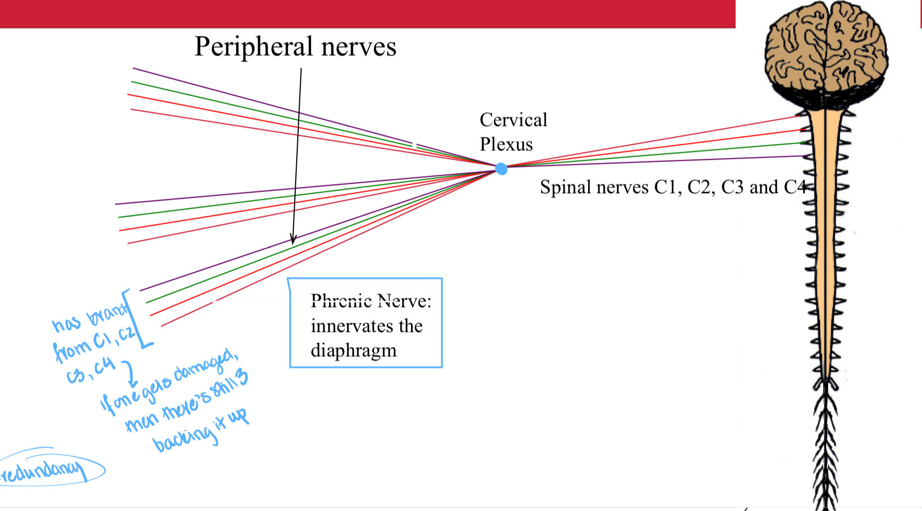
* in the pic, the cervical plexus is a junction of spinal nerves C1,2,3,4 -- for every peripheral nerve, there's a little innervation from each -- if one happens to get damaged, ur still gonna be innervated by smthn!
54
cervical plexus
- forms from anterior rami C1-4, deep to sternocleidomastoid
- sensory innervation of the neck
- motor innervation of severeal local muscles
- most significant nerve = phrenic nerve (innervates the diaphragm!)
- sensory innervation of the neck
- motor innervation of severeal local muscles
- most significant nerve = phrenic nerve (innervates the diaphragm!)
55
brachial plexus
- forms from anterior rami C5-T1, deep to clavicle
- sensory and motor innervation of the axilla and arms
- significant nerves include: musculotaneous, median, radial, ulnar
- sensory and motor innervation of the axilla and arms
- significant nerves include: musculotaneous, median, radial, ulnar
56
lumbar plexus
- forms from anterior rami of L1-4 (with contributions from T12)
- embedded within the psoas muscle
- sensory and motor innervation to the abdominal wall muscles, ant and medial thigh muscles
- significant nerves: iliohypogastric, ilioinguinial, femoral, genitofemoral, obturator
- embedded within the psoas muscle
- sensory and motor innervation to the abdominal wall muscles, ant and medial thigh muscles
- significant nerves: iliohypogastric, ilioinguinial, femoral, genitofemoral, obturator
57
sacral plexus
- forms from anterior rami of L4-S4, is found embedded in psoas muscle
- sensory and motor innervation to the genitals, post thigh muscles, gluteals, and most of lower leg and foot
- significant nerves: sciatic, pudendal
- sensory and motor innervation to the genitals, post thigh muscles, gluteals, and most of lower leg and foot
- significant nerves: sciatic, pudendal
58
dermatomes and myotomes
gives u a map of which spinal nerves innervate what regions
-- dermatomes = sensory
-- myotomes = motor
* super useful bc they can help us figure out the causes of nervous injury (like it can help narrow down if it's more peripheral or central)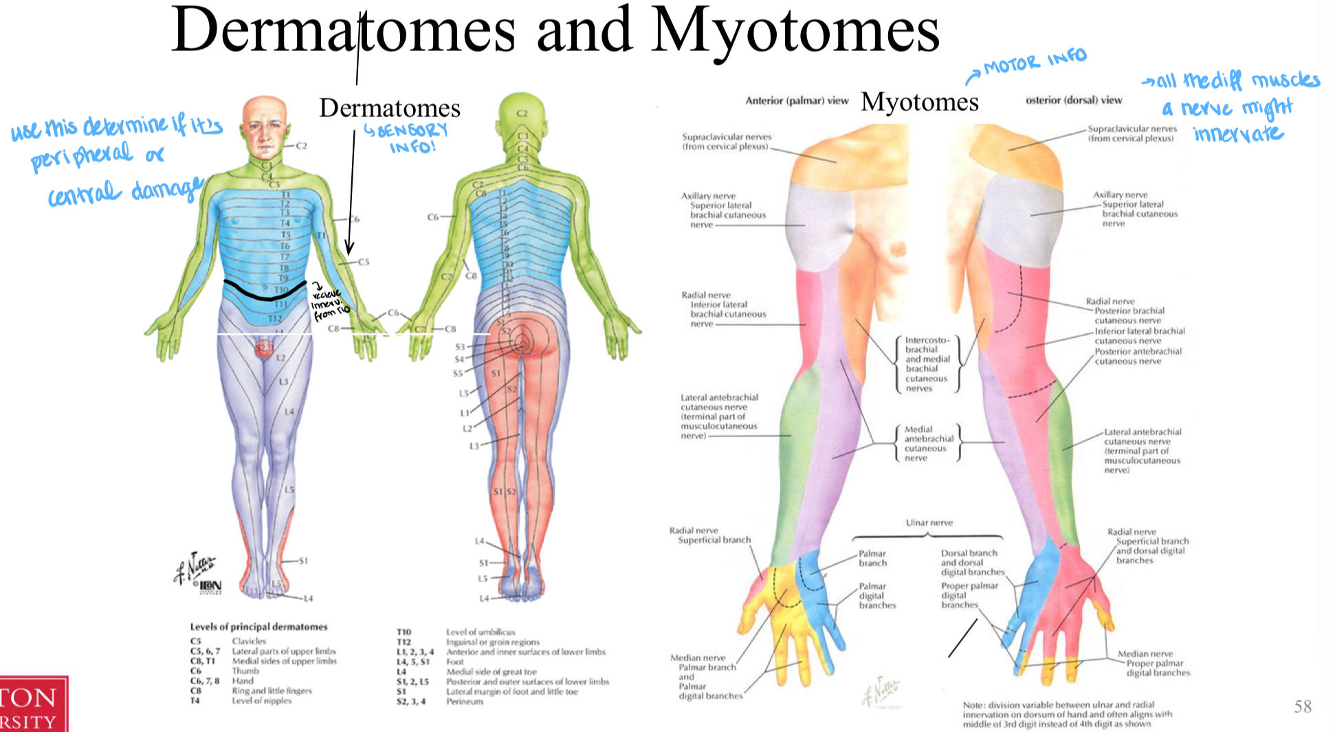
-- dermatomes = sensory
-- myotomes = motor
* super useful bc they can help us figure out the causes of nervous injury (like it can help narrow down if it's more peripheral or central)
59
cranial nerves (CN)
- there are 12 (CN1-12)
- innervate ur eyes and face bookie
- come directly from the brain!
- innervate ur eyes and face bookie
- come directly from the brain!
60
challenge! list all 12 cranial nerves!
1. olfactory
2. optic
3. oculomotor
4. trochlear
5. trigeminal
6. abducens
7. facial
8. vestibulocochlear
9. glossopharyngeal
10. vagus
11. accessory
12. hypoglossal
2. optic
3. oculomotor
4. trochlear
5. trigeminal
6. abducens
7. facial
8. vestibulocochlear
9. glossopharyngeal
10. vagus
11. accessory
12. hypoglossal