Facial Recognition
Study these flashcardsFree · spaced repetition · iPhone, Android, Mac & web
All cards
1
Define prosopagnosia
Specific inability to recognise faces
Can sometimes result from brain damage
Can sometimes result from brain damage
2
6 types of evidence for the brain having a dedicated neural system for facial recognition
Selective losses after brain damage
fMRI
Heritability of individual differences
Single-unit recording
Developmental issues
Selective adaptation to properties of faces
fMRI
Heritability of individual differences
Single-unit recording
Developmental issues
Selective adaptation to properties of faces
3
Which brain area is acquired prosopagnosia associated with?
Lesions of the fusiform gyrus, especially right hemisphere
4
Example of acquired prosopagnosia
Wada and Yamamoto 2001
Man had brain haemorrhage
Could not recognise family/himself
Famours person recognition test: 8/38 (mean control = 36)
BUT normal recognition of objects, normal colour vision
Lesion to right inferior occipital region at level of fusiform gyrus and inferior temporal gyrus
Man had brain haemorrhage
Could not recognise family/himself
Famours person recognition test: 8/38 (mean control = 36)
BUT normal recognition of objects, normal colour vision
Lesion to right inferior occipital region at level of fusiform gyrus and inferior temporal gyrus
5
Define double dissociation
When two related mental processes are shown to function independently of each other
Eg. speech production and speech comprehension, occupy different brain areas
Eg. speech production and speech comprehension, occupy different brain areas
6
Evidence of double dissociation
CK case study
Moscovitch et al, 1997
Head injury
Assoicative visual agnosia (can't visually recognise common objects)
Severely alexic (can't read)
BUT can still identify faces, unless faces are inverted (including cartoons)
May be because most ppl solve this task by using processes that are also used in object recognition
So understanding non-inverted and inverted faces use different processes, despite seeming linked
Moscovitch et al, 1997
Head injury
Assoicative visual agnosia (can't visually recognise common objects)
Severely alexic (can't read)
BUT can still identify faces, unless faces are inverted (including cartoons)
May be because most ppl solve this task by using processes that are also used in object recognition
So understanding non-inverted and inverted faces use different processes, despite seeming linked
7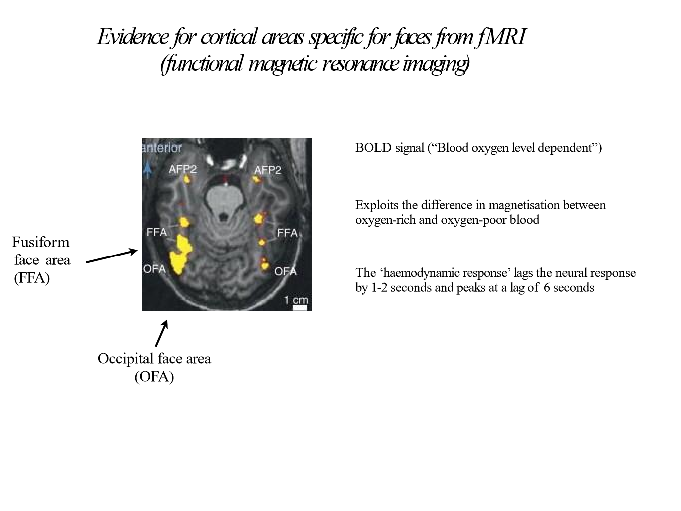
Image occlusion: name the hidden labels
8
What did Kanwisher, McDermott and Chun find and when?
1997
fMRI
Identified a restricted cortical area in right hemisphere that responds differently to faces than to objects (FFA)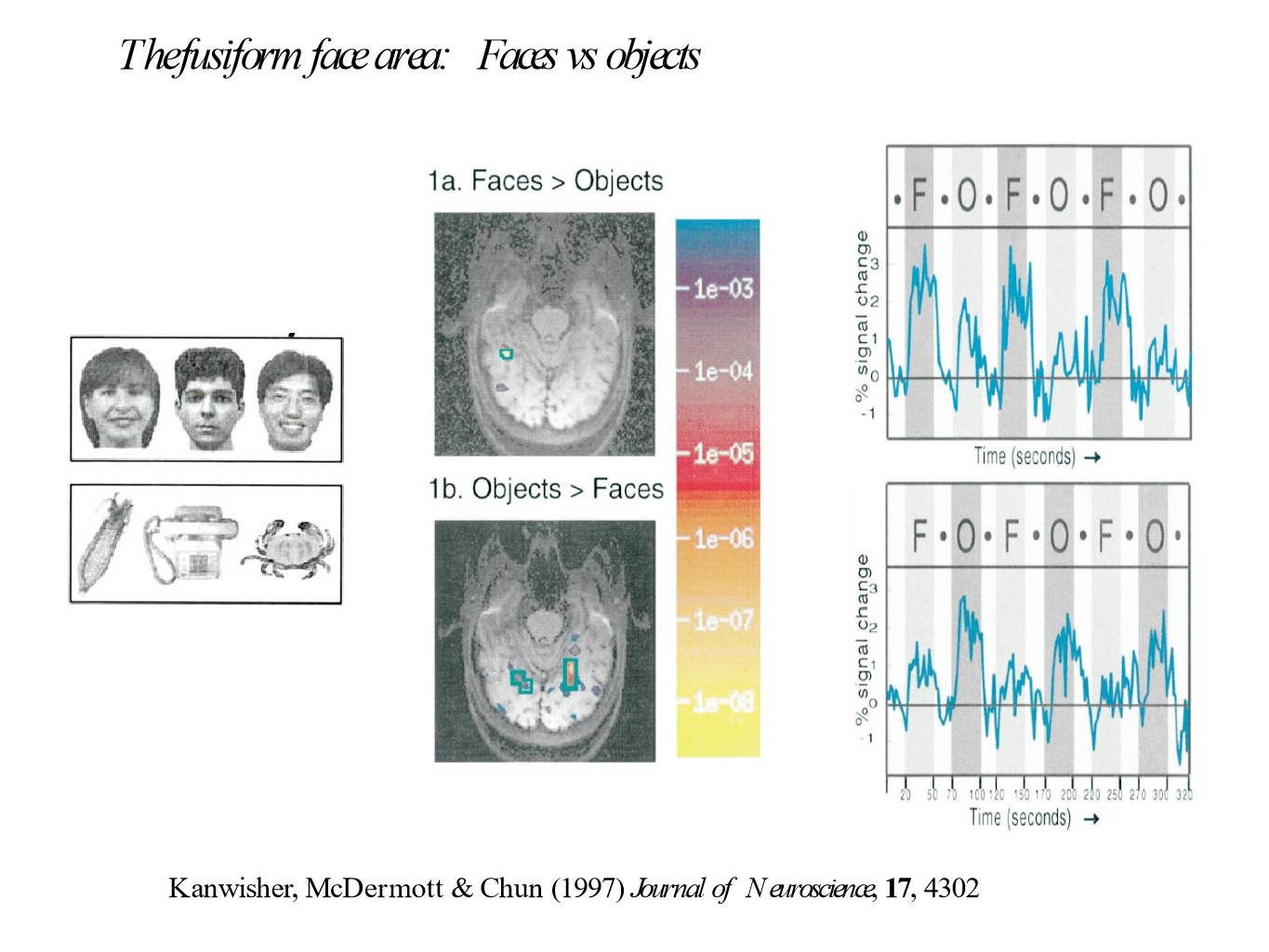
fMRI
Identified a restricted cortical area in right hemisphere that responds differently to faces than to objects (FFA)
9
Alternative explanations to Kanwisher, McDermott and Chun's findings
Images of faces and objects may differ in low-level features (contrast, texture)
FFA may respond more strongly when discriminating between similar stimuli (faces) than between different categories (objects)
FFA may respond to any part of an animate object
Could be because we pay more attention to faces > objects
Kanwisher et al ruled these explanations out with further tests
FFA may respond more strongly when discriminating between similar stimuli (faces) than between different categories (objects)
FFA may respond to any part of an animate object
Could be because we pay more attention to faces > objects
Kanwisher et al ruled these explanations out with further tests
10
What did Lafer-Sousa find and when?
2016
Findings suggest more than 1 face-specific region of temporal lobe
Findings suggest more than 1 face-specific region of temporal lobe
11
Define heritability
Measure of current variance in a trait in a population that can be attributed to genetic variations within the population
12
What did Wilmer et al do and when?
2010
MZ and DZ twins tested
Cambridge Face Memory Test (CFMT)
Abstract Art Memory Test
Verbal Paired Associates Memory Test
MZ correlations on CFMT = 0.70
DZ = 0.29
Suggests high heritability for face recognition
Only small correlations between CFMT and the 2 other tests
No significant relationship between facial recognition and IQ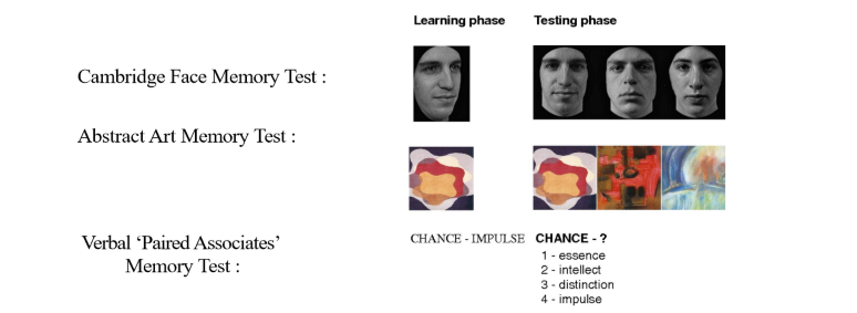
MZ and DZ twins tested
Cambridge Face Memory Test (CFMT)
Abstract Art Memory Test
Verbal Paired Associates Memory Test
MZ correlations on CFMT = 0.70
DZ = 0.29
Suggests high heritability for face recognition
Only small correlations between CFMT and the 2 other tests
No significant relationship between facial recognition and IQ
13
Define congenital prosopagnosia
Genetically determined, life-long difficulty in recognising faces
BUT normal intelligence, low-level visual processing and social cognition
BUT normal intelligence, low-level visual processing and social cognition
14
Opposite of prosopagnosia
Super-recognisers
15
What did Tsao do and when?
2006
fMRI in macaque monkeys to localise face specific areas
Then recorded from these areas with microelectrodes (single neuron recordings)
Found 97% of visually responsive neurons responded more to faces > objects
Most cells responded to human, monkey and schematic faces
Thought to be mokey homologue of human FFA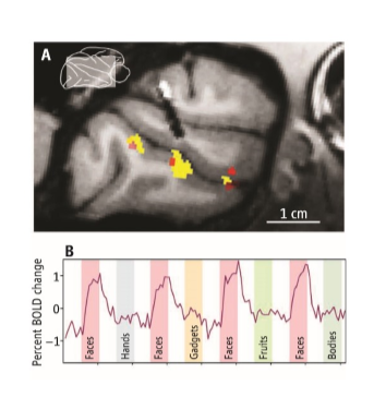
fMRI in macaque monkeys to localise face specific areas
Then recorded from these areas with microelectrodes (single neuron recordings)
Found 97% of visually responsive neurons responded more to faces > objects
Most cells responded to human, monkey and schematic faces
Thought to be mokey homologue of human FFA
16
What did Tsao find in 2016?
Several face patches identified in macaque cortex strongly interconnected to subcortical structures (eg. amygdala)
Strangely, little connection to prefrontal cortex
Strangely, little connection to prefrontal cortex
17
What did McMahon et al do and when?
2014
Recorded specific cells in superior temporal sulcus in macaques
Easy to find cells which respond to specific stimuli - ergo, many thousands of cells must be responding to the same stimulus
Long latencies (~200ms) so responses not an early stage of perceptual analysis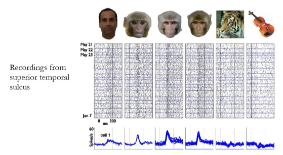
Recorded specific cells in superior temporal sulcus in macaques
Easy to find cells which respond to specific stimuli - ergo, many thousands of cells must be responding to the same stimulus
Long latencies (~200ms) so responses not an early stage of perceptual analysis
18
What did Quiroga et al do and when?
2005
Electrodes implanted into 8 pps with epilepsy (not successful treated with drugs) for 7-10 days
In medial temporal lobe
Specific cells responded to specific ppl
Latency ~300ms so not part of primary face-perception system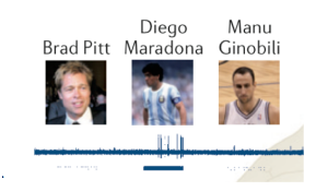
Electrodes implanted into 8 pps with epilepsy (not successful treated with drugs) for 7-10 days
In medial temporal lobe
Specific cells responded to specific ppl
Latency ~300ms so not part of primary face-perception system
19
Criticism of Quiroga et al's study
- Suggests only one cell responds to each familiar person BUT very small sample size as there are millions of neurons just within medial temporal lobe. Other cells may be responding
20
What did Johnson et al do and when?
1991
24 neonates
Schematic = ~ 41 degree head movement
Scrambled = ~31
Blank = ~16
All results statistically significant
Double blind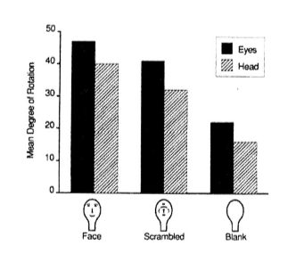
24 neonates
Schematic = ~ 41 degree head movement
Scrambled = ~31
Blank = ~16
All results statistically significant
Double blind
21
What is a popular hypothesis for neonate's responses to face-like stimuli?
Likely to be mediated by sub-cortical pathways (eg. amygdala)
22
What did Reif et al do and when?
2017
Stimulus presented to foetuses
More head turns towards face-like stimuli
BUT many showed no response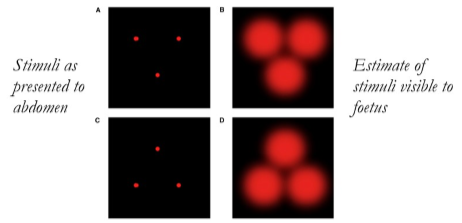
Stimulus presented to foetuses
More head turns towards face-like stimuli
BUT many showed no response
23
What did Webster do and when?
2004
Used adaptation to examine perceptual processes
Pps adapted to extreme (eg. male)
Then shown morphed neutral face (male/female)
Pps distinctly saw the opposite to the face they were adapted to before (eg. see only female in neutral condition)
Effects appear to not depend on adaptation to low-level visual features
There is transfer of the after-effects across size, retinal position and orientation
Eg. adapt to face tilted 45 degrees left that has been distorted, tested with face tilted 45 degrees right = after effect along face axis that was adapted, not the axis of retinotopic space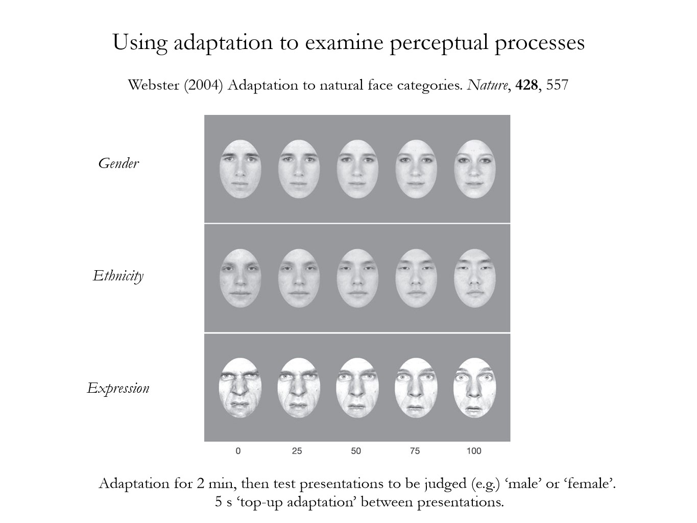
Used adaptation to examine perceptual processes
Pps adapted to extreme (eg. male)
Then shown morphed neutral face (male/female)
Pps distinctly saw the opposite to the face they were adapted to before (eg. see only female in neutral condition)
Effects appear to not depend on adaptation to low-level visual features
There is transfer of the after-effects across size, retinal position and orientation
Eg. adapt to face tilted 45 degrees left that has been distorted, tested with face tilted 45 degrees right = after effect along face axis that was adapted, not the axis of retinotopic space
24
What conclusions can be drawn from Webster's study?
Don't exactly know what is being adapted within the brain or where it is located
Existence of after effects suggest we have a neural representation specific for classes of this stimuli (faces)
Existence of after effects suggest we have a neural representation specific for classes of this stimuli (faces)