Brain Imaging
Study these flashcardsFree · spaced repetition · iPhone, Android, Mac & web
All cards
1
Difference between MRI and fMRI
MRI studies brain antomy
fMRI studies brain function
fMRI studies brain function
2
What does fMRI measure?
BOLD (blood oxygen level dependent signal)
More neural activity = more energy needs = more O2 needed = more blood flow = higher fMRI signal
More neural activity = more energy needs = more O2 needed = more blood flow = higher fMRI signal
3
What does BOLD measure and what does it not measure?
Metabolic demands of neurons
Not neural activity
Neural activity is infered
Not neural activity
Neural activity is infered
4
Evaluate fMRI
+ High spatial resolution, very precise (although low spatial resolution in comparison to invasive techniques used in non-human animals)
+ Non invasive
+ Can look at large structure of the brain
+ Whole brain coverage to uncover networks in health and disease
- Low temporal resolution, 2-6 seconds (a lot slower than the brain works), not the technique which is slow, but the variable which is being measured
- Indirect measure of neural activity
- Correlational not causal relationship between BOLD and behaviour
+ Non invasive
+ Can look at large structure of the brain
+ Whole brain coverage to uncover networks in health and disease
- Low temporal resolution, 2-6 seconds (a lot slower than the brain works), not the technique which is slow, but the variable which is being measured
- Indirect measure of neural activity
- Correlational not causal relationship between BOLD and behaviour
5
Define local field potential (LFP)
Slower neuron signal that reflects post-synaptic potentials, and reflects info that comes from nearby neurons (population activity)
6
Define multi-unit activity
Reflects AP
Similar to what most electrophysiology measures
Similar to what most electrophysiology measures
7
Study which looked at correlation between BOLD activity and neuronal activity
Logothetis et al 2001
Combined BOLD fMRI and electrophysiological recordings
BOLD correlates with spike output (exact info the neuron is currently processing)
BUT correlates better with local field potentials (LFP) compared to multi-unit activity (MUA)
Combined BOLD fMRI and electrophysiological recordings
BOLD correlates with spike output (exact info the neuron is currently processing)
BUT correlates better with local field potentials (LFP) compared to multi-unit activity (MUA)
8
What does BOLD reflect and what are the implications?
AP
Excitatory/inhibitry input to an area
Inputs from same layer
Feedforward/feedback projections
Small change in neural activity of many neurons may show larger activation differences than large change in only a few neurons
Ergo, fMRI results may not always match with neurophysiological results
Excitatory/inhibitry input to an area
Inputs from same layer
Feedforward/feedback projections
Small change in neural activity of many neurons may show larger activation differences than large change in only a few neurons
Ergo, fMRI results may not always match with neurophysiological results
9
How does fMRI work?
Protons in body line up with magnetic field and spin
Protons in deoxy blood not as well organised with their spinning
When brain area is active = AP = increased blood flow and O2 = displacement of deoxyHb from capillaries, venules, small veins
MRI scanner measure difference in proton spinning bewteen oxy and deoxy blood
Protons in deoxy blood not as well organised with their spinning
When brain area is active = AP = increased blood flow and O2 = displacement of deoxyHb from capillaries, venules, small veins
MRI scanner measure difference in proton spinning bewteen oxy and deoxy blood
10
Name 5 other brain imaging methods
Diffusion tensor imaging (DTI)
Electroencephalogram (EEG)
Magnetoencephalography (MEG)
Brain lesions
Transcranial magnetic stimulation (TMS)
Electroencephalogram (EEG)
Magnetoencephalography (MEG)
Brain lesions
Transcranial magnetic stimulation (TMS)
11
Outline DTI
Measures anatomical connectivity between brain regions
Provides info about location and orientation of white matter tracts based on magnitude and direction of molecular diffusion
No restrictions = diffuse equally in all directions (isotropic)
Restrictions = diffusion may occur along one axis (anisotropic)
Provides info about location and orientation of white matter tracts based on magnitude and direction of molecular diffusion
No restrictions = diffuse equally in all directions (isotropic)
Restrictions = diffusion may occur along one axis (anisotropic)
12
Outline EEG
Measures electrical potentials of neurons through electrodes placed on scalp
13
Difference between evoked and event-related potentials in EEGs
Evoked = field potentials that occur in response to sensory stimulus
Event-related = field potentials that occur in response to perceptual/cognitive event
Event-related = field potentials that occur in response to perceptual/cognitive event
14
Evaluate EEGs
+ Non-invasive
+ High temporal resolution
+ Allows study of interactions between cortical circuits
- Low spatial resolution
- Correlational not causal relationship between potentials and behaviour
+ High temporal resolution
+ Allows study of interactions between cortical circuits
- Low spatial resolution
- Correlational not causal relationship between potentials and behaviour
15
Outline MEG
Measures very small changes in magnetic field caused by electrical activity using superconducting coils
16
Evaluate MEG
+ Non-invasive
+ High temporal resolution
+ Higher spatial resolution than EEG, as magnetic fields are unimpeded by high resistance of skull
- Inverse problem: which neural population is related to the behaviour?
- Only detects current tangential to the surface of the skull
- Correlational not causal relationship between potentials and behaviour
+ High temporal resolution
+ Higher spatial resolution than EEG, as magnetic fields are unimpeded by high resistance of skull
- Inverse problem: which neural population is related to the behaviour?
- Only detects current tangential to the surface of the skull
- Correlational not causal relationship between potentials and behaviour
17
Outline brain lesions
Provides info about necessity of brain region for a particular function
When a process evokes fMRI activity in brain region and the process is disrupted when the same brain area is lesioned, potentially conclude that execution of the process relies on that brain region
When a process evokes fMRI activity in brain region and the process is disrupted when the same brain area is lesioned, potentially conclude that execution of the process relies on that brain region
18
Evaluate brain lesions
+ Useful for large scale systems
+ Can be used to investigate plasticity and recovery of function
- Lesions likely to be large and extensive rather than specific to one area
- Large variability across patients
+ Can be used to investigate plasticity and recovery of function
- Lesions likely to be large and extensive rather than specific to one area
- Large variability across patients
19
Outline TMS
Uses electromagnetic coil close to scalp
Current passes through coil, generating magnetic field in nearby brain tissue producing localised electrical currents
Creates temporary lesions on the brain
Disrupts function of a specific brain region
Current passes through coil, generating magnetic field in nearby brain tissue producing localised electrical currents
Creates temporary lesions on the brain
Disrupts function of a specific brain region
20
Purpose of TMS
Determine whether regions activated in fMRI are essential for task performace (cause and effect)
Potential treatment of disorders (eg. increase dopamine levels in frontal cortex to treat depression)
Potential treatment of disorders (eg. increase dopamine levels in frontal cortex to treat depression)
21
Define detection
Determining whether activity of a given voxel (region) changes in response to experimental manipulation
22
Define estimation
Measuring the time course within an active voxel (region) in response to the experimental manipulation
23
What are the 4 design types for fMRI experiments?
Block design
Slow event-related (ER) design
Rapid counterbalanced ER design
Rapid jittered ER design
Slow event-related (ER) design
Rapid counterbalanced ER design
Rapid jittered ER design
24
Outline block design
Trials from different conditions grouped into separate blocks
Allows fMRI signal to accumulate over trials of the same kind (same condition)
Used when network of areas involved in a task is unknown and whole brain needs to be searched to identify them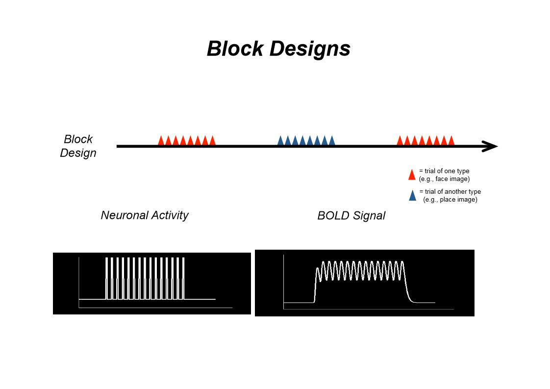
Allows fMRI signal to accumulate over trials of the same kind (same condition)
Used when network of areas involved in a task is unknown and whole brain needs to be searched to identify them
25
Evaluate block design
+ High detection power
+ Most widely used approach for fMRI studies
- Poor estimation power (only knows what happens in this block, not in every single presentation)
- Subjects get into a mental set for a block, brain may adapt resulting in decreased fMRI signals due to repetition of stimuli presentation
- Very predictable for subject
- Can't look at effects of single events (ie. presentation of single stimuli)
+ Most widely used approach for fMRI studies
- Poor estimation power (only knows what happens in this block, not in every single presentation)
- Subjects get into a mental set for a block, brain may adapt resulting in decreased fMRI signals due to repetition of stimuli presentation
- Very predictable for subject
- Can't look at effects of single events (ie. presentation of single stimuli)
26
Outline slow ER design
Trials from different conditions randomised across whole experiment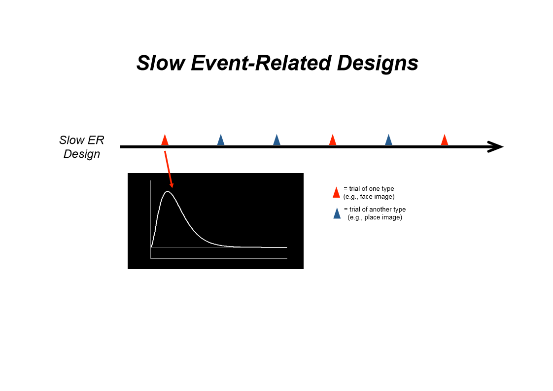
27
When are fast ER designs used?
When areas involved in a task have been identified
Want to explore functions of a brain region in more detail
Want to explore functions of a brain region in more detail
28
What does it mean if the trials are jittered?
Blank intervals of variable duration introduced to avoid overlapping signals between trials of different conditions cancelling each other out
29
What do you have to do for fast ER designs?
Control trial history by making it the same for all trial types
Jitter the trials
Jitter the trials
30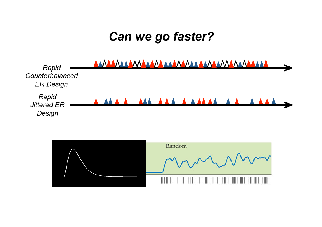
Image occlusion: name the hidden labels
31
What is the problem with trial history?
The peak of 1 and the trough of another can cancel each other out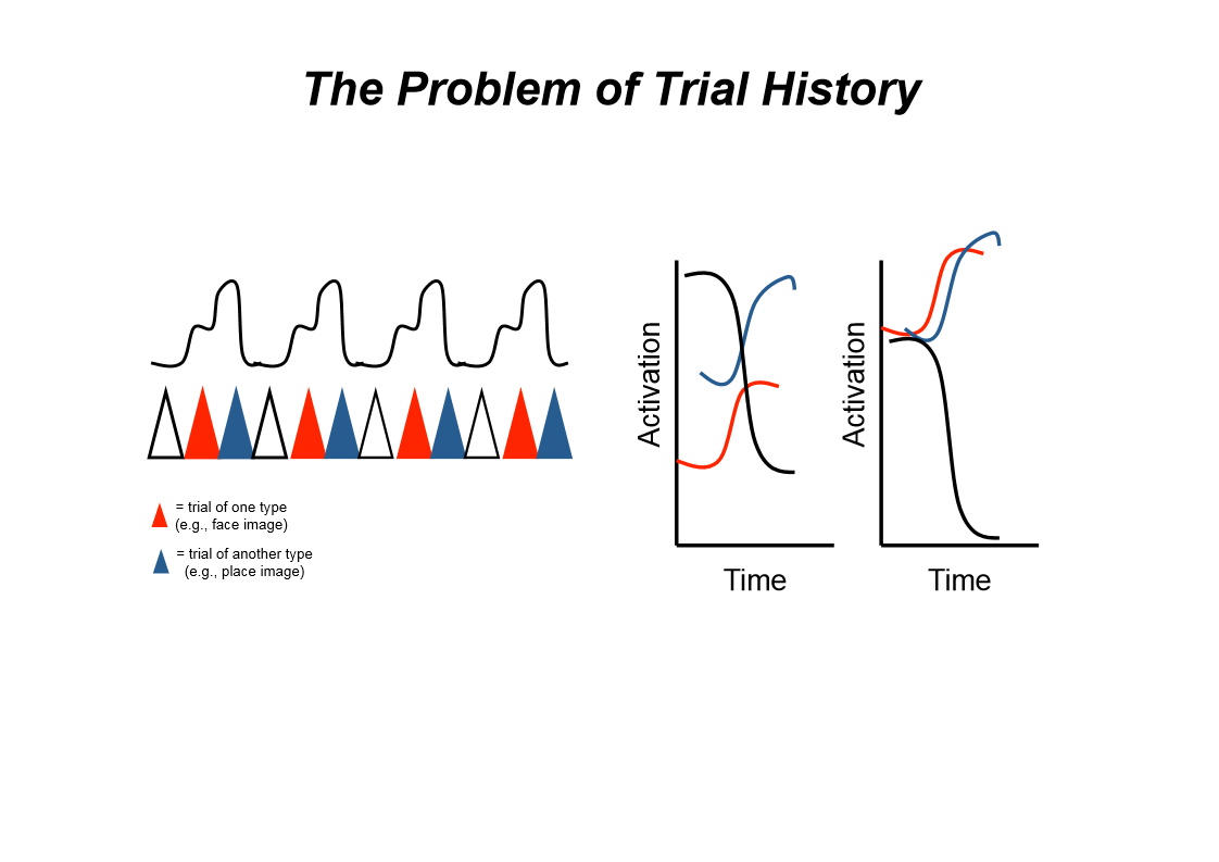
32
Evaluate fast ER designs
+ Can have many trial types in an unpredictable order
+ Can do post hoc sorting of trial type (eg. correct vs incorrect, remembered vs forgotten)
+ Better estimation power than block
- Reduced detection compared to block
- Accurate detection requires accurate modelling
+ Can do post hoc sorting of trial type (eg. correct vs incorrect, remembered vs forgotten)
+ Better estimation power than block
- Reduced detection compared to block
- Accurate detection requires accurate modelling
33
Difference in uses between block and ER designs
Block = don't know where to look in the brain
ER = when looking at how the brain processes things, compares different stimuli, know where it is but want to understand the processes involved
ER = when looking at how the brain processes things, compares different stimuli, know where it is but want to understand the processes involved
34
State the 2 basic statistical approaches
T tests
Correlations, GLM (general linear models)
Correlations, GLM (general linear models)
35
Outline t-tests
Compare activation levels between 2 conditions
Uses a time shift to account for haemodynamic lag
Uses a time shift to account for haemodynamic lag
36
What are correlations used for?
Model activation and see whether any areas show a similar pattern
37
What are the 2 main data analyses methods?
Voxelwise analysis
Region of interest (ROI) analysis
Region of interest (ROI) analysis
38
Outline voxelwise analysis
Search whole brain for voxels (regions) that show significantly higher responses to a task/stimulus compaed to control conditions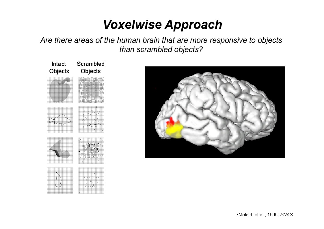
39
Evaluate voxelwise approach
+Requires no prior knowledge about role of different brain regions, searches whole brain
- Neglects individual differences
- Can result in compromised spatial resolution due to intersubject averaging
- Neglects individual differences
- Can result in compromised spatial resolution due to intersubject averaging
40
Outline ROI approach
Region selected from previous studies using other techniques
fMRI used to only study signals from voxels in this region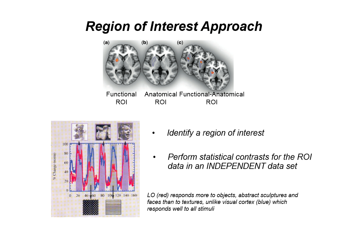
fMRI used to only study signals from voxels in this region
41
Evaluate ROI approach
+ Reduced multiple comparison problems seen in whole brain analyses (only studies one region), fewer degrees of freedom, fewer t-tests needed
+ Higher statistical power
+ Avoids problems with functional mislocalisation due to intersubject averaging
- Neglects other areas that could also play a fundamental role in the task
+ Higher statistical power
+ Avoids problems with functional mislocalisation due to intersubject averaging
- Neglects other areas that could also play a fundamental role in the task
42
Difference between grey and white matter
Grey = synapses and dendrites
White = axons
White = axons
43
3 reasons for the noise recorded in fMRI
Partial volume effects
Head motion
Vessels
Head motion
Vessels
44
Reasons for fMRI noise: partial volume effects
Larger voxels = stronger signal to noise ratio (SNR) (high SNR means signal higher than noise, so clearer image), except when activated region doesn't fill entire voxel - do not refer to image
fMRI signal often occurs in grey matter
If voxel includes white matter/fluid/space outside brain, signal is decreased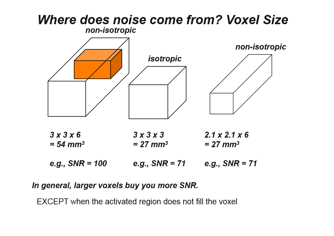
fMRI signal often occurs in grey matter
If voxel includes white matter/fluid/space outside brain, signal is decreased
45
How to mediate partial volume effects
Scan at higher resolution (smaller voxels)
46
How does head motion affect the results?
Results in activations shifting over time
Difficult to align brain images
Difficult to align brain images
47
How might vessels affect fMRI results?
Large vessels line sulci
Produce BOLD activation further from true site of activation (where has the activity come from?)
Even bigger problem when comparing between pps (may have different vasculature)
Produce BOLD activation further from true site of activation (where has the activity come from?)
Even bigger problem when comparing between pps (may have different vasculature)
48
2 ways of avoiding confounds
Choose appropriate control condition
Avoid statistical errors
Avoid statistical errors
49
How to choose appropriate control condition?
Can't compare a task to nothing (eg. blank screen) - activation in test condition could be due to increased attention rather than the stimulus itself
Also need to make sure task difficulty is equal across controls and trials
Also need to make sure task difficulty is equal across controls and trials
50
What is a non-independence error?
Occurs when stats tests performed are not independent from the means used to select the brain region
Selected the brain area than did stats test using that brain area
Selected the brain area than did stats test using that brain area
51
How to avoid stats errors (non-independence errors)
Use independent data from the original data (used to identify the brain areas involved) when performing stats analyses
Allows conclusions to be made about how this area processes info etc.
Allows conclusions to be made about how this area processes info etc.