Brain Anatomy
Study these flashcardsFree · spaced repetition · iPhone, Android, Mac & web
All cards
1
What are the 3 techniques used to study brain function?
Study of neurological patients
Brain imaging
Animal models
Brain imaging
Animal models
2
Strengths and limitations of studying neurological patients (case study)
+ Detailed analysis of single subject
+ Study of human behaviour
+ Can potentially relate to brain function
- Limited generalisation
- Rare
- Low stats power (i.e. n=1)
- Lack of control over extent of damage
+ Study of human behaviour
+ Can potentially relate to brain function
- Limited generalisation
- Rare
- Low stats power (i.e. n=1)
- Lack of control over extent of damage
3
3 types of brain imaging
EEG
MEG
fMRI
MEG
fMRI
4
Strengths + limitations of EEG
+ Good temporal resolution
- Bad spatial resolution
- Bad spatial resolution
5
Strengths + limitations of fMRI
+ Better spatial resolution
- Measures haemodynamic response (blood O2 level dependent response) so activity is inferred
- Lower temporal resolution than EEG (counter by looking at specific areas of the brain, faster)
- Measures haemodynamic response (blood O2 level dependent response) so activity is inferred
- Lower temporal resolution than EEG (counter by looking at specific areas of the brain, faster)
6
Strengths + limitations of MEG
+ Better temporal and spatial resolution than EEG
- Expensive
- Very few mobile systems
- Expensive
- Very few mobile systems
7
Limitations of measuring brain activity
Correlative results
Can't show causation
Very little control of pps outside of the lab
Can't show causation
Very little control of pps outside of the lab
8
Strengths + limitations of using animal models
+ Can conduct experiments including invasive procedures
+ Can monitor over long periods of time
+ Greater control over experimental situation
+ Similar brain anatomy and neurochemical systems (depends on animal)
- Difficult to translate tasks between humans and animals (human tasks are often language based eg. questionnaires and interviews)
- Differences in specific brain regions (eg. rats and mice may not even have a prefrontal cortex)
+ Can monitor over long periods of time
+ Greater control over experimental situation
+ Similar brain anatomy and neurochemical systems (depends on animal)
- Difficult to translate tasks between humans and animals (human tasks are often language based eg. questionnaires and interviews)
- Differences in specific brain regions (eg. rats and mice may not even have a prefrontal cortex)
9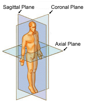
Image occlusion: name the hidden labels
10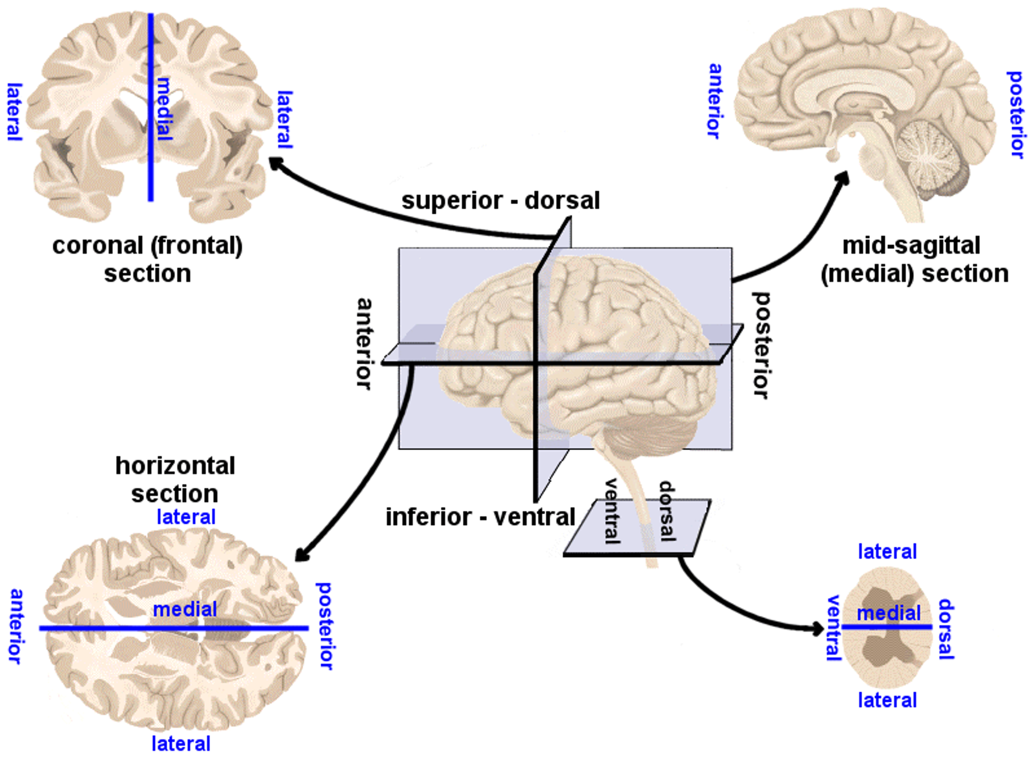
Image occlusion: name the hidden labels
11
Image occlusion: name the hidden labels
12
Image occlusion: name the hidden labels
13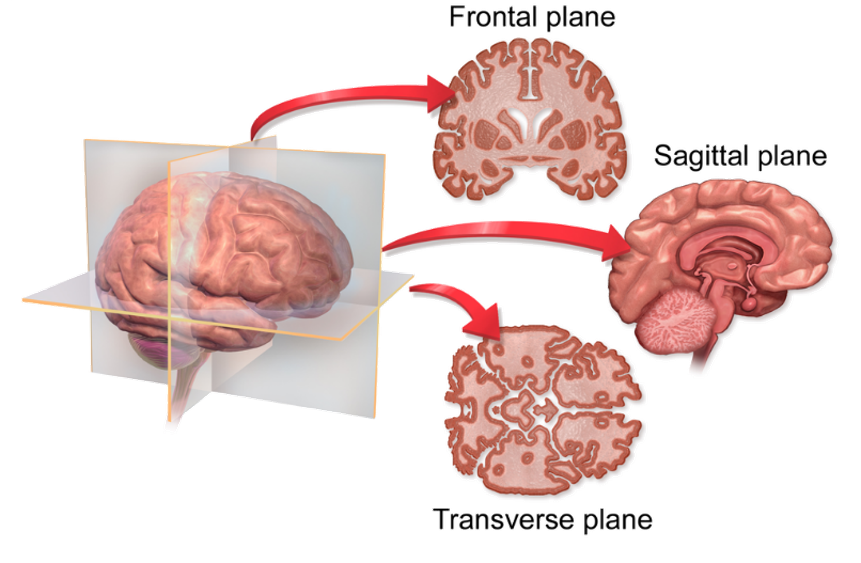
Image occlusion: name the hidden labels
14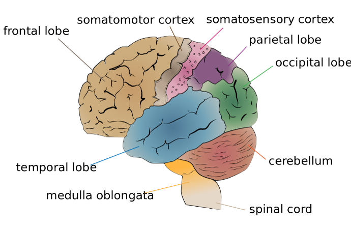
Image occlusion: name the hidden labels
15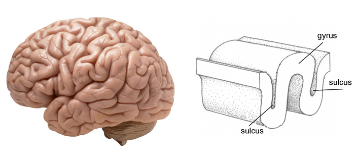
Image occlusion: name the hidden labels
16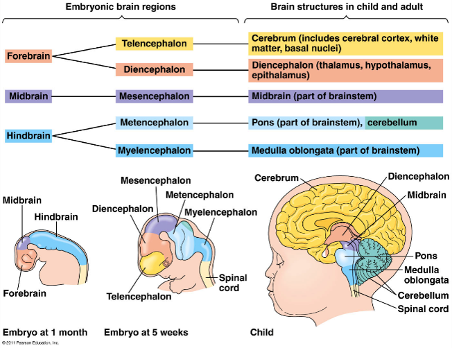
Image occlusion: name the hidden labels
17
Image occlusion: name the hidden labels
18
Image occlusion: name the hidden labels
19
What do neurones consist of?
Soma (cell body)
Dendrites
Axon hillock
Axon
Axon terminal
Dendrites
Axon hillock
Axon
Axon terminal
20
What are glia?
Support cells that make up 50% of the brain
21
What are the 3 different types of glia cells?
Astrocytes
Microglia
Oligodendrocytes
Microglia
Oligodendrocytes
22
Describe function of astrocytes
Anchor neurons to blood supply
Clear neurotransmitters from synaptic cleft
Clear neurotransmitters from synaptic cleft
23
Describe microglia
Specialised macrophages (white blood cell) activated by brain damage
24
Describe function of oligodendrocytes
Form insulating sheaths around neurons to improve conduction speed
25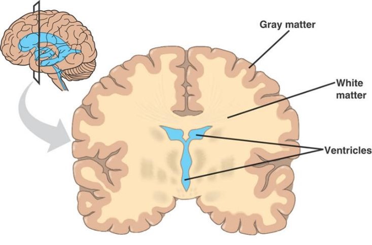
Image occlusion: name the hidden labels
26
Name the 5 major lobes of the brain
Occipital
Temporal
Parietal
Frontal
Cerebellum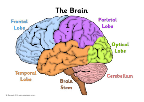
Temporal
Parietal
Frontal
Cerebellum
27
Describe the occipital lobe
Primary visual cortex of brain (V1)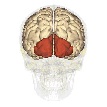
28
What can damage to the occipital lobe cause?
Blindsight
Consciously blind in affected visual field
Can 'guess' correctly when asked about stimuli in the 'blind' part of their visual field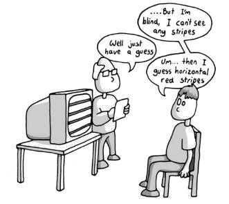
Consciously blind in affected visual field
Can 'guess' correctly when asked about stimuli in the 'blind' part of their visual field
29
Describe the temporal lobe
Involved in:
Auditory perception
Understanding language
Memory formation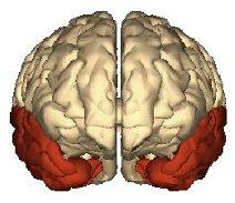
Auditory perception
Understanding language
Memory formation
30
What can damage to the temporal lobe cause?
Visual agnosia
Can still visually perceive objects but can't relate visual input to the object (can with other modes, eg. touch)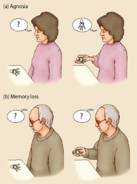
Can still visually perceive objects but can't relate visual input to the object (can with other modes, eg. touch)
31
Describe the parietal lobe
Integrates info from different senses to give us a coherent view of the world
Heavily implicated in functions like attention
Updates representation of where things are predicted to be as a result of movements you are about to make (predicts)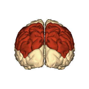
Heavily implicated in functions like attention
Updates representation of where things are predicted to be as a result of movements you are about to make (predicts)
32
What can damage to the parietal lobe cause?
Unilateral spatial neglect
One side of space no longer consciously perceived
Occors across all senses and affects memory
One side of space no longer consciously perceived
Occors across all senses and affects memory
33
Describe the frontal lobe
Supports higher cognitive functions including:
Voluntary attention
Decisions making
Long term planning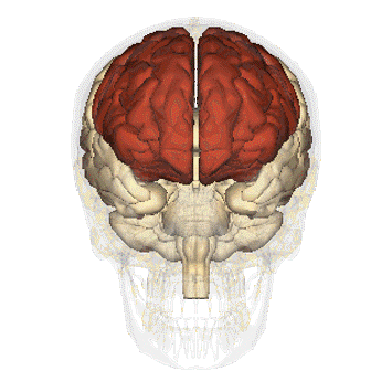
Voluntary attention
Decisions making
Long term planning
34
Example of when frontal lobe was damaged
Prefrontal lobotomis in 1940s and 1950s
Rosemary Kennedy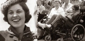
Rosemary Kennedy
35
Describe the cerebellum
Necessary for motor coordination and motor learning
Involved in cognitive functions
Contains >50% brain's neurons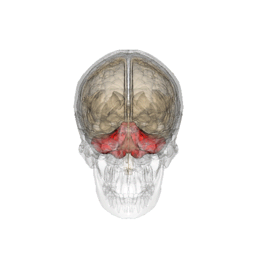
Involved in cognitive functions
Contains >50% brain's neurons
36
What can damage to the cerebellum cause?
Ataxia and other movement errors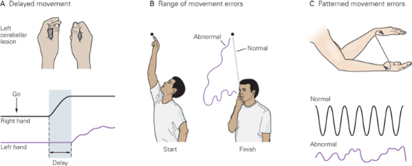
37
Name the 6 major subcortical regions of the brain
Brainstem
Hypothalamus
Thalamus
Hippocampus
Amygdala
Striatum
Hypothalamus
Thalamus
Hippocampus
Amygdala
Striatum
38
Describe the brain stem
Includes pons, medulla and midbrain
Maintains autonomic functions (eg. breathing and heart rate)
Coordinates simple motor behaviours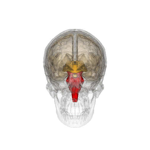
Maintains autonomic functions (eg. breathing and heart rate)
Coordinates simple motor behaviours
39
What does the midbrain do?
Contains a number of nuclei which release chemical NETs that modulate cortical function
40
What can damage to the brainstem cause?
Often fatal
Survivors = locked in syndrome
Survivors = locked in syndrome
41
Describe the hypothalamus
Regulates motivated behaviour
Integrates info from brain and body, and coordinating neuroendocrine, autonomic and behavioural responses
Made up of many different nuclei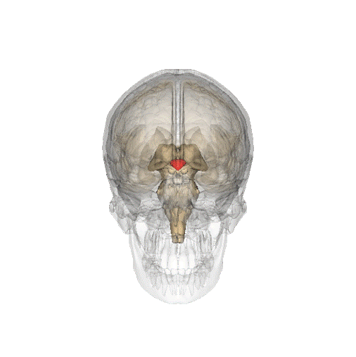
Integrates info from brain and body, and coordinating neuroendocrine, autonomic and behavioural responses
Made up of many different nuclei
42
What can damage to the hypothalamus cause?
Kallman syndrome (puberty doesn't begin)
43
Describe the thalamus
Connects sensory systems to cortical regions processing info
Different nuclei process info from different sensory systems
Other nuclei process movement info and communicate this to spinal cord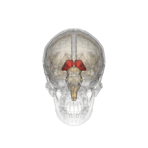
Different nuclei process info from different sensory systems
Other nuclei process movement info and communicate this to spinal cord
44
What can damage to the thalamus cause?
Range of disorders
Eg. auditory-tactile synaesthesia
Eg. auditory-tactile synaesthesia
45
Describe the hippocampus
Encodes spatial info
Stores episodic memory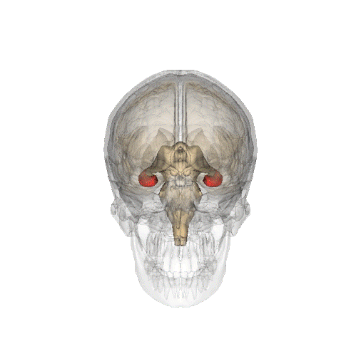
Stores episodic memory
46
What can damage to the hippocampus cause?
HM
Hippocampi bilaterally removed
No longer formed episodic memories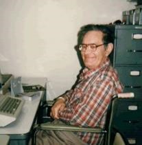
Hippocampi bilaterally removed
No longer formed episodic memories
47
Describe the amygdala
Relates enviro cues with their emotional and motivational significance
Eg. fear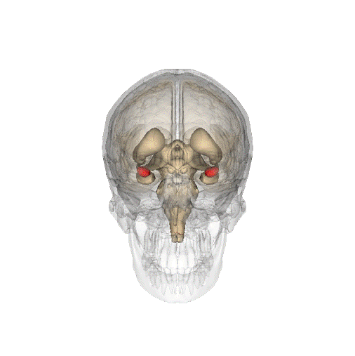
Eg. fear
48
What can damage to the amygdala cause?
Patient SM
Lost amygdala bilaterally
Doesn't feel fear
Unable to draw fear in faces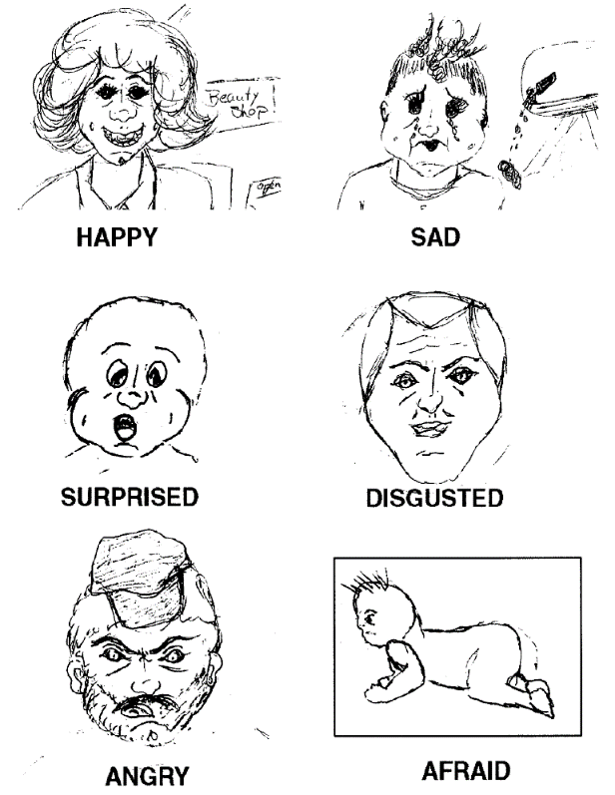
Lost amygdala bilaterally
Doesn't feel fear
Unable to draw fear in faces
49
Describe the striatum
Consist of the caudate and putamen
Found within basal ganglia
Basal ganglia contributes to action selection (voluntary, such as voluntary movement of arm)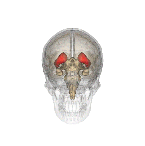
Found within basal ganglia
Basal ganglia contributes to action selection (voluntary, such as voluntary movement of arm)
50
What can damage to the striatum cause?
Parkinson's disease (loss of movement) - pic
Huntington's Disease (excessive movement)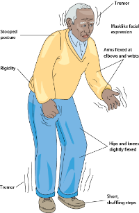
Huntington's Disease (excessive movement)
51
What makes white matter and grey matter different?
Cell body is grey
Myelin sheat is white
Grey matter = not insulated cell bodies
White matter = insulated axons with myelin sheath
Myelin sheat is white
Grey matter = not insulated cell bodies
White matter = insulated axons with myelin sheath
52
Name for where the optic nerves cross
Optic chiasm
53
Outline the 2 visual streams
Dorsal stream - where, vision for action (goes towards motor cortex)
Ventral stream - what, vision for perception
Ventral stream - what, vision for perception
54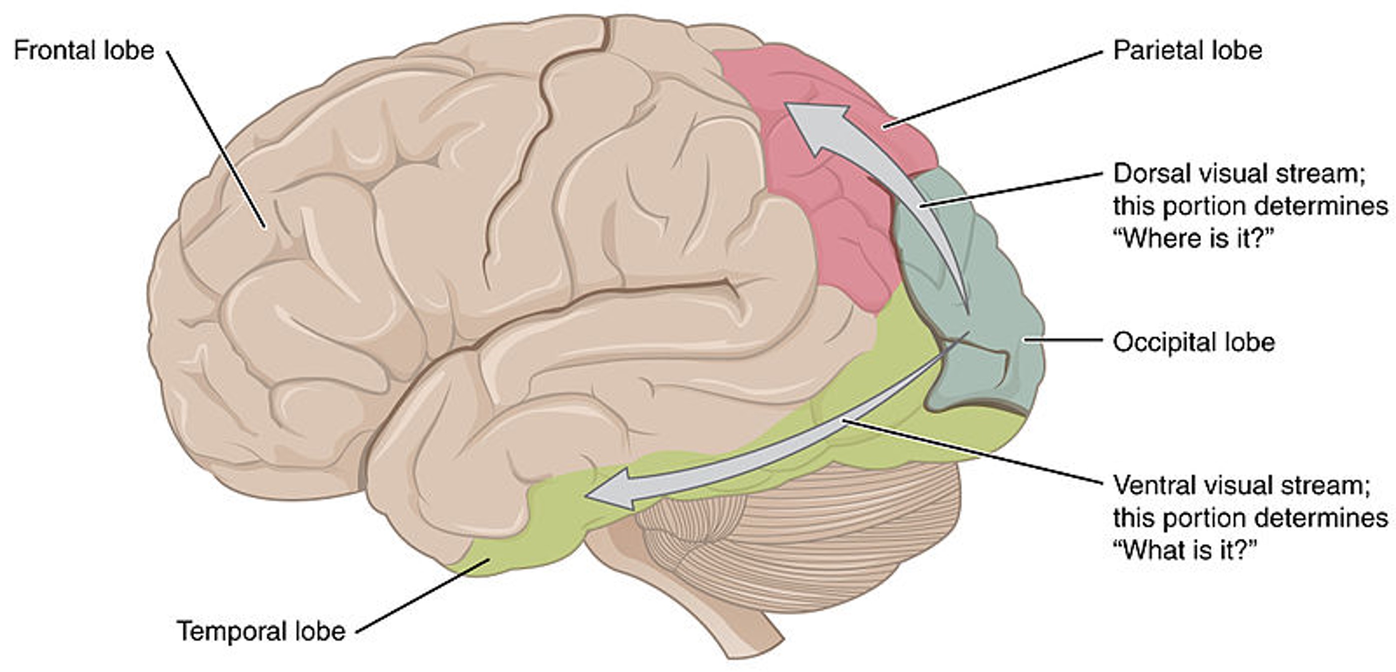
Image occlusion: name the hidden labels
55
Name 3 disorders of the dorsal visual stream
Hemispatial / unilateral spatial neglect
Simultagnosia
Optic ataxia
Simultagnosia
Optic ataxia
56
Name 3 disorders of the ventral visual stream
Prosopagnosia
Visual agnosia
Cerebral achromatopsia
Visual agnosia
Cerebral achromatopsia
57
What happens in V1?
Representation of info from fovea goes here
Retinotopic
Encodes colour, edges etc.
Retinotopic
Encodes colour, edges etc.
58
How is the auditory cortex arranged?
Tonotopically
59
How does the primary auditory cortex receive info?
From basilar membraine in cochlea, via medial geniculate nucleus of the thalamus
60
What happens in the cochlea?
High frequency sounds vibrate basilar membrane at base
Low frequency sounds vibrate basilar membrane at apex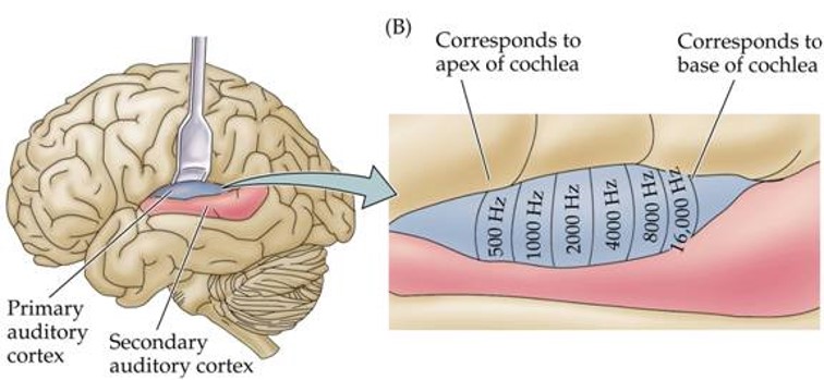
Low frequency sounds vibrate basilar membrane at apex
61
How is the somatosensory cortex organised?
Somatotopically
BUT all somatotopic representations are plastic to some extent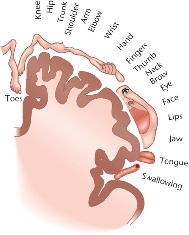
BUT all somatotopic representations are plastic to some extent
62
How is the primary motor cortex organised?
Somatotopically
BUT all somatotopic representations are plastic to some extent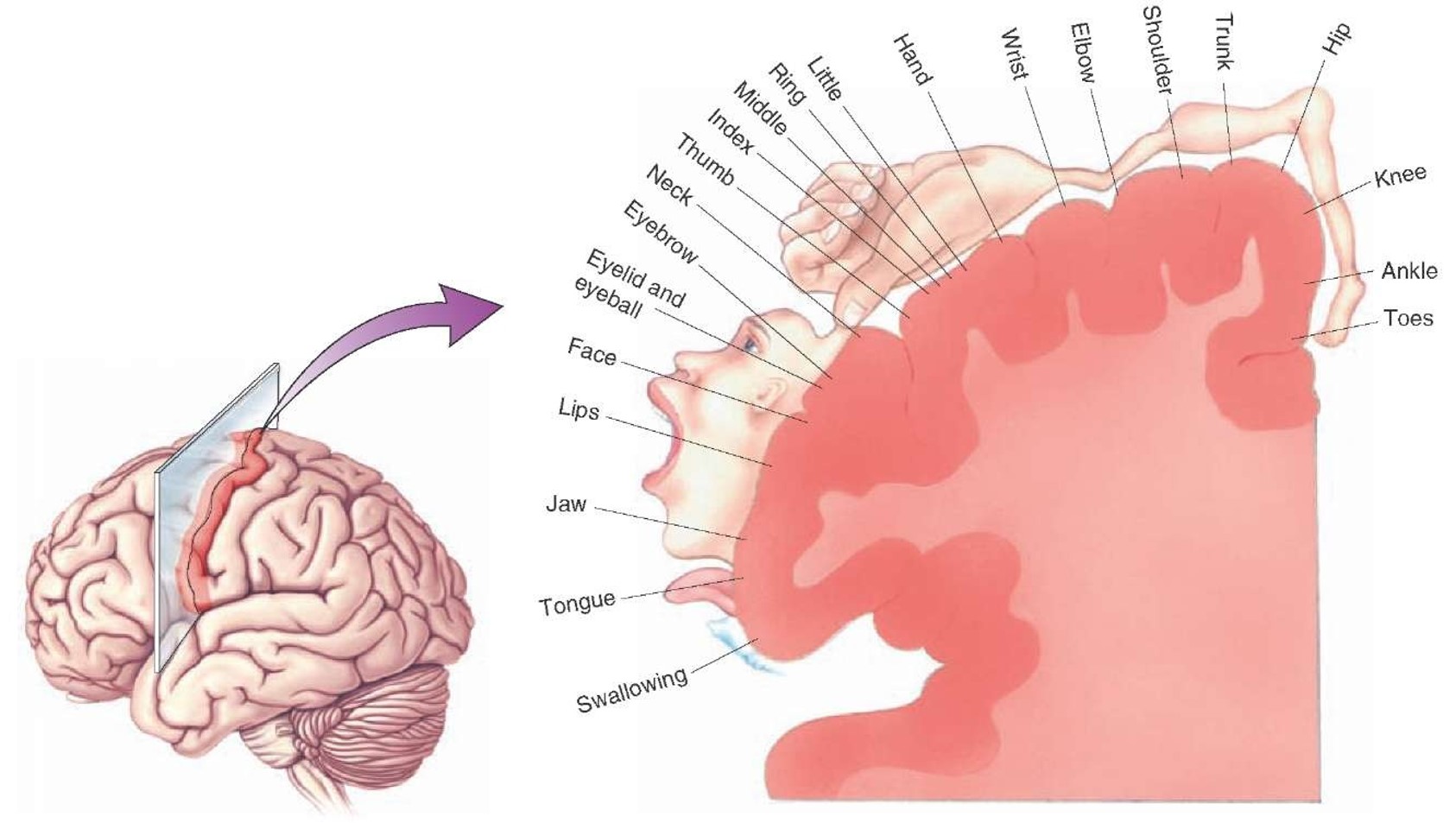
BUT all somatotopic representations are plastic to some extent
63
Outline different parts of the motor system
Primary motor cortex (has somatotopic representations)
Cerebellum (has somatotopic representations)
Parietal cortex
Prefrontal cortex (goal representation)
Cerebellum (has somatotopic representations)
Parietal cortex
Prefrontal cortex (goal representation)
64
Outline brain regions involved in smooth, goal-directed voluntary movement and effect of damage in each area
Primary motor cortex (difficulty executing movement)
Premotor areas (difficulty planning movement)
Posterior parietal cortex (difficulty planning movement)
Basal ganglia (difficulty in movement selection - too much/too little)
Cerebellum (difficulty coordinating movement)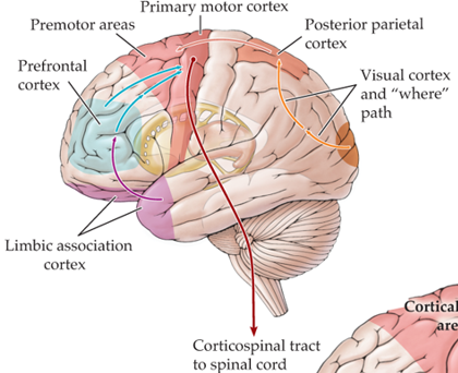
Premotor areas (difficulty planning movement)
Posterior parietal cortex (difficulty planning movement)
Basal ganglia (difficulty in movement selection - too much/too little)
Cerebellum (difficulty coordinating movement)
65
Which regions are involved in motivation?
Hypothalamus
Rest of the body - blood sugar levels can affect motivation to eat
Rest of the body - blood sugar levels can affect motivation to eat
66
Which regions are involved in emotion?
Amygdala (Patient SM)
Prefrontal cortex (Phineas Gage)
Hypothalamus
Prefrontal cortex (Phineas Gage)
Hypothalamus
67
Which regions are involved in language?
Broca's area (speech production)
Wernicke's area (speech comprehension)
Usually in left hemisphere
Wernicke's area (speech comprehension)
Usually in left hemisphere
68
2 types of long term memory
Explicit (declarative)
Implicit (non-declarative)
Implicit (non-declarative)
69
Explicit LTM
Episodic
Semantic
Semantic
70
Implicit LTM
Procedural
Conditioning (emotional)
Conditioning (emotional)
71
Which regions are involved in memory?
Prefrontal cortex - working memory
Striatum - procedural memory
Neocortex - perceptual memory, semantic memory, priming
Amygdala - emotional memory
Cerebellum - conditioning
Hippocampus - declarative memory (episodic, semantic)
Striatum - procedural memory
Neocortex - perceptual memory, semantic memory, priming
Amygdala - emotional memory
Cerebellum - conditioning
Hippocampus - declarative memory (episodic, semantic)
72
Describe executive function (7)
Involved in:
Planning
Decision-making
Error correction/trouble shooting
Novel sequences of actions
Strong habitual responses/suppressing temptations
Working memory
Problem solving
Planning
Decision-making
Error correction/trouble shooting
Novel sequences of actions
Strong habitual responses/suppressing temptations
Working memory
Problem solving