anatomy of mediastinum
Study these flashcardsFree · spaced repetition · iPhone, Android, Mac & web
All cards
1
where does superior mediastinum and inferior mediastinum divided?
(at transverse plane view)
extends from sternal angle to the IV disc bertween T4 & T5 vertebrae
extends from sternal angle to the IV disc bertween T4 & T5 vertebrae
2
Image occlusion: name the hidden labels
3
Image occlusion: name the hidden labels
4
Image occlusion: name the hidden labels
5
Image occlusion: name the hidden labels
6
Image occlusion: name the hidden labels
7
Image occlusion: name the hidden labels
8
Image occlusion: name the hidden labels
9
Image occlusion: name the hidden labels
10
Image occlusion: name the hidden labels
11
Image occlusion: name the hidden labels
12
Image occlusion: name the hidden labels
13
Image occlusion: name the hidden labels
14
Image occlusion: name the hidden labels
15
Image occlusion: name the hidden labels
16
Image occlusion: name the hidden labels
17
Image occlusion: name the hidden labels
18
Image occlusion: name the hidden labels
19
Image occlusion: name the hidden labels
20
Image occlusion: name the hidden labels
21
Image occlusion: name the hidden labels
22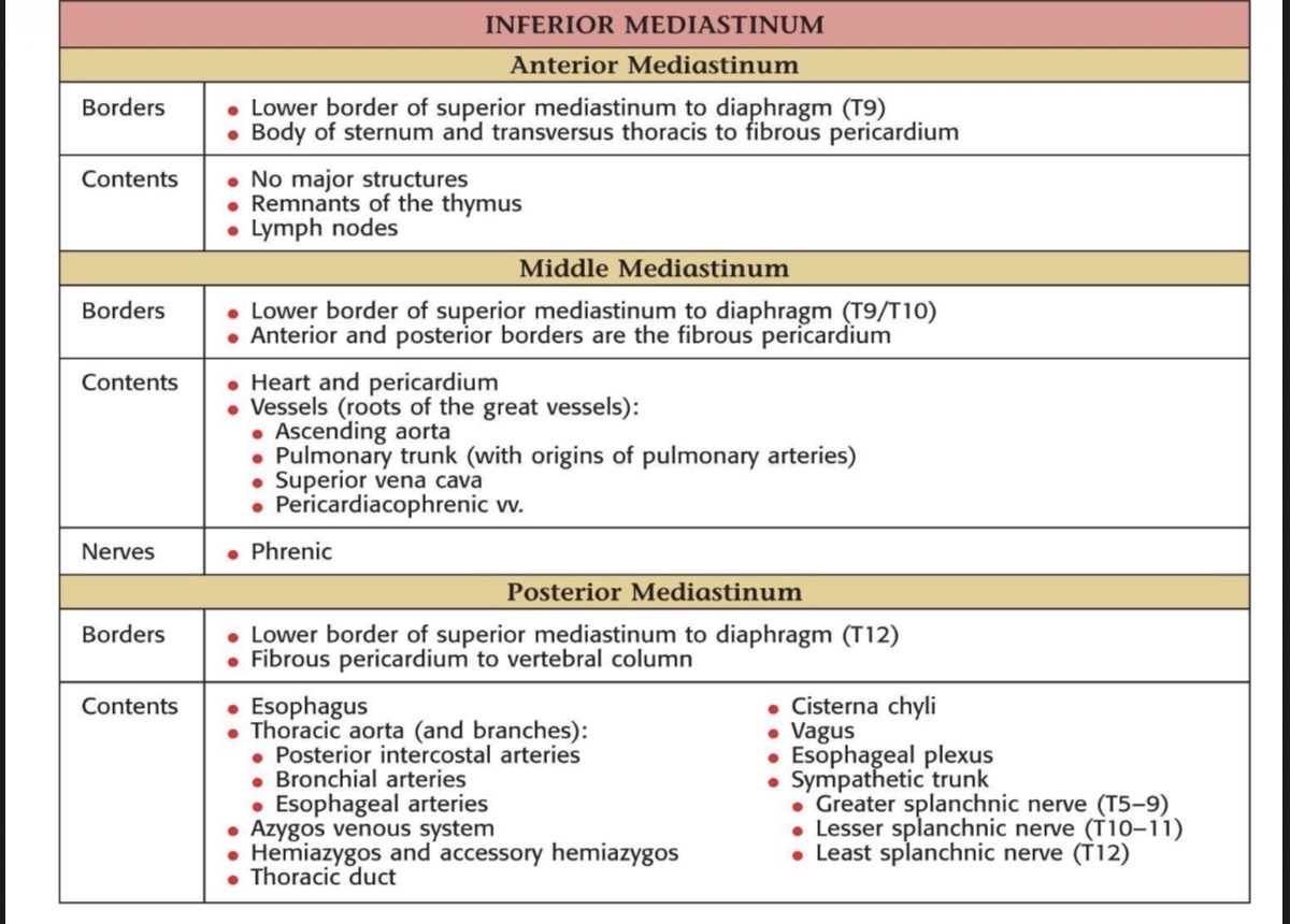
Image occlusion: name the hidden labels
23
Image occlusion: name the hidden labels
24
Image occlusion: name the hidden labels
25
Image occlusion: name the hidden labels
26
Image occlusion: name the hidden labels
27
Image occlusion: name the hidden labels
28
Image occlusion: name the hidden labels
29
Image occlusion: name the hidden labels
30
Image occlusion: name the hidden labels
31
Image occlusion: name the hidden labels
32
Image occlusion: name the hidden labels
33
Image occlusion: name the hidden labels
34
Image occlusion: name the hidden labels
35
Image occlusion: name the hidden labels
36
Image occlusion: name the hidden labels
37
Image occlusion: name the hidden labels
38
Image occlusion: name the hidden labels
39
Image occlusion: name the hidden labels
40
Image occlusion: name the hidden labels
41
Image occlusion: name the hidden labels
42
Image occlusion: name the hidden labels
43
Image occlusion: name the hidden labels
44
Image occlusion: name the hidden labels
45
Image occlusion: name the hidden labels
46
Image occlusion: name the hidden labels
47
Image occlusion: name the hidden labels
48
Image occlusion: name the hidden labels
49
Image occlusion: name the hidden labels
50
Image occlusion: name the hidden labels
51
Image occlusion: name the hidden labels
52
Image occlusion: name the hidden labels
53
Image occlusion: name the hidden labels
54
Image occlusion: name the hidden labels
55
Image occlusion: name the hidden labels
56
Image occlusion: name the hidden labels
57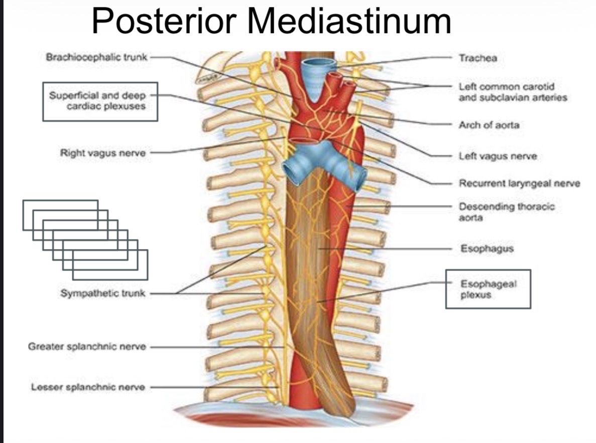
Image occlusion: name the hidden labels
58
Image occlusion: name the hidden labels
59
Image occlusion: name the hidden labels
60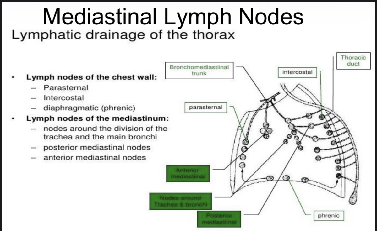
Image occlusion: name the hidden labels
61
Image occlusion: name the hidden labels
62
Image occlusion: name the hidden labels
63
Image occlusion: name the hidden labels
64
Image occlusion: name the hidden labels
65
Image occlusion: name the hidden labels
66
Image occlusion: name the hidden labels
67
Image occlusion: name the hidden labels
68
Image occlusion: name the hidden labels
69
What is Superior vena cava syndrome (SVC) ?
Obstruction (blockage) of SVC
70
Why does SVC blockage can cause syndrome?
Because the SVC drains blood from above the diaphragm
(head, neck, face, arms)
When blocked → pressure builds up upstream.
(head, neck, face, arms)
When blocked → pressure builds up upstream.
71
SVC obstruction : Engorgement of veins (big, visible veins)
Blood can’t drain → pressure builds →
👉 Neck veins (JVD) and chest veins become swollen
👉 Body creates collateral veins to bypass the blockage
👉 Neck veins (JVD) and chest veins become swollen
👉 Body creates collateral veins to bypass the blockage
72
SVC obstruction : Facial swelling / facial plethora
Blood pools in the face →
👉 Face looks puffy, red, bluish, worse when lying down
👉 Face looks puffy, red, bluish, worse when lying down
73
SVC obstruction : Dyspnea (shortness of breath)
Why breathing becomes hard:
• Swollen veins compress the airway
• Mediastinal mass may directly press on the trachea
• Venous congestion causes airway edema
👉 That’s why patients may have stridor or cough too
• Swollen veins compress the airway
• Mediastinal mass may directly press on the trachea
• Venous congestion causes airway edema
👉 That’s why patients may have stridor or cough too
74
SVC obstruction : Dysphagia (difficulty swallowing)
The esophagus runs through the mediastinum
Compression or congestion → swallowing feels difficult
Compression or congestion → swallowing feels difficult
75
SVC obstruction : Hoarseness of voice
The recurrent laryngeal nerve runs in the mediastinum
Compression → hoarse voice
Compression → hoarse voice
76
SVC obstruction : Cerebral edema (dangerous part)
Blood from the brain can’t drain properly →
👉 Increased intracranial pressure
👉 Headache, confusion, drowsiness
👉 In severe cases: loss of consciousness
👉 Increased intracranial pressure
👉 Headache, confusion, drowsiness
👉 In severe cases: loss of consciousness
77
Pemberton's maneuver test
What the test does:
• Patient raises both arms for ~1 minute
What happens physiologically:
• This further narrows venous drainage at the thoracic inlet
• If SVC is already partially blocked → congestion worsens
Positive test =
• Facial flushing
• Bluish discoloration
• Difficulty breathing
👉 This strongly suggests SVC obstruction
• Patient raises both arms for ~1 minute
What happens physiologically:
• This further narrows venous drainage at the thoracic inlet
• If SVC is already partially blocked → congestion worsens
Positive test =
• Facial flushing
• Bluish discoloration
• Difficulty breathing
👉 This strongly suggests SVC obstruction
78
Mediastinitis is inflammation/infection of the mediastinum, usually spreading from nearby structures.
Causes (HIGH-YIELD)
• Deep neck infections
→ spread downward into mediastinum via loose connective tissue
• Esophageal injury
• Penetrating chest wound
• Esophageal perforation
• Air leakage from esophagus
→ air tracks up → subcutaneous emphysema
Key anatomical reason (exam favourite)
• Pretracheal space
• Lies anterior to trachea
• Infection spreads → mediastinitis
• Fascia continuous with:
• Pericardium → pericardial effusion
• Parietal pleura → empyema
👉 Buzzwords to drop: pretracheal space, loose fascia, empyema, pericardial effusion
Mediastinitis
Mediastinitis is inflammation/infection of the mediastinum, usually spreading from nearby structures.
Causes (HIGH-YIELD)
• Deep neck infections
→ spread downward into mediastinum via loose connective tissue
• Esophageal injury
• Penetrating chest wound
• Esophageal perforation
• Air leakage from esophagus
→ air tracks up → subcutaneous emphysema
Key anatomical reason (exam favourite)
• Pretracheal space
• Lies anterior to trachea
• Infection spreads → mediastinitis
• Fascia continuous with:
• Pericardium → pericardial effusion
• Parietal pleura → empyema
👉 Buzzwords to drop: pretracheal space, loose fascia, empyema, pericardial effusion
79
medistinal tumors/ cyst
Important pressure effects
• Left lung tumour
→ mediastinal lymph nodes enlarge
→ compress left recurrent laryngeal nerve
→ hoarseness of voice
• Tumour / cyst compressing SVC
→ SVC syndrome
→ facial swelling, dilated upper limb veins
• Other structures compressed:
• Sympathetic trunk
• Phrenic nerve
• Trachea
• Main bronchi
• Esophagus
• Left lung tumour
→ mediastinal lymph nodes enlarge
→ compress left recurrent laryngeal nerve
→ hoarseness of voice
• Tumour / cyst compressing SVC
→ SVC syndrome
→ facial swelling, dilated upper limb veins
• Other structures compressed:
• Sympathetic trunk
• Phrenic nerve
• Trachea
• Main bronchi
• Esophagus
80
anterior mediastinal mass
Think “4 Ts”
• Thymus
• Thymoma
• Thymic cyst
• Enlarged thymus
• Tumour
• Germ cell tumour
• Teratoma
• Tissue (Lymphoid)
• Lymphoma
• Leukemia
• TB
📌 If exam says anterior mediastinum → lymphoma / thymus / germ cell
• Thymus
• Thymoma
• Thymic cyst
• Enlarged thymus
• Tumour
• Germ cell tumour
• Teratoma
• Tissue (Lymphoid)
• Lymphoma
• Leukemia
• TB
📌 If exam says anterior mediastinum → lymphoma / thymus / germ cell
81
middle mediastinal mass
Think airways + nodes
• Foregut duplication cysts
• Bronchogenic cyst
• Lymphadenopathy
• Tuberculosis
• Fungal infection
• Vascular malformation
📌 Middle = cysts + lymph nodes
• Foregut duplication cysts
• Bronchogenic cyst
• Lymphadenopathy
• Tuberculosis
• Fungal infection
• Vascular malformation
📌 Middle = cysts + lymph nodes
82
posterior mediastinal mass
Think nerves & spine
• Neurogenic tumours
• Thoracic neuroblastoma
• Neurofibroma
• Extra-medullary hematopoiesis
• Vascular malformation
📌 Posterior = neurogenic
• Neurogenic tumours
• Thoracic neuroblastoma
• Neurofibroma
• Extra-medullary hematopoiesis
• Vascular malformation
📌 Posterior = neurogenic