All cards
1
How many bones are in the skull
22
2
Blue half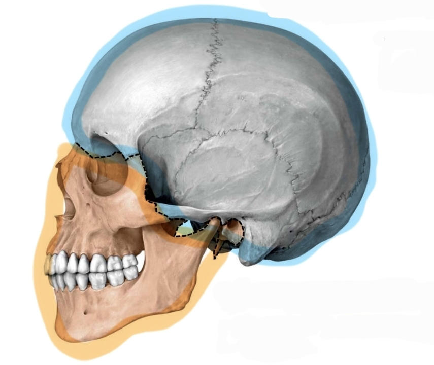
Neurocranium
3
Neurocranium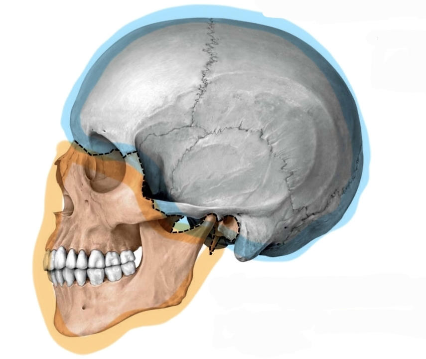
Blue half
4
Orange half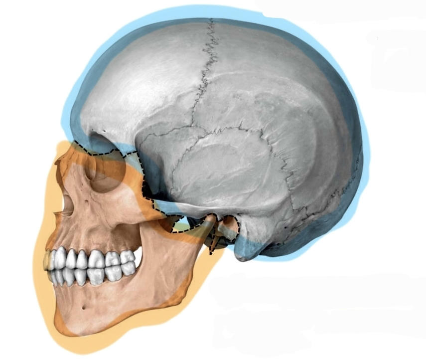
Viscerocranium
5
Viscerocranium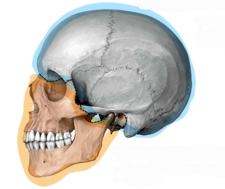
Orange half
6
Pink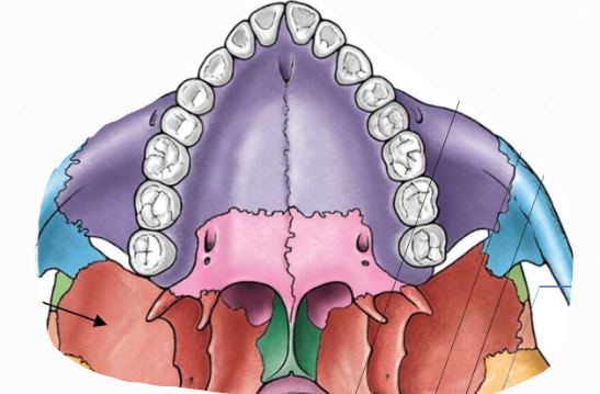
Palatine
7
Palatine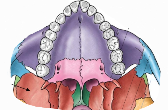
Pink
8
Bone 1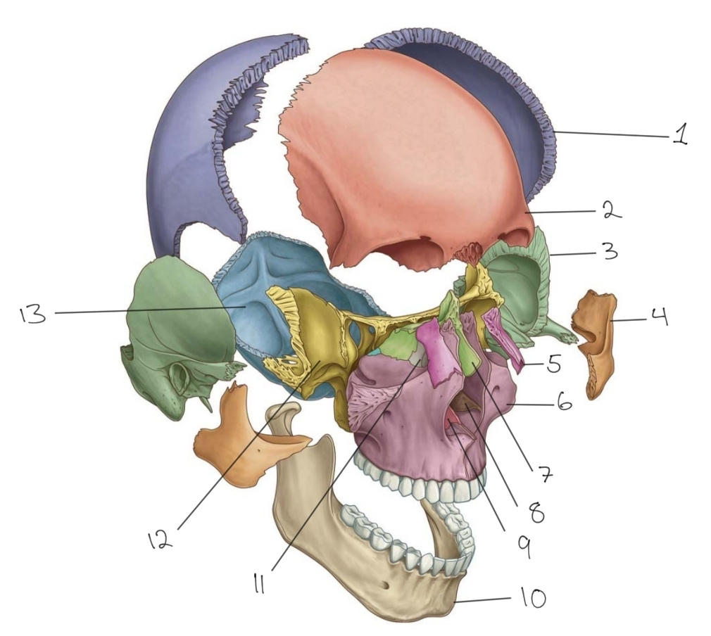
Parietal
9
Parietal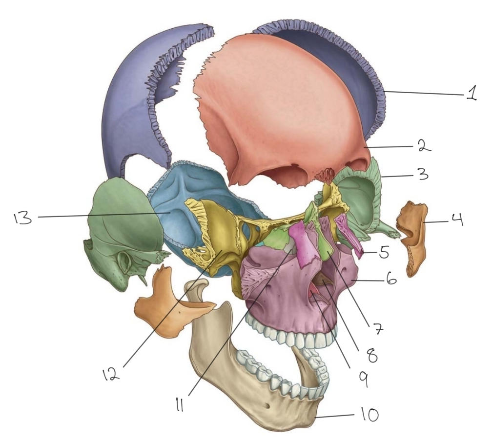
Bone 1
10
Bone 2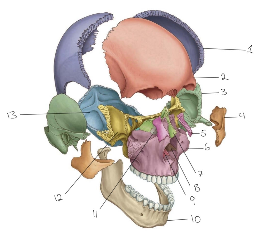
Frontal
11
Frontal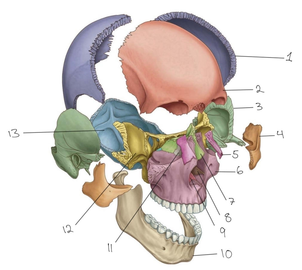
Bone 2
12
Bone 3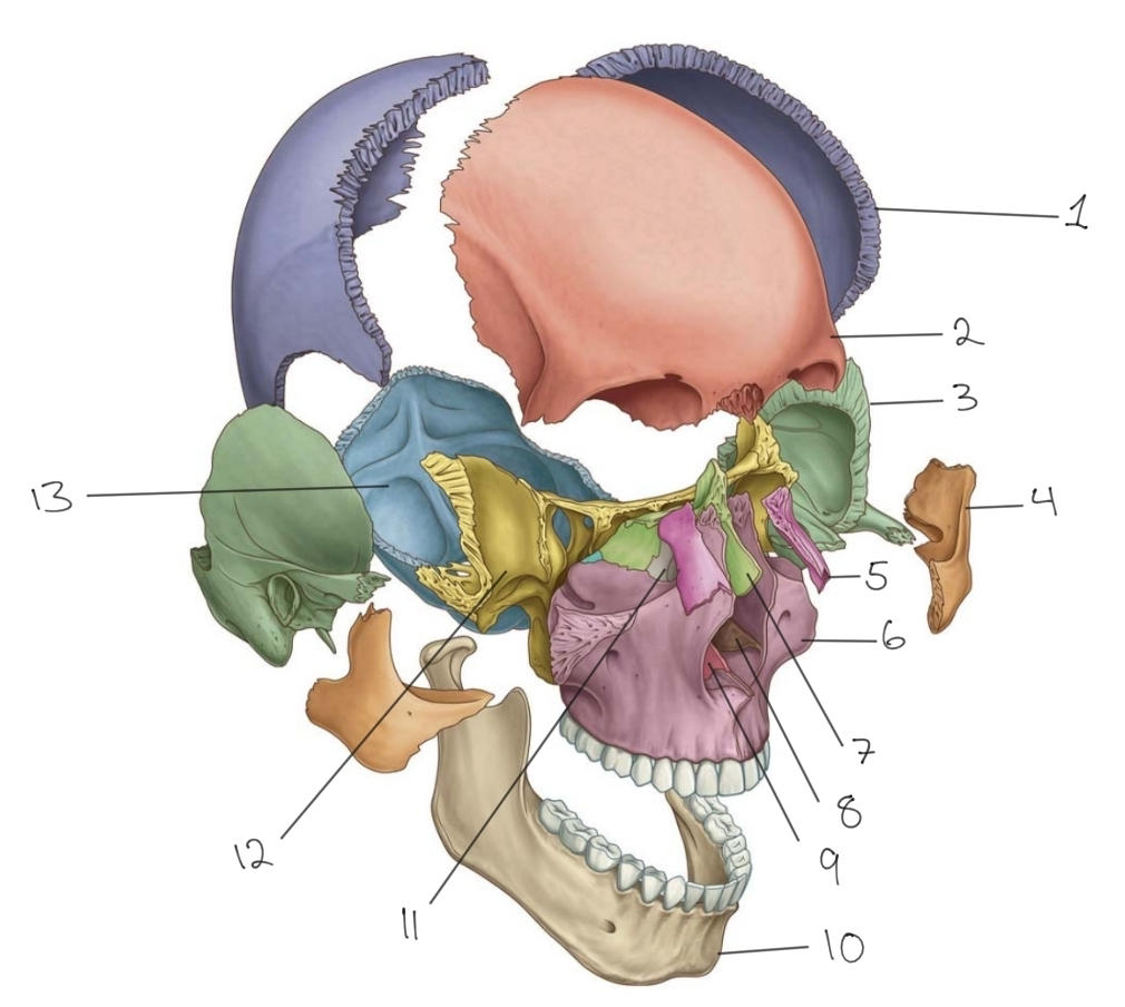
Temporal
13
Temporal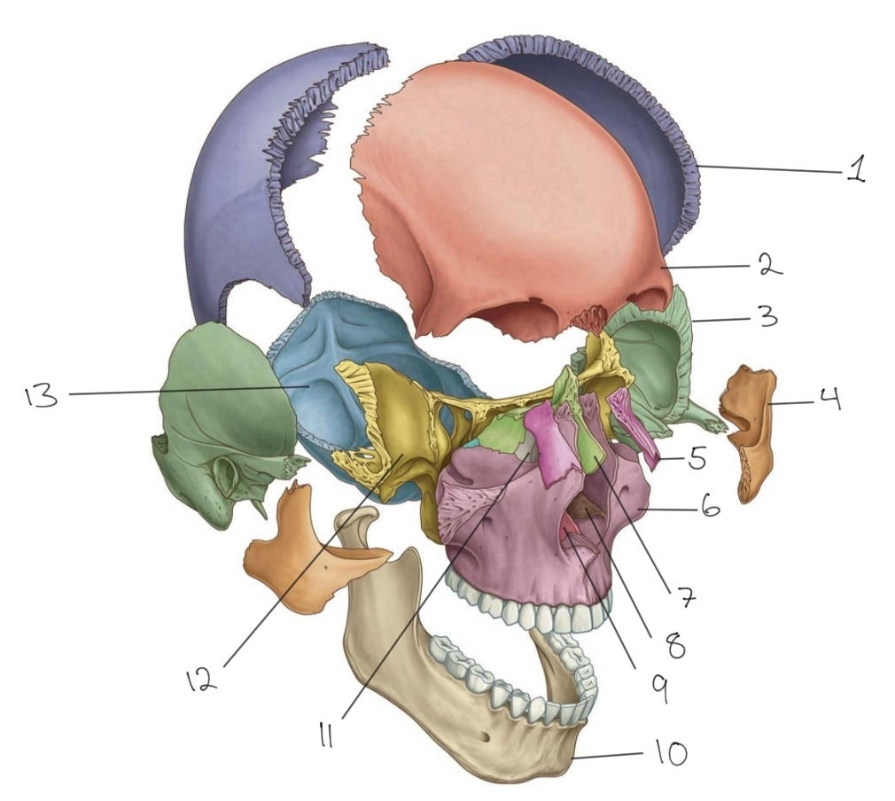
Bone 3
14
Bone 4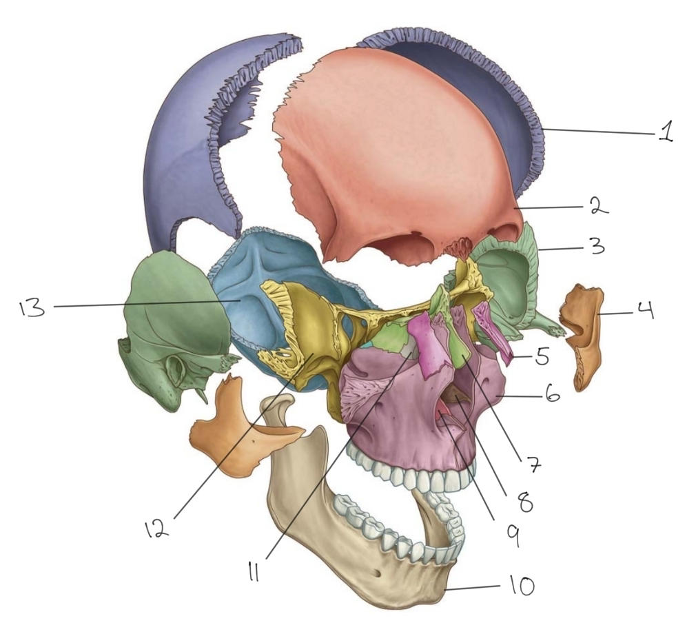
Zygomatic
15
Zygomatic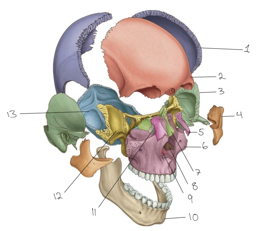
Bone 4
16
Bone 5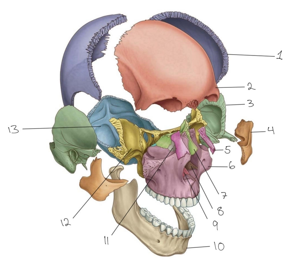
Nasal
17
Nasal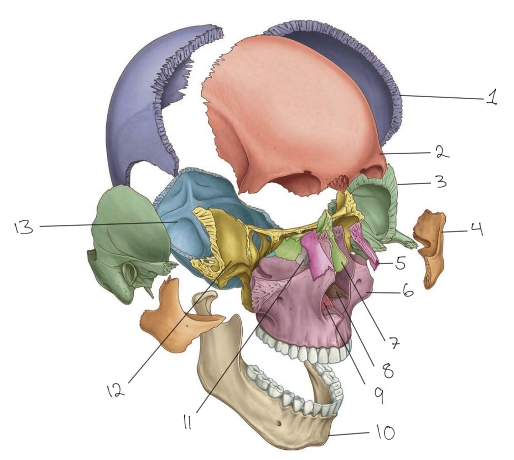
Bone 5
18
Bone 6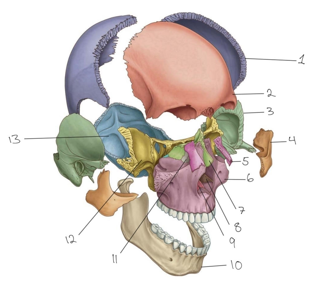
Maxilla
19
Maxilla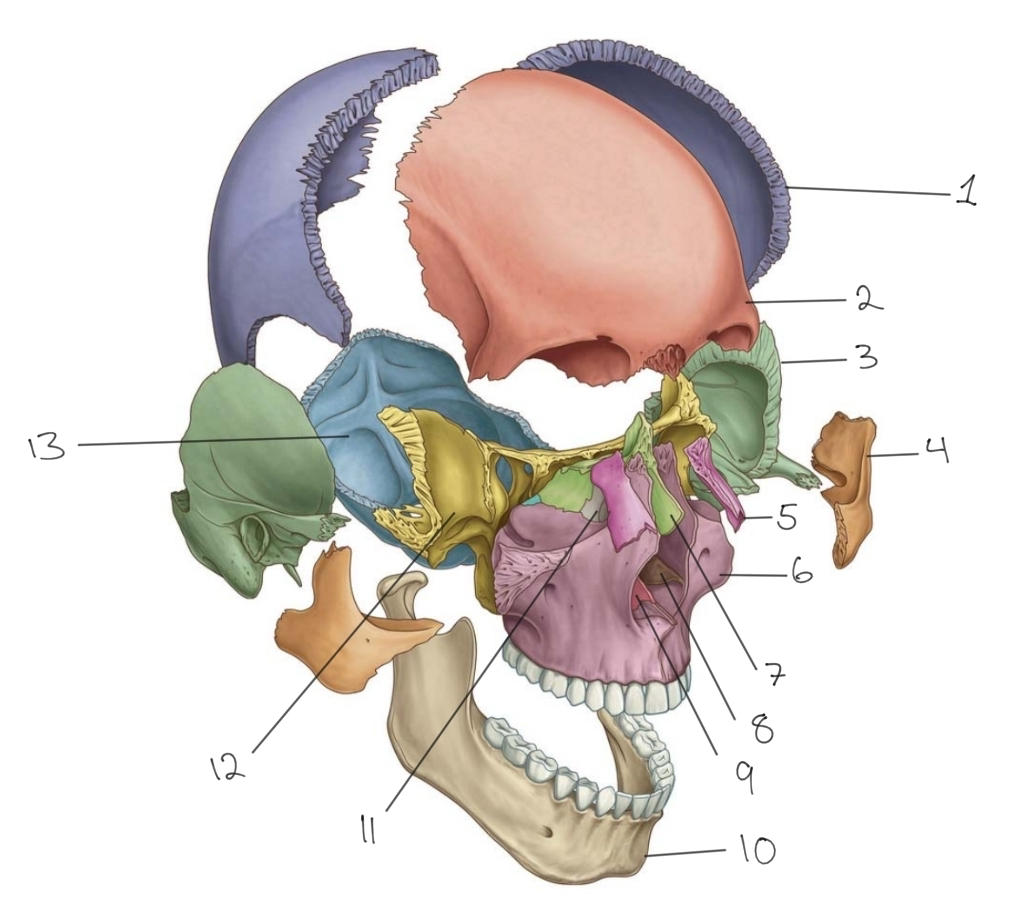
Bone 6
20
Bone 7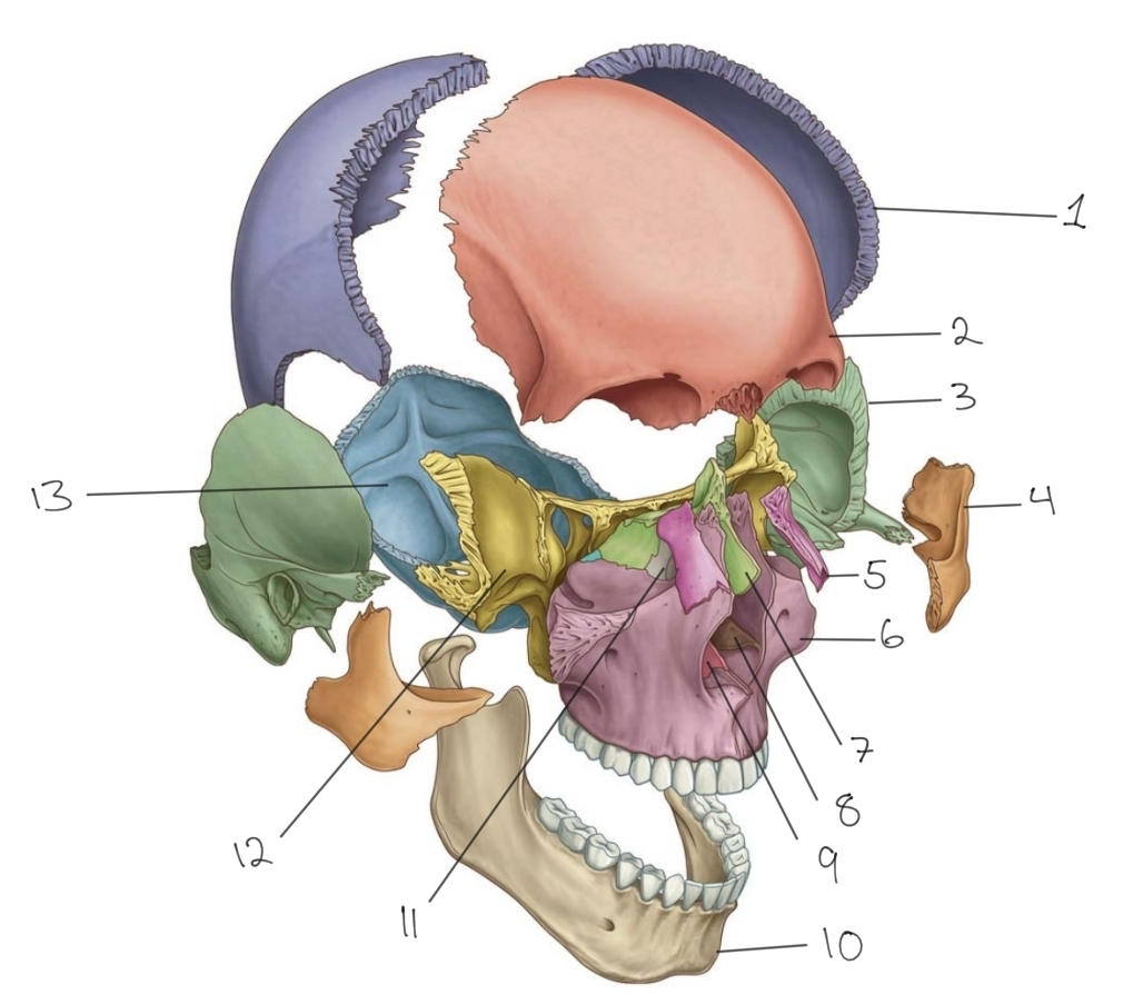
Ethmoid
21
Ethmoid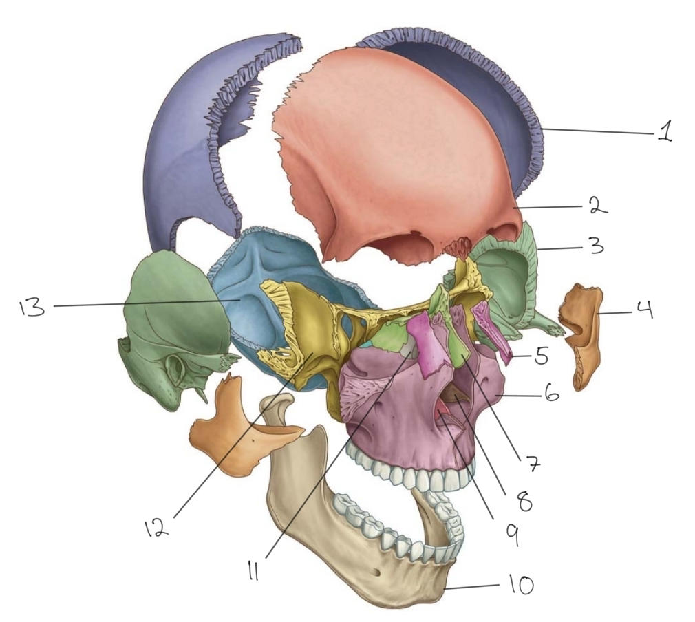
Bone 7
22
Bone 8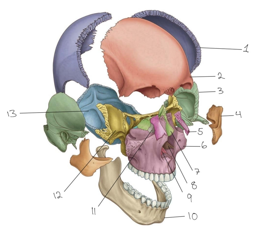
Inferior nasal concha (INC)
23
Inferior nasal concha (INC)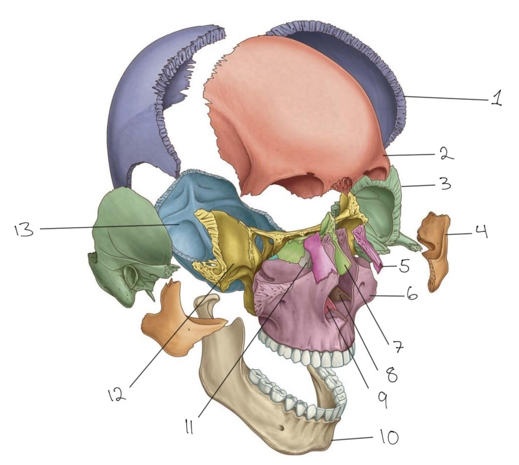
Bone 8
24
Bone 9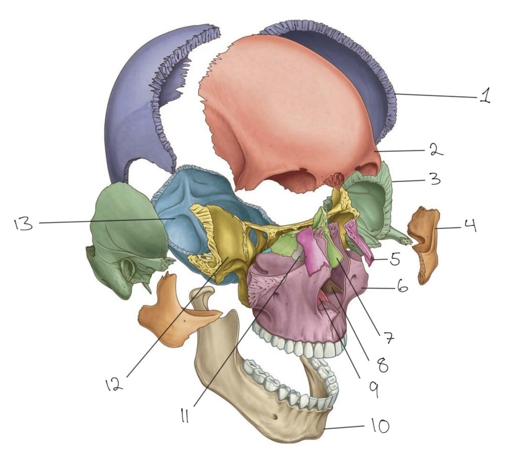
Vomer
25
Vomer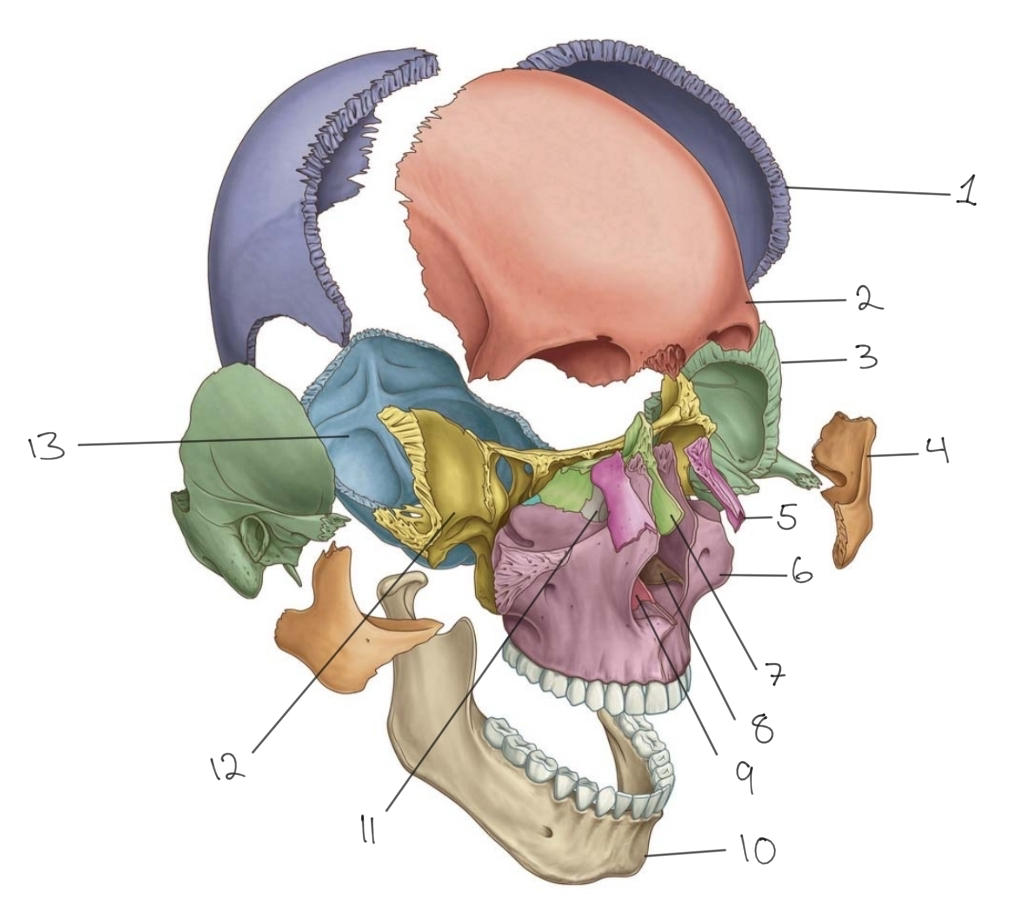
Bone 9
26
Bone 10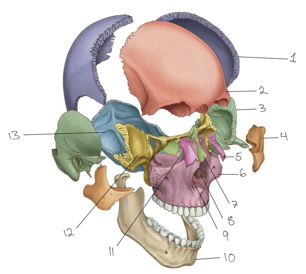
Mandible
27
Mandible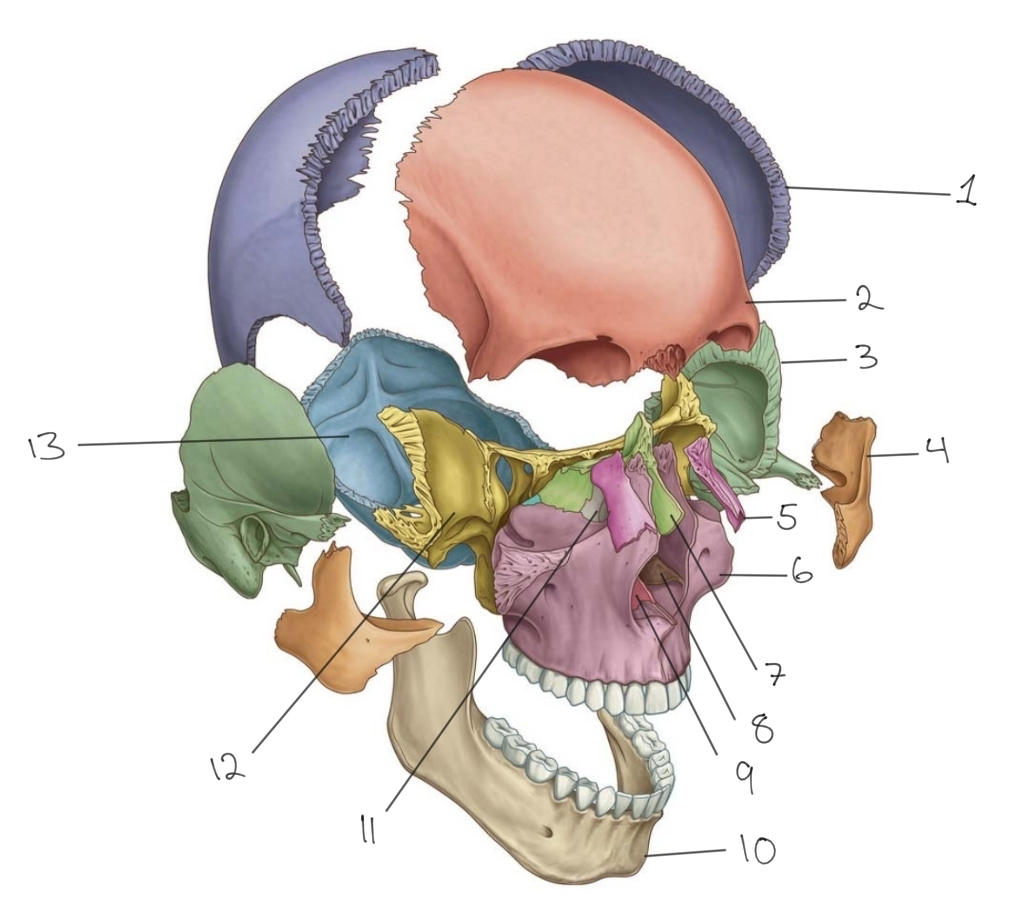
Bone 10
28
Bone 11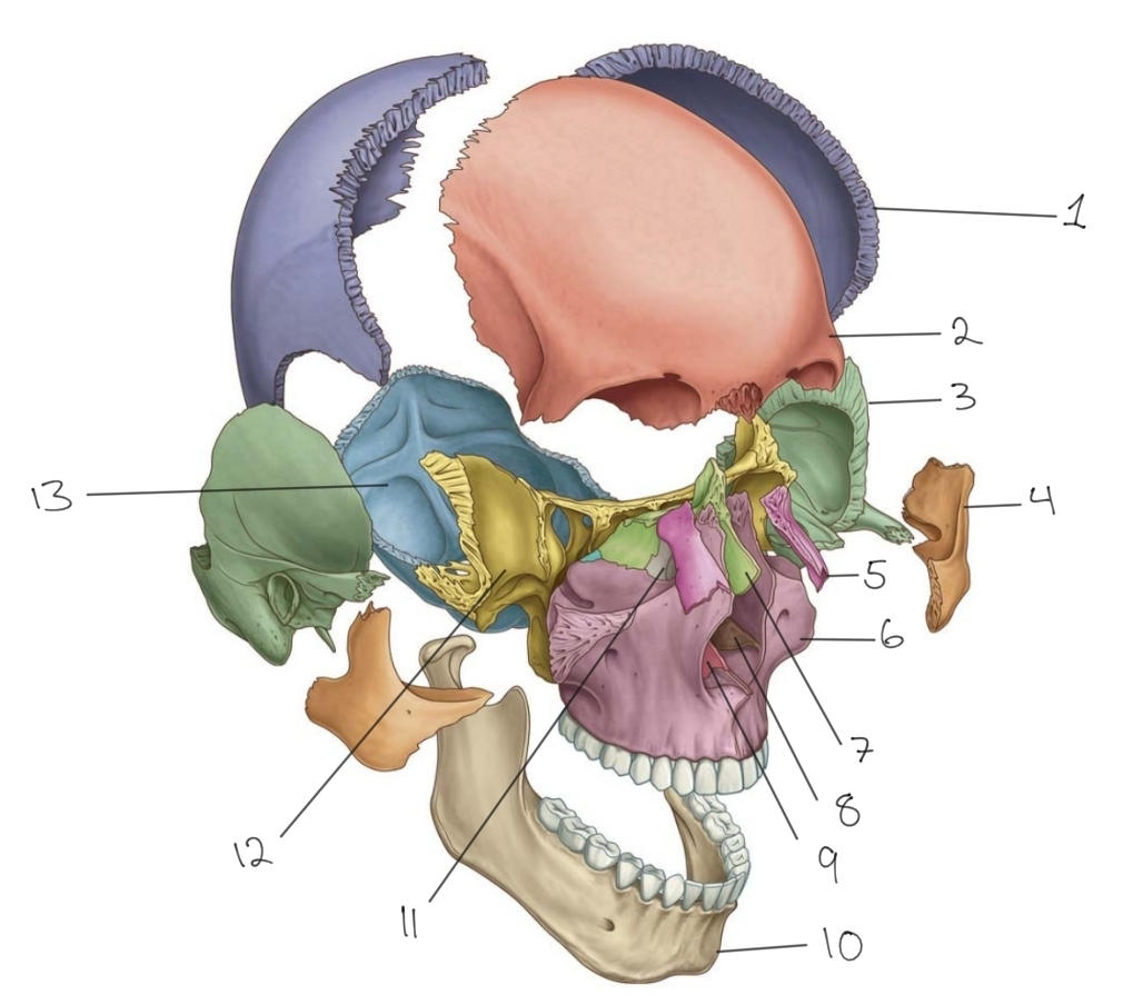
Lacrimal
29
Lacrimal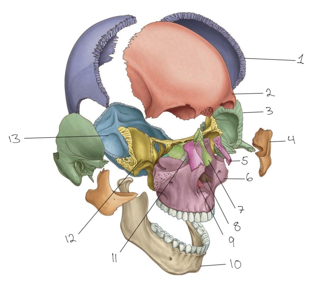
Bone 11
30
Bone 12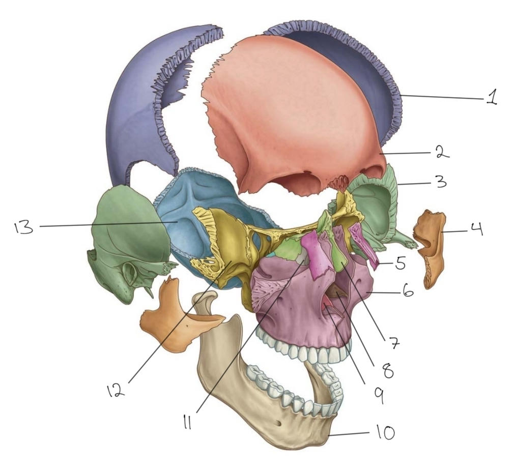
Sphenoid
31
Bone 13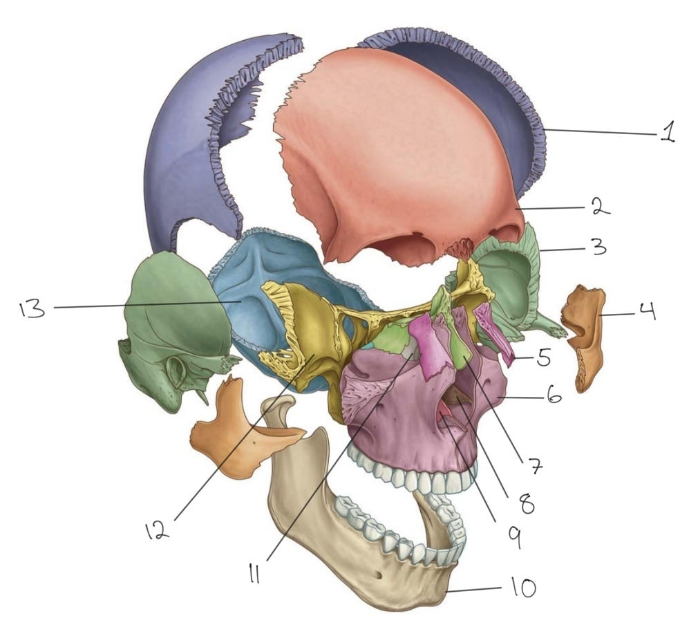
Occipital
32
Occipital 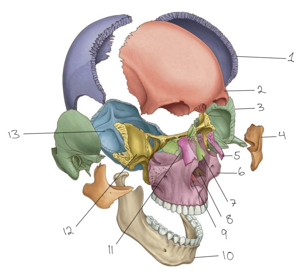
Bone 13
33
Sphenoid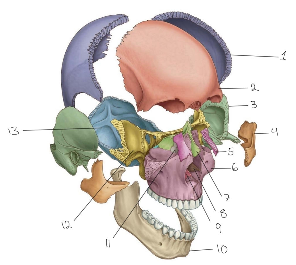
Bone 12
34
Identify the cranial fossa and distinguish the different bones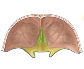
Anterior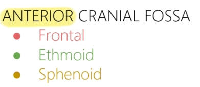
35
Identify the cranial fossa and distinguish the different bones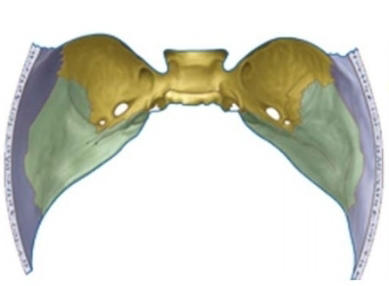
Middle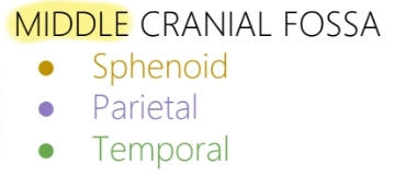
36
Identify the cranial fossa and distinguish the different bones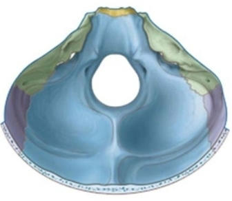
Posterior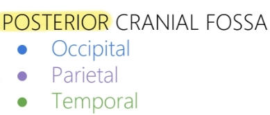
37
Name suture 1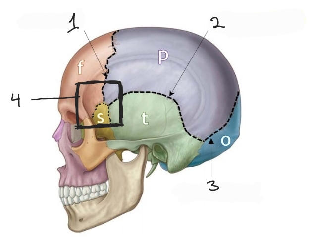
Coronal
38
Name suture 2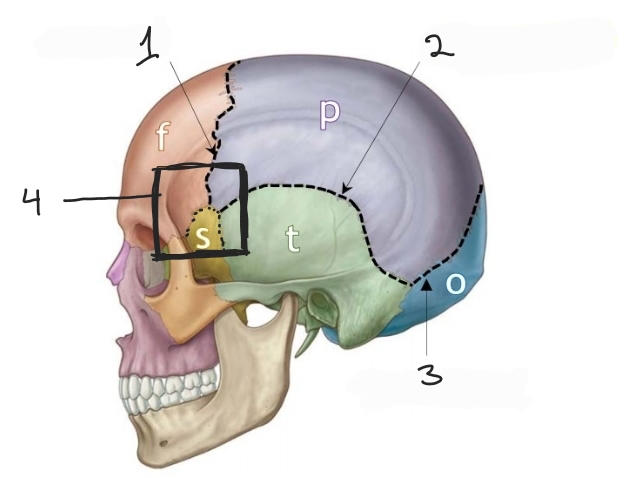
Squamosal
39
Name suture 4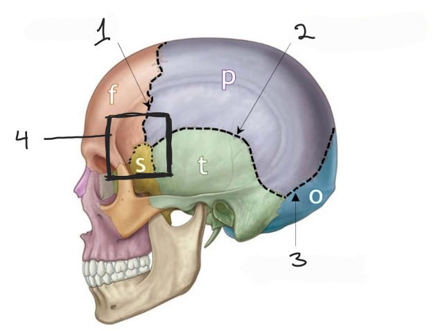
Pterion
40
Name suture 1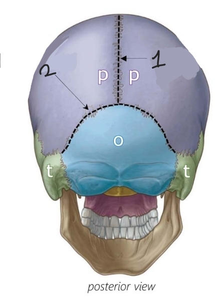
Sagittal
41
Name suture 2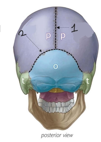
Lamboid
42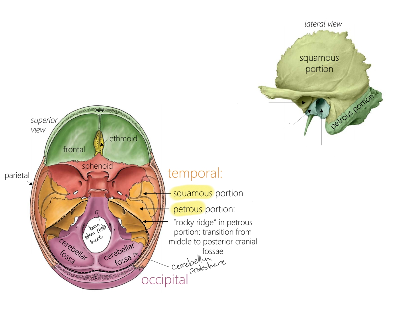
Where is the squamous portion of the temporal bone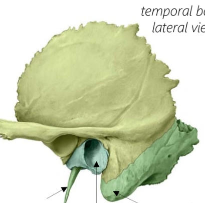
43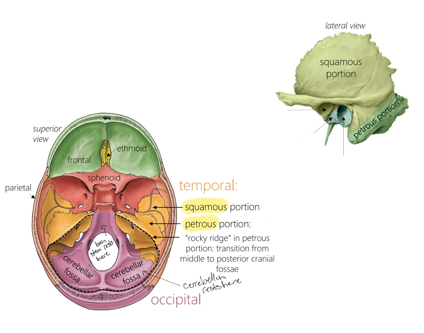
Where is the petrous portion of the temporal bone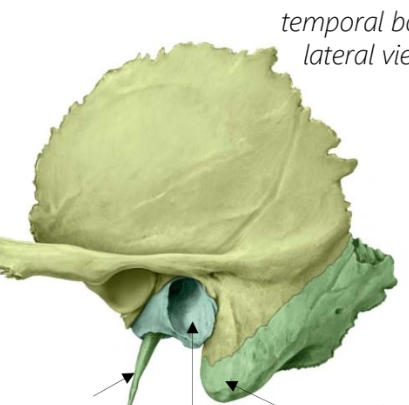
44
Which portion of the temporal bone is in light green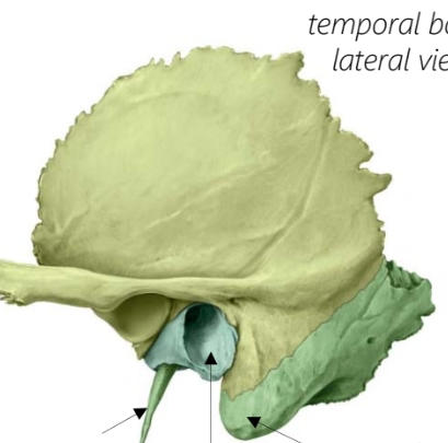
Squamous
45
Which portion of the temporal bone is in dark green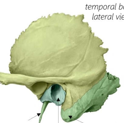
Petrous
46
Name foramen 1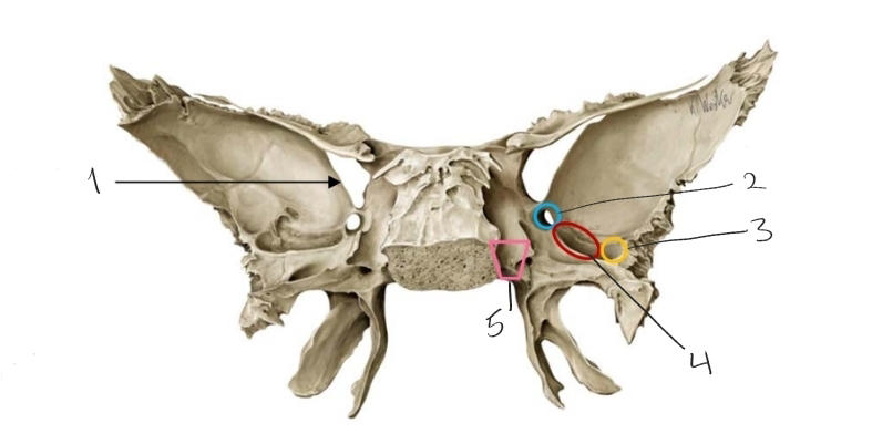
Superior orbital fissure
47
Name foramen 2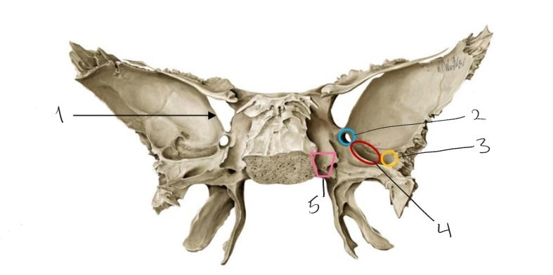
Foramen rotundum
48
Name foramen 3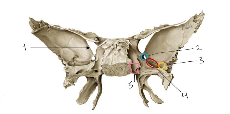
Foramen spinosum
49
Name foramen 4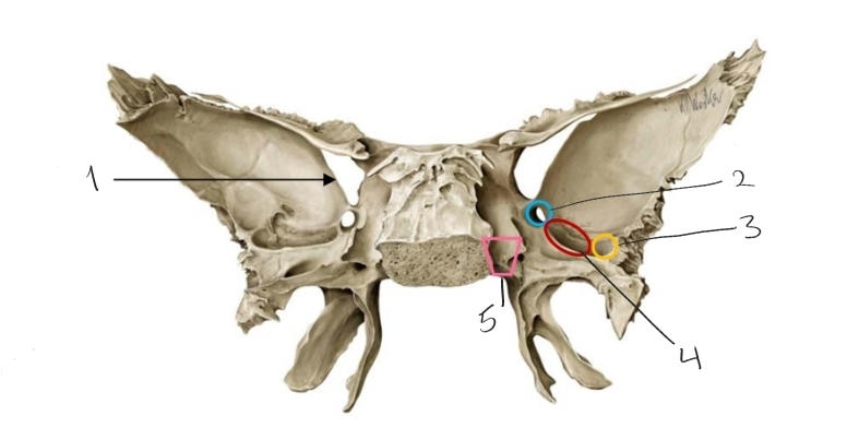
Foramen ovale
50
Name foramen 5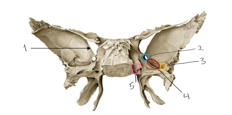
Foramen lacerum
51
Name the foramen in blue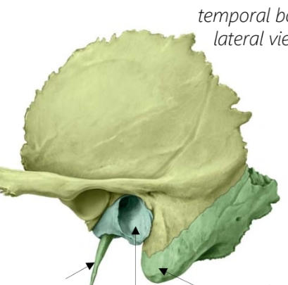
External acoustic meatus
52
Name foramen 1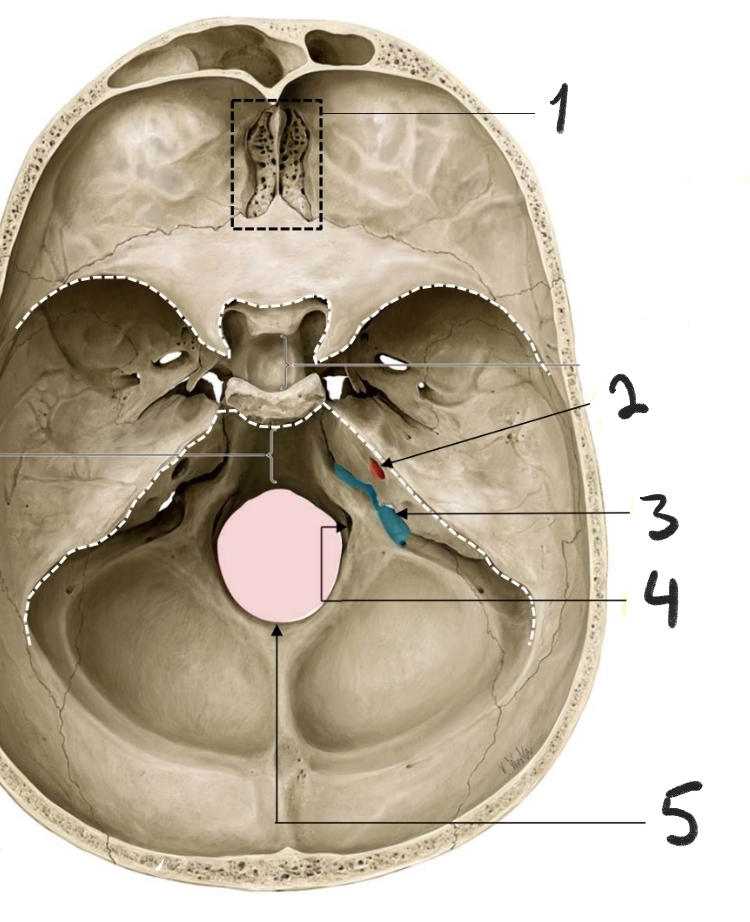
Cribriform foramina
53
Name foramen 2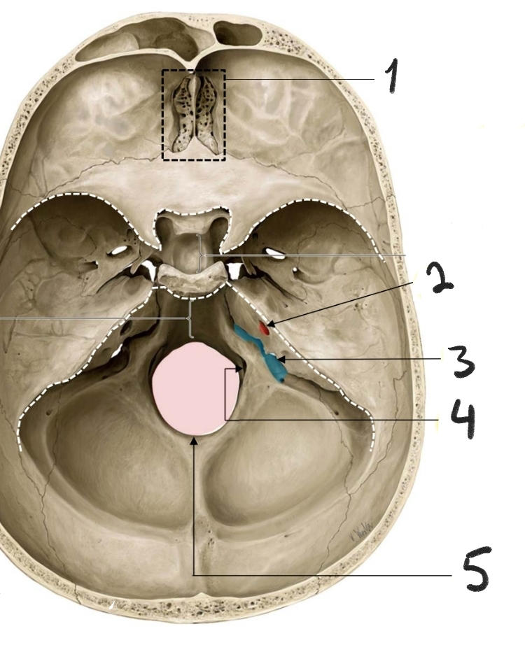
Internal acoustic meatus
54
Name foramen 3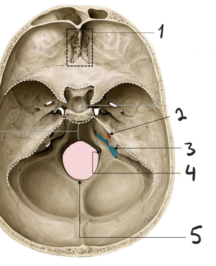
Jugular foramen
55
Name foramen 4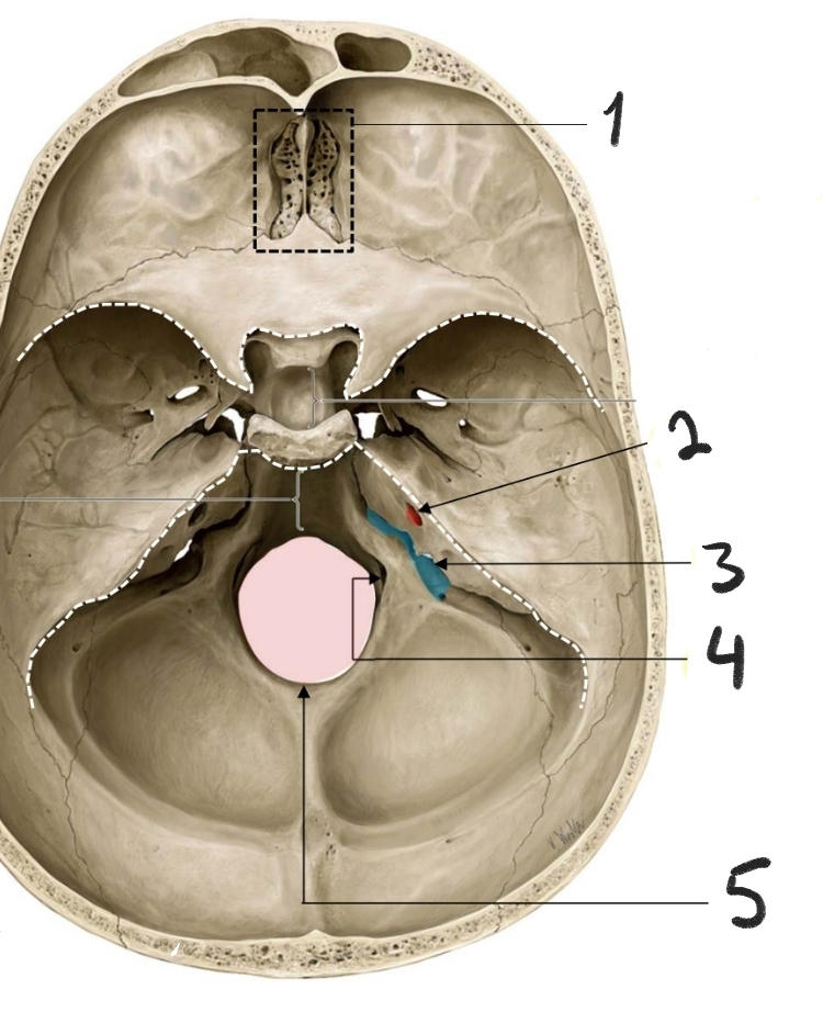
Hypoglossal canal
56
Name foramen 5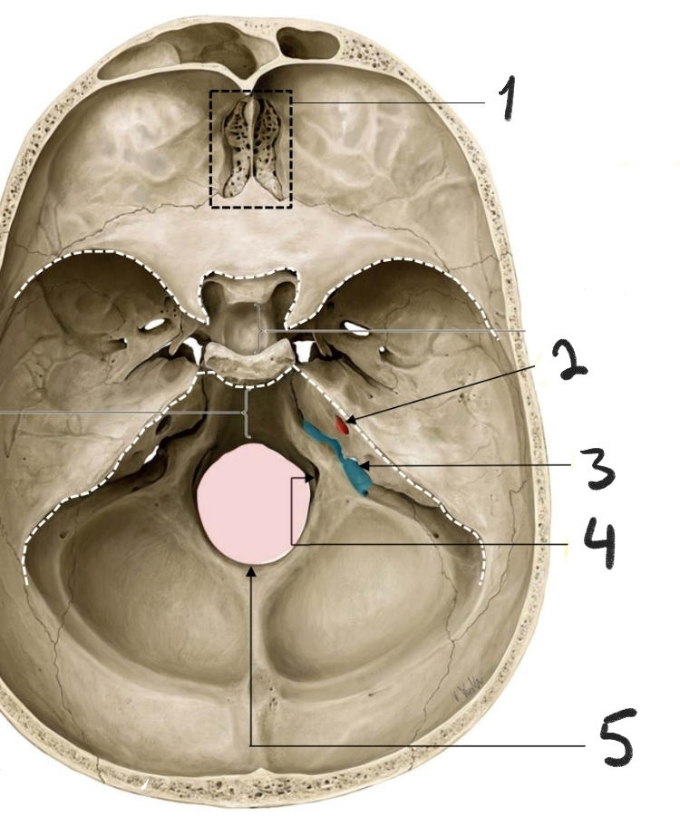
Foramen magnum
57
What 4 types of info do cranial nerves carry +1 ex each
1-general somatic (skeletal muscles in head and neck, skin) 2-general visceral (blood vessels,salivary glands, intraocular muscles) 3-special somatic (vision and hearing, balance) 4-special visceral (taste, smell)
58
Name the cranial nerves
I Olfactory II Optic III Oculomotor IV Trochlear V Trigeminal VI Abducens VII Facial VIII Vestibulocochlear IX Glossopharangeal X Vagus XI Spinal accessory XII Hypoglossal
59
Cranial nerve info mnemonic
Some Say Marry Money But My Brother Says Big Brains Matter More
60
What foramen does the Olfactory nerve pass through
Cribriform foramina
61
What info does the Olfactory nerve relay to the brain
Special sense: Olfactory (smell)
62
What are the three branches of the Trigeminal nerve and what kind of info do they relay
Ophthalmic (sensory), Maxillary (sensory), Mandibular (sensory and motor)
63
What three pieces of info does the facial nerve relay
Special sensory: anterior 2/3 of tongue, Motor: facial expression muscles, Parasympathetic: lacrimal, nasal, salivary and palatial glands
64
Which foramen does the facial nerve pass through
IAM then Stylomastoid foramen
65
Which foramen does the spinal accessory nerve enter and exit
Enters foramen magnum and exits jugular foramen
66
What info does the spinal accessory nerve relay
Motor: trapezius and sternocleidomastoid muscles
67
Where does the vagus nerve exit the skull
Jugular foramen
68
What info does the vagus nerve relay
Motor: pharynx and larynx, Sensory: larynx, Parasympathetic: lungs, heart, esophagus, most of the gut
69
Which foramen does the glossopharyngeal nerve pass through
Jugular foramen
70
What info does the glossopharyngeal nerve relay
Sensory: posterior 1/3 of tongue,carotid body and pharynx, Parasympathetic: salivary and parotid glands, Motor: pharynx
71
What are the three parts of the Opthalmic nerve
Nasociliary, Frontal, Lacrimal (NFL)
72
Which foramen does the ophthalmic nerve pass through
Superior orbital fissure
73
What is the role of the ophthalmic nerve
Sensory: skin of upper face
74
Which foramen does the maxillary nerve pass through around
Infraorbital fissure
75
What is the role of the maxillary nerve and what nerve branches off
Sensory: skin of mid face, upper teeth and upper lip Nerve: superior alveolar nerve
76
Which foramen does the mandibular nerve pass through
Foramen ovale
77
What info does the mandibular nerve carry and what nerves branch off
Motor: muscles of mastication, Sensory: Lower skin, lower teeth and anterior 2/3 of tongue Nerves: Inferior alveolar nerve, lingual nerve
78
Name 1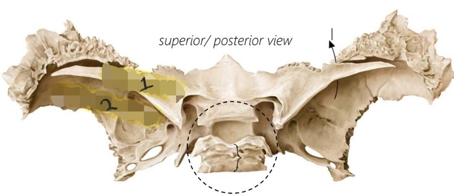
Lesser wing
79
Name 2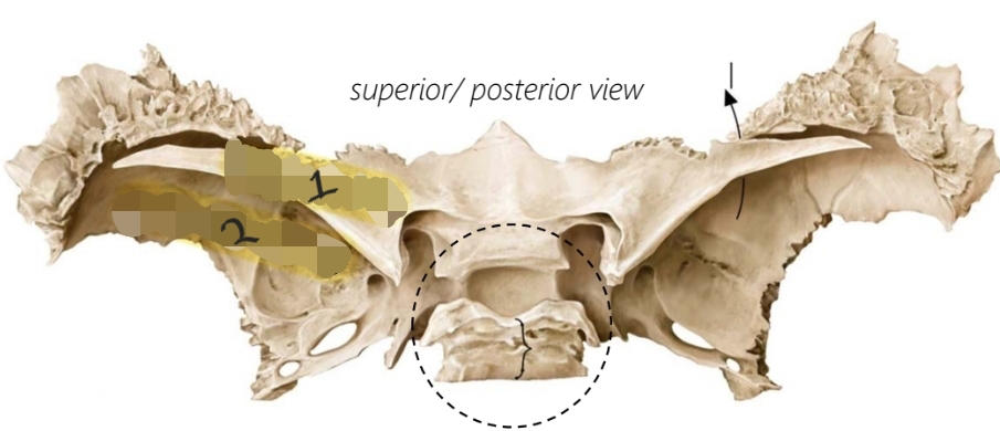
Greater wing
80
Name circled region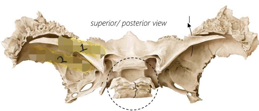
Sella turcica
81
Name the highlighted region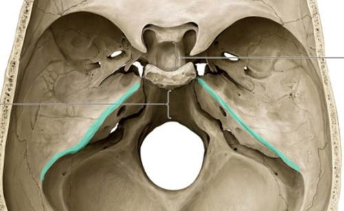
Rocky ridge
82
Name the processes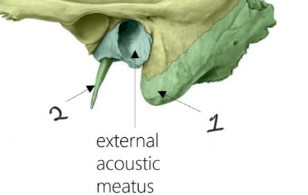
1- Mastoid process 2- Styloid process
83
Name this and the processes that make it up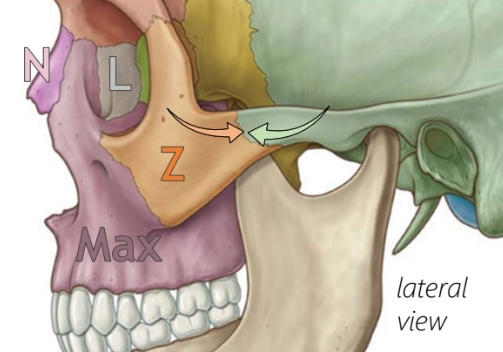
Zygomatic arch, temporal process of the zygomatic bone, zygomatic process of the temporal bone
84
Name 1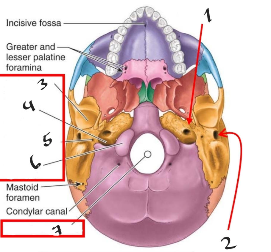
Carotid canal
85
Name 2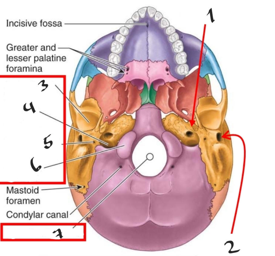
External acoustic meatus
86
Name 3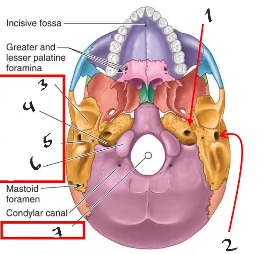
Mandibular fossa
87
Name 4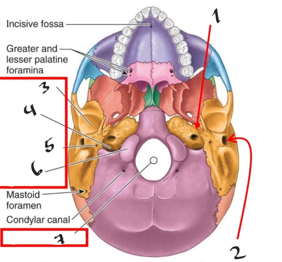
Jugular foramen
88
Name 5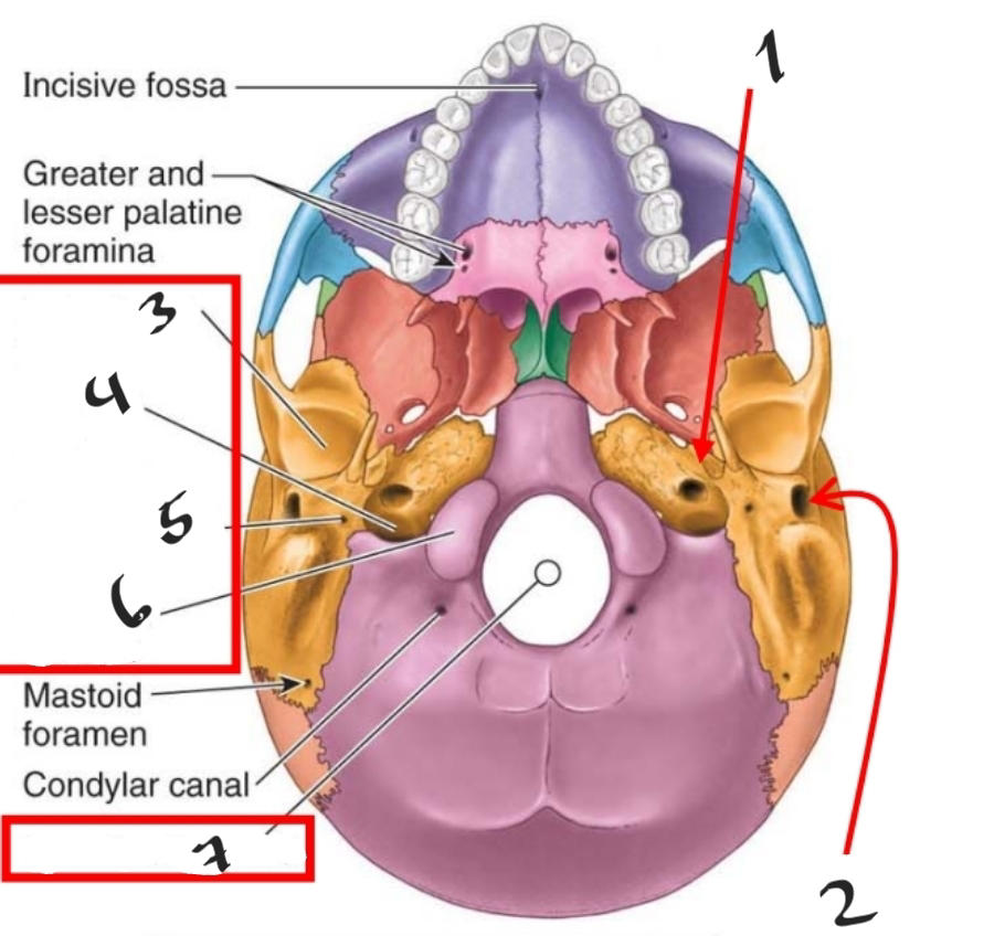
Stylomastoid process
89
Name 6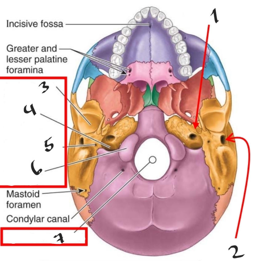
Occipital condyle
90
Name 7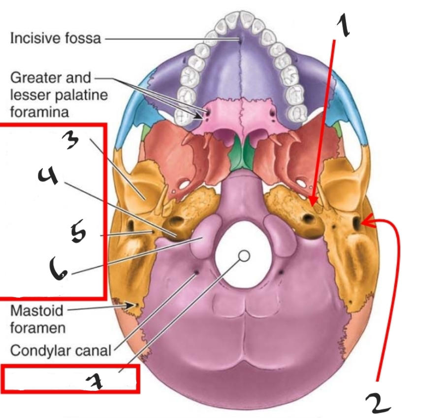
Foramen magnum
91
Which two cranial nerves originate in the brain
Olfactory and optic
92
What foramen does the Optic nerve pass through
Optic canal
93
What foramen does the Oculomotor nerve pass through
Superior orbital fissure
94
What foramen does the Trochlear nerve pass through
Superior orbital fissure
95
What foramen does the Abducens nerve pass through
Superior orbital fissure
96
What foramen does the Vestibulocochlear nerve pass through
Internal acoustic meatus
97
What is the function of the Optic nerve
Sensory: vision
98
What is the function of the Oculomotor nerve
Motor: Extraocular eye muscles (all but superior oblique and lateral rectus plus levator palpebrae superioris) Parasympathetic: Pupil constriction
99
What is the function of the Trochlear nerve
Motor: Extraocular muscle (superior oblique)
100
What is the function of the Abducens nerve
Motor: Extraocular muscle (lateral rectus)
101
What is the function of the Vestibulocochlear nerve
Sensory: hearing, balance
102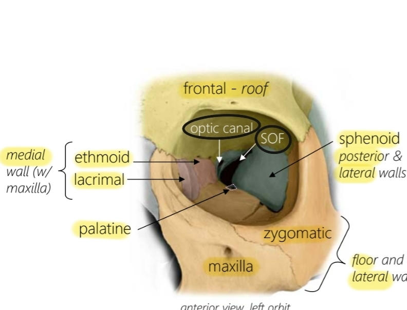
Name the bones and foramina that make up the orbit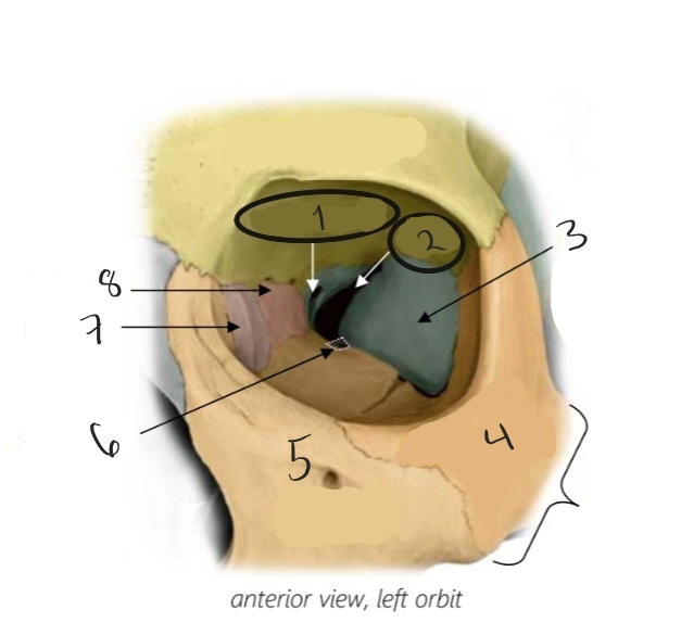
103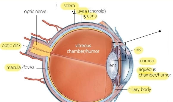
Name the structures of the eye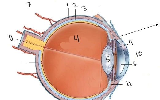
104
What tissue type makes up the sclera
Fibrous, dense connective
105
What kind of cells are in the retina
photoreceptor cells
106
What is the Optic disk
Where all the neurons converge on the Optic nerve (No photo receptors, blind spot)
107
What is the macula of the retina
The area with the most photoreceptors
108
What are the three components of the uvea
Choroid: Vascular layer, Ciliary body: Maintains/ changes shape of lense, Iris: Made up of 2 muscles pupillary sphincter and pupillary dialator
109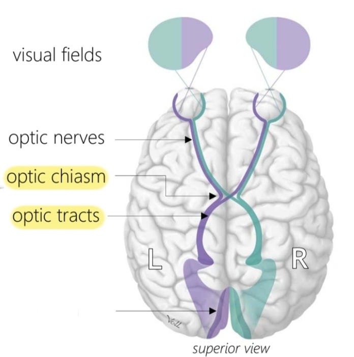
Name 1,2 and 3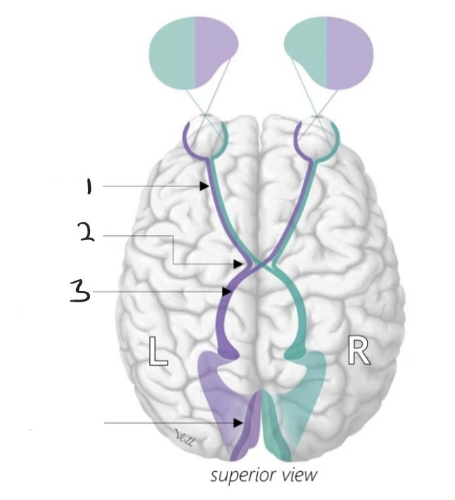
110
Name the 6 extraocular muscles and their fuction
Inferior (depresses and adducts), superior (elevates and adducts), lateral (lateral rotation) and medial (medial rotation) rectus. Superior (depresses and abducts) and inferior (elevates and abducts) oblique.
111
Where do the extraocular muscles originate
Rectus muscles: common tendinous ring around Optic nerve, Superior oblique: common tendinous ring, through trochlea, Inferior oblique: infero-medial orbital wall
112
What is the external ear called? Which foramen makes up the canal? Where does it end?
Auricle, external auditory meatus, timpanic membrane
113
What is the middle ear for? What are the names of the bones? Where does it end? What does it open into?
To convert waves to mechanical vibrations, reduce energy loss. Malleus, incus, stapes. Oval window. Auditory tube.
114
Where is the bony labyrinth, what are its two parts, what makes up the two parts
In the inner in (petrous portion of temporal bone). Vestibular aparatus (3 semicircular canals and vestibule), cochlea (membrous labyrinth:surrounded by perilymph and filled with edolymph, scala vestibuli: ascending, scala tympani: descending).
115
What is the thick base of the semicircular canals called
Ampulla
116
What do the cells in the ampulla do
Detect movement of endolymph (interpreted as head rotation)
117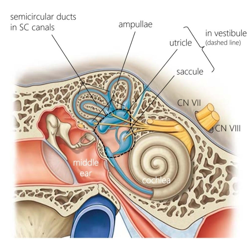
Name these structures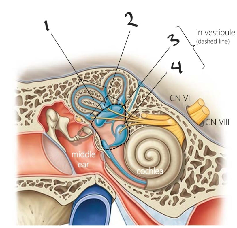
118
What do the utricle and saccule do and how
Detect linear acceleration and head position. The hair cells in gel pushed or pulled by gravity (ex telling brain you're upside down)
119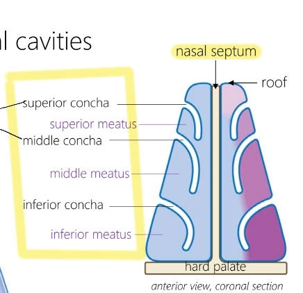
Name these structures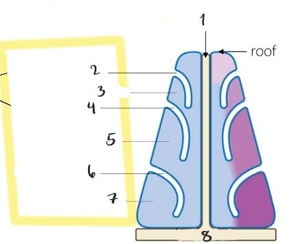
120
Name the openings to the nasal cavity
Exterior: nostrils, Interior: Choanae
121
What is the role of the conchae
To slow and warm air, the air is thus exposed to more surface area of mucosa and for longer (helps with warming air and filtering particles
122
What is the ostium
opening to eustachian tube that helps to equalize pressure
123
Name the sinuses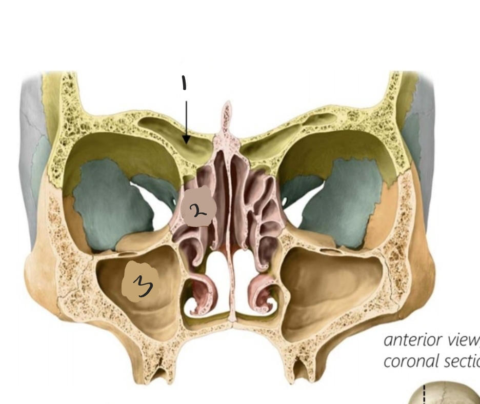
1: Frontal sinuses 2: Ethmoid sinuses 3: Maxillary sinuses
124
Name the sinus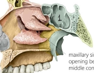
1: Sphenoid sinus
125
Functions of sinuses
immune, warming air, altering voice, scaling skull size while keeping it light
126
Where does the sphenoid sinus drain to
Above the superior concha into the sphenoethmoidal recess
127
Where do the frontal, ethmoid and maxillary sinuses drain to
Below the middle concha into the middle meatus
128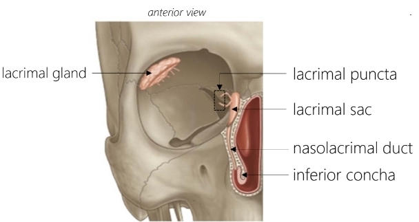
Name these structures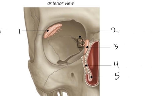
129
What are the two branches of the carotid artery
External carotid, internal carotid
130
What does the internal carotid artery supply blood to
The brain, first branch= ophthalmic (to eye)
131
What does the external carotid artery supply blood to
Neck, face, scalp, oral cavity, nasal cavity
132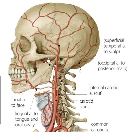
Name the artery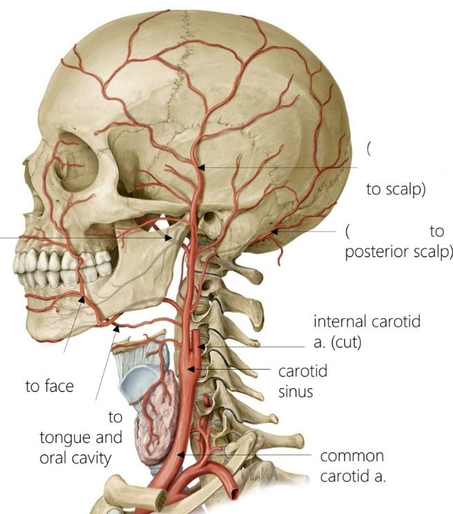
133
Which two arteries supply blood to the nasal cavity
Sphenopalatine, ethmoidal (branch of opthalmic)
134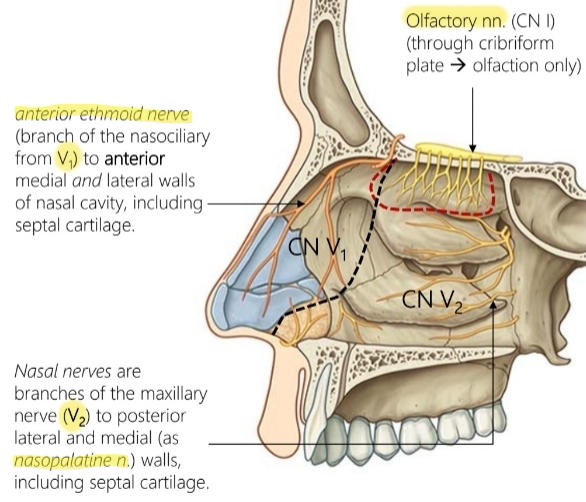
Name the nerves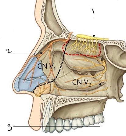
135
What is the posterior opening to the oral cavity called
Oropharyngeal isthmus
136
What muscles make up the floor of the oral cavity
Hyoid muscles (mylohyloid and geniohyoid)
137
What muscle makes up the anterior wall of the oral cavity
Orbicularis Oris (lips)
138
What muscle makes up the lateral wall of the oral cavity
Buccinator
139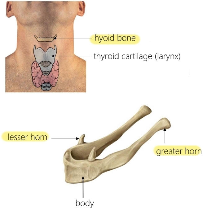
Name these structure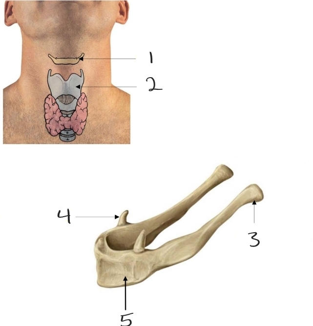
140
Name these muscles and where they attatch and their function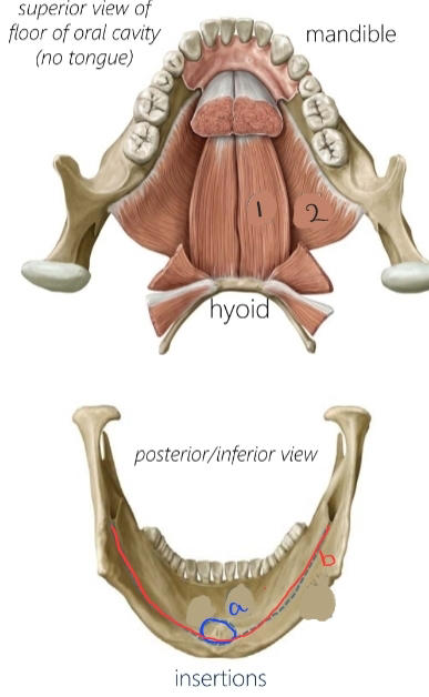
Elevate and bring forward hyloid and tongue when swallowing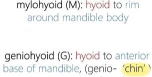
141
What is the division between the anterior and posterior of the tongue called
Terminal sulcus
142
What is the structure down the middle of the tongue called
Lingual septum
143
What are the three components of the intrinsic muscles of the tongue and what are their function
Longitudinal, transverse and vertical. Fine motor control.
144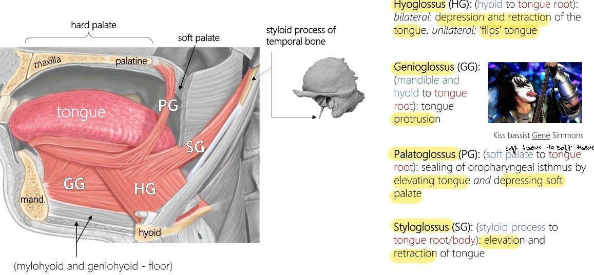
Name the extrinsic muscles of the tongue and the functions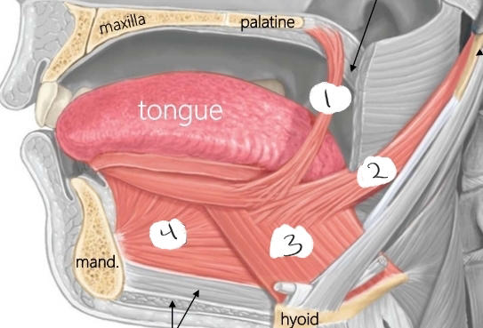
145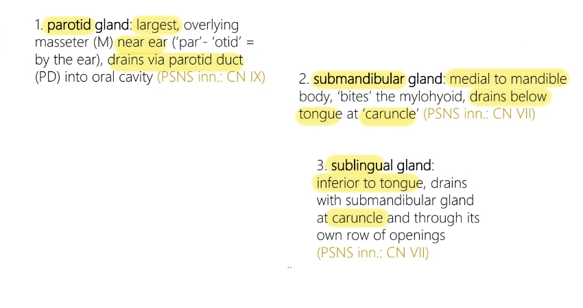
Name the three salivary glands, their location and where they drain to
146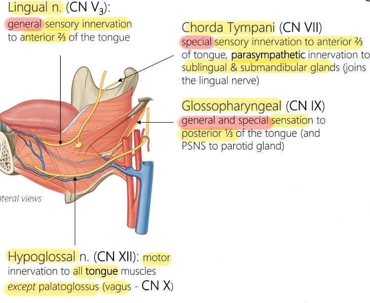
Name the 4 nerves that inervate the tongue and their functions
147
What is the main artery to the tongue
Lingual artery
148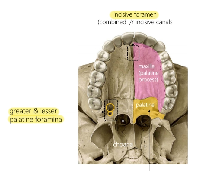
Name these structures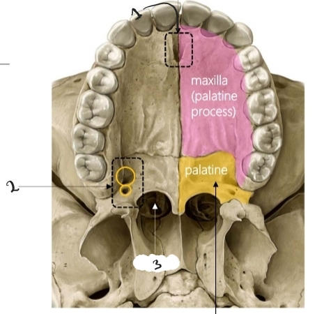
149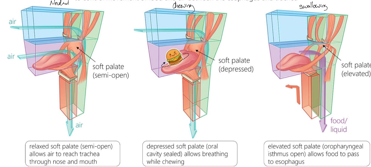
Describe what the soft palate does in a neutral position, when chewing and when swallowing
150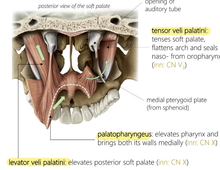
Name the muscles of the soft palate and their functions
151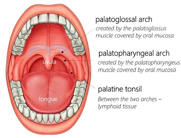
Name these structures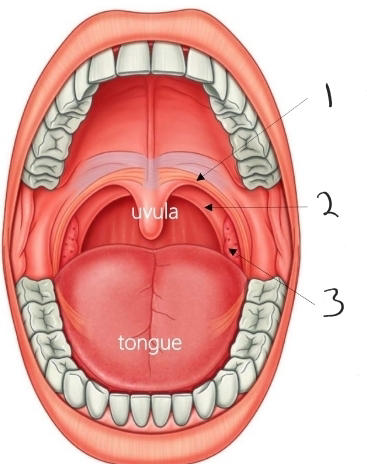
152
What contribution does the nasal cavity make to the oral cavity and through where
Sphenopalatine artery, Nasopalatine nerve, through incisive canal
153
What neurovasculature goes through the lesser and greater palatine canal
Greater and lesser palatine nerve, greater and lesser palatine artery
154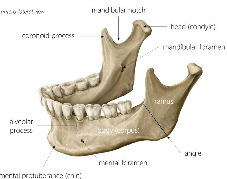
Name the structures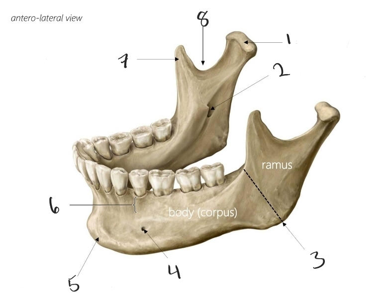
155
Name 1 and 2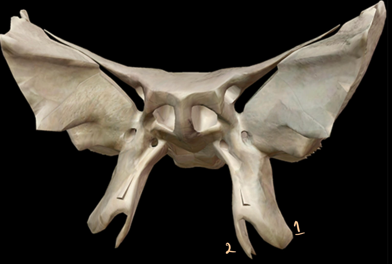
1: lateral pterygoid plate 2: medial pterygoid plate
156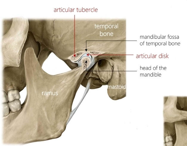
Name these structures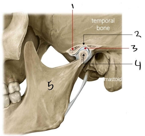
157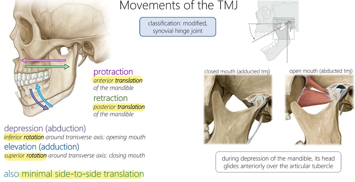
Describe the movement of the TMJ
158
What is this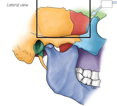
Temporal fossa
159
What's this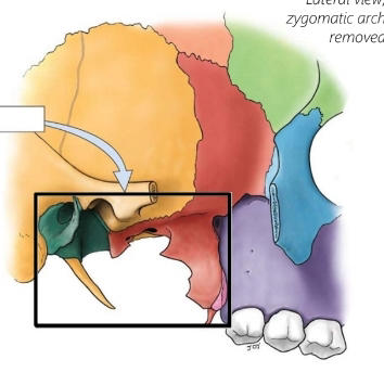
Infratemporal fossa
160
Name the muscles of mastication, where they attatch and their function
1)Temporalis, temporal fossa to coronoid process, elevation and retraction 2)Masseter, zygomatic arch to lateral ramus and angle, elevation and protraction 3)Medial pterygoid, maxilla/palatine and medial of lateral pterygoid plate to medial ramus and angle, elevation 4)Lateral pterygoid, greater wing and lateral side of lateral pterygoid plate to articular disk and condyle, bilateral: protraction, unilateral: translation
161
Which nerve is responsible for motor inervation to the muscles of mastication
Mandibular nerve
162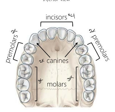
Name the teeth
163
Name the neurovasculature to the teeth
Top teeth: maxillary nerve branches to superior alveolar nerve, superioralveolar artery. Bottom teeth: mandibular nerve branches to inferior alveolar nerves, inferior alveolar artery
164
What compartment of the neck contains the pharynx and larynx
Visceral
165
What compartment of the neck contains the vasculature
Carotid
166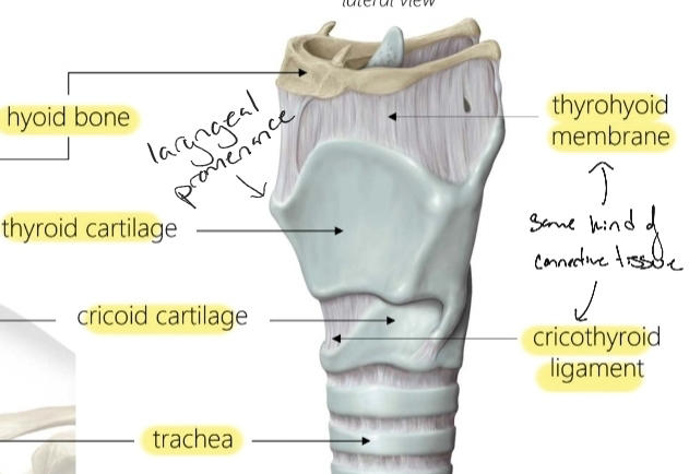
Name these structures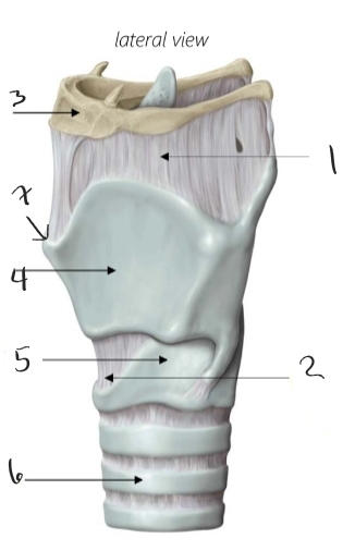
167
Name these muscles, where they attach and their function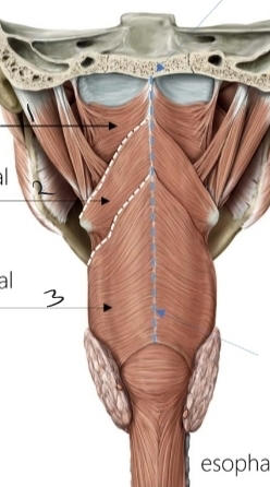
Peristalsis: contract above bolus, relax below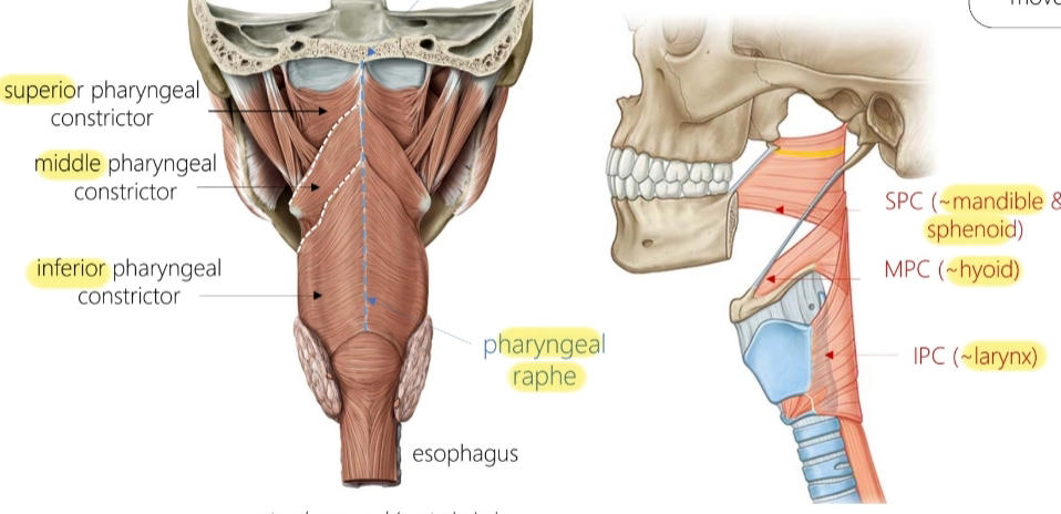
168
Name these muscles, where they attach and their function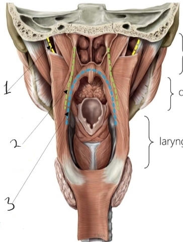
Elevation of soft palate, elevates and shortens pharynx ahead of bolus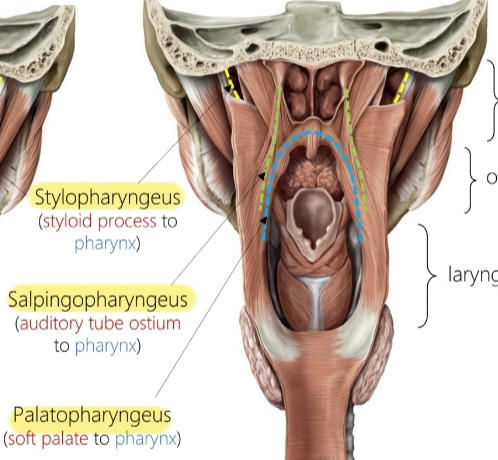
169
What nerves innervate the muscles of the pharynx
Vagus, except stylopharangeas is innervated by the glossopharngeal
170
What are these structures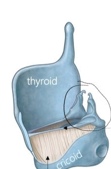
Arytenoids
171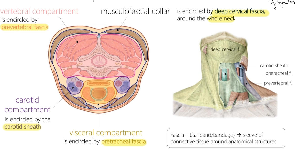
Name the fascia and what compartment they enclose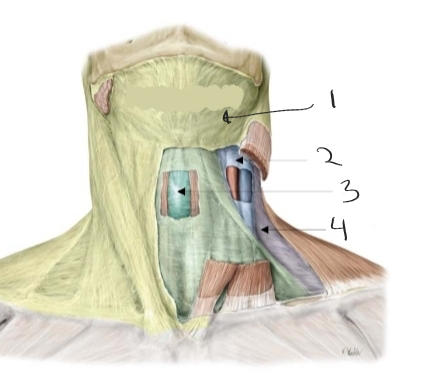
172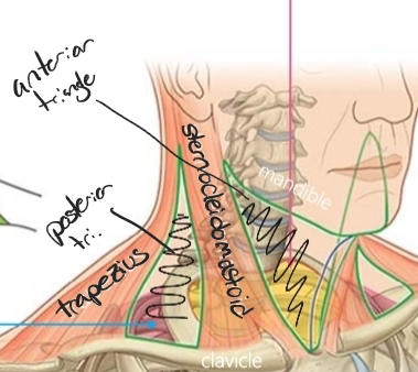
Name these 4 structures 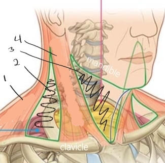
173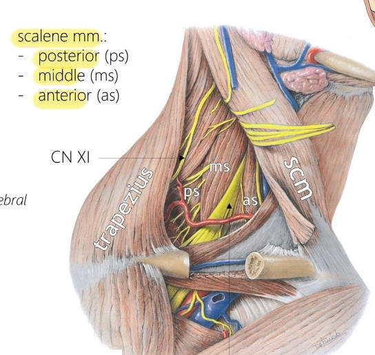
Name the muscles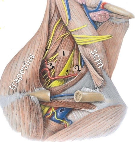
174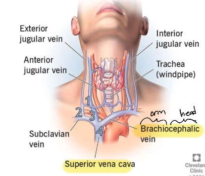
Name the vasculature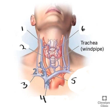
175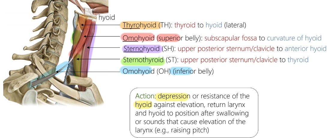
Name the infrahyoid muscles, their location and function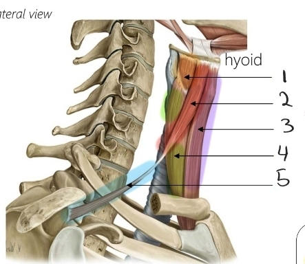
176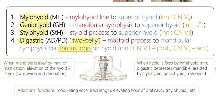
Name the suprahyoid muscles, their location and function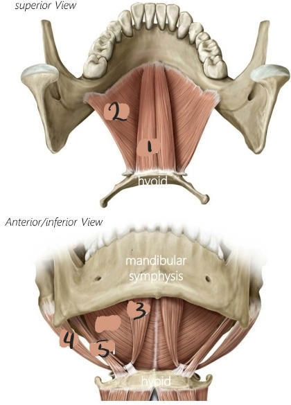
177
Name the triangles and some important structures in each triangle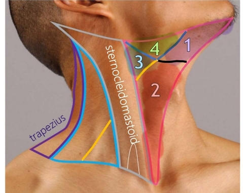
178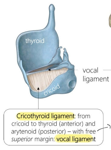
Name these structures 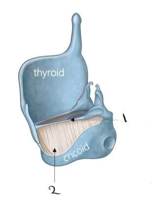
179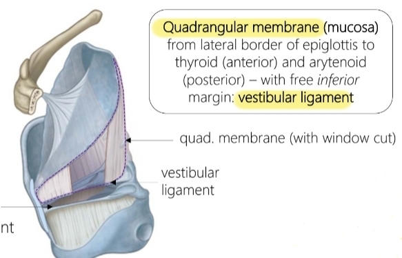
Name these structures 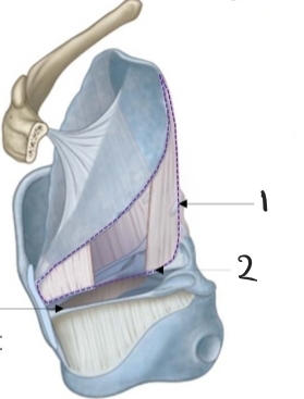
180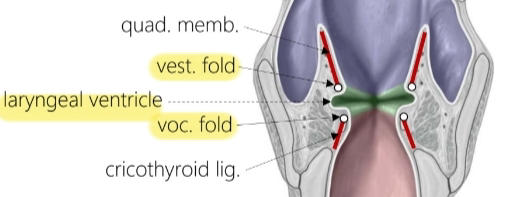
Name these structures 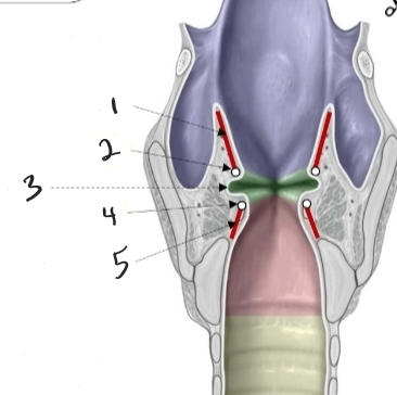
181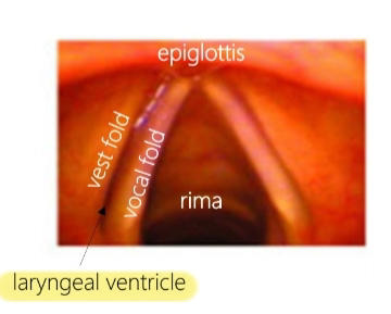
Name the structures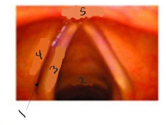
182
What muscle is in orange and what is there function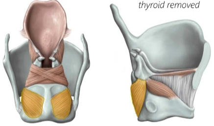
Posterior cricoarytenoids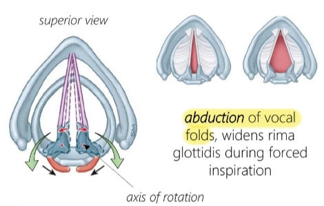
183
What muscle is in red and what is there function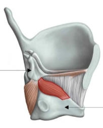
Lateral cricoarytenoids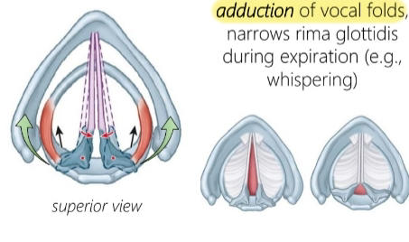
184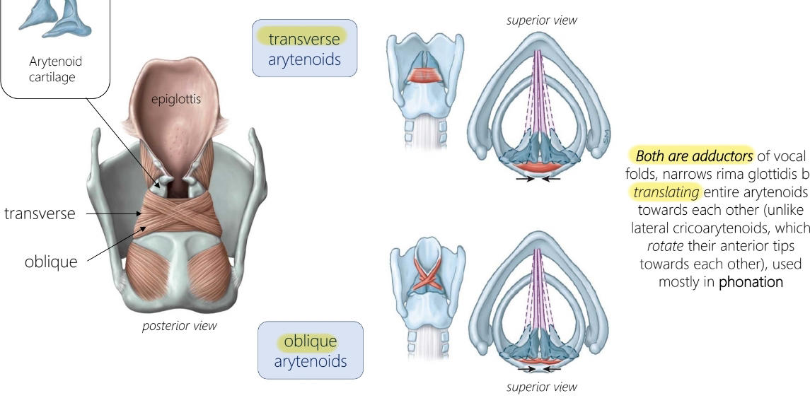
Name these muscles and their function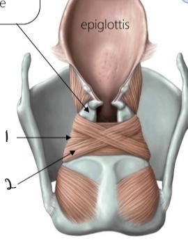
185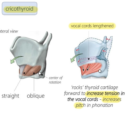
What is this muscle and what does it do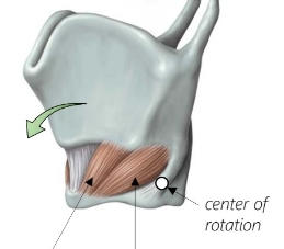
186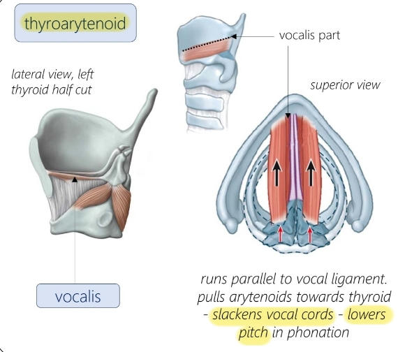
What is this muscle and what does it do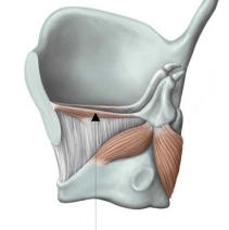
187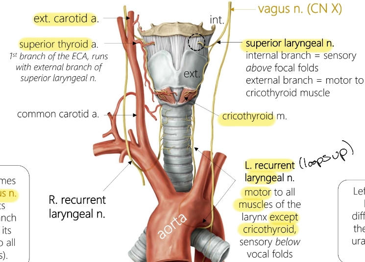
Name the neurovasculature of the larynx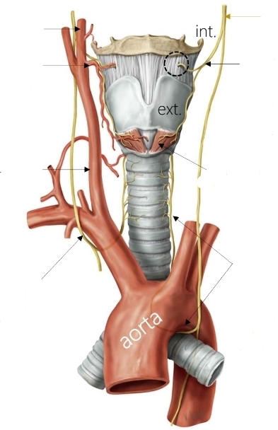
188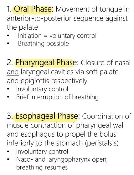
Talk about the phases of deglutition
189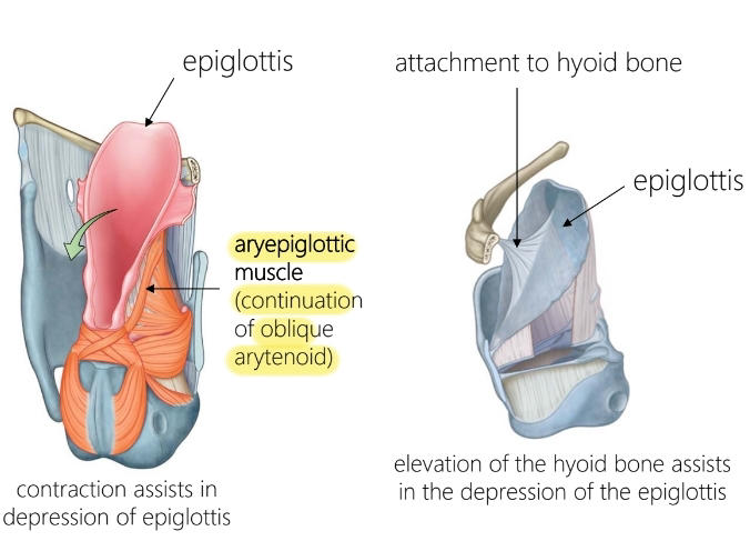
What is this muscle, what is its role and what is the role of the attachment to the hyoid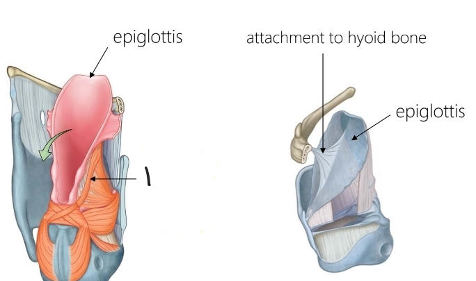
190
Name the portion in pink and it's contents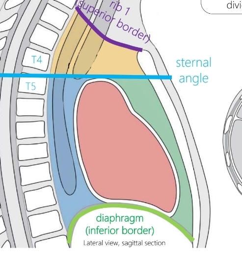
Middle mediastinum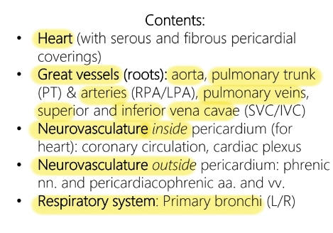
191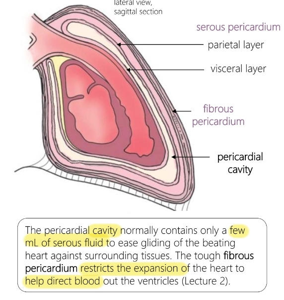
Name the pericardial layers and their function
192
What is it called when there is an excess buildup of fluid in the pericardial cavity
Pericardial effusion
193
What happens when pericardial effusion happens too quickly
Inc. heart rate! heart may stop (too much stress):Cardiactamponade
194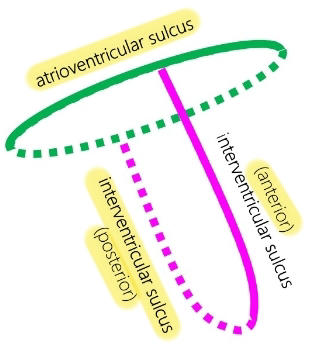
Name the structures in pink and green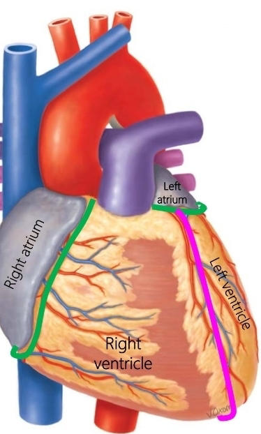
195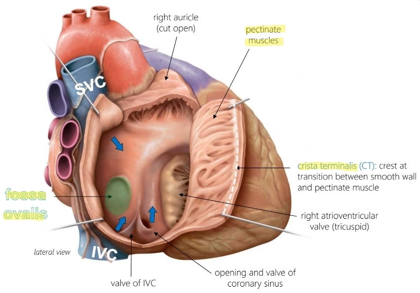
Name the structures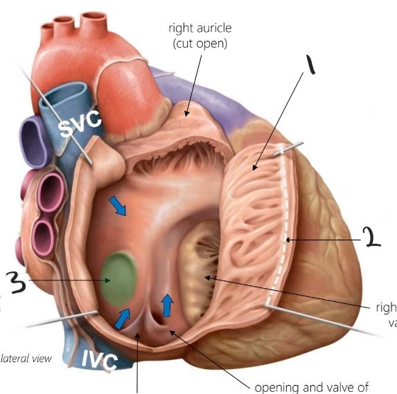
196
Name the prenatal shunts and what they do
Bypass pulmonary circulation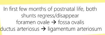
197
What makes the 'lub' 'dub' sounds
'Lub' :during systole, AV valves close. 'Dub' :during diastole, Pulmonary and Aortic valves close
198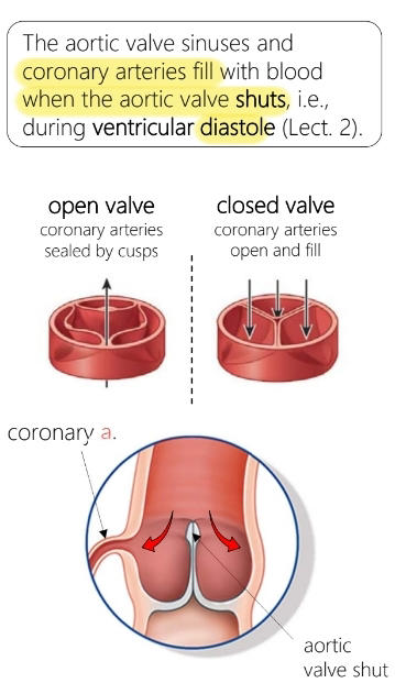
How do the coronary arteries get blood
199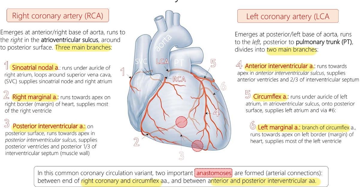
Name the coronary arteries and where they supply blood to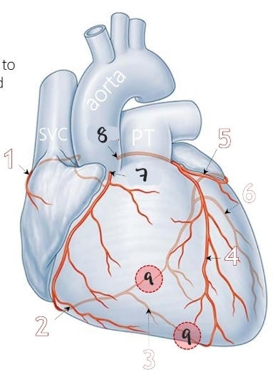
200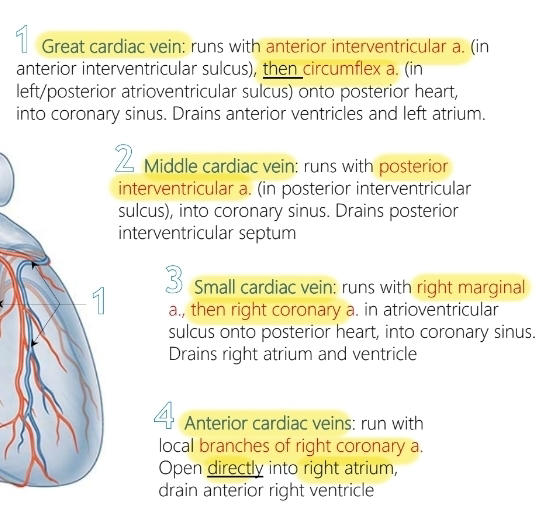
Name the venous drainage of the heart and which part of the heart they drain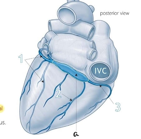
201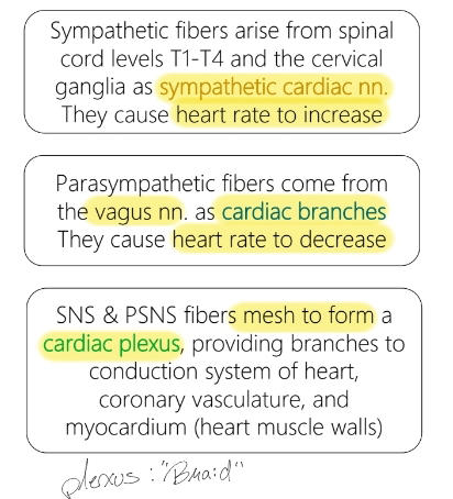
Describe the innervation of the heart (ANS)
202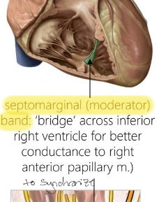
What structure is in green and what does it do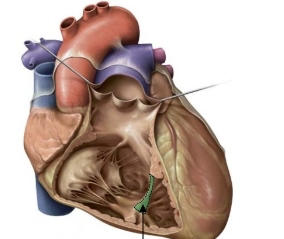
203
Name the portion in yellow and it's contents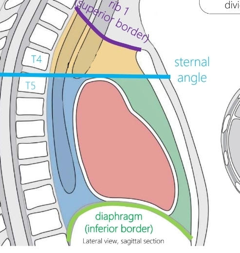
Superior mediastinum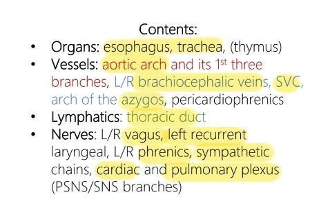
204
Name the portion in green and it's contents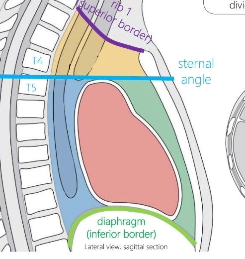
Anterior mediastinum, thymus gland
205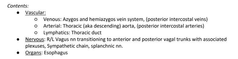
Name the portion in blue and it's contents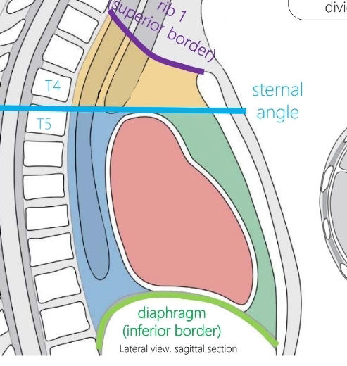
206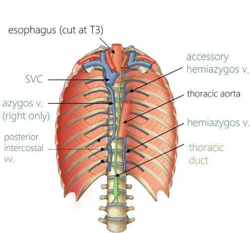
What are the azygos and hemiazygos veins
207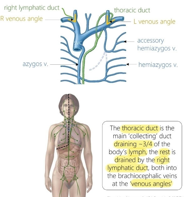
Thoracic duct vs right lymphatic duct
208
How does the esophagus move through the posterior mediastinum
Right of aorta to anterior of aorta
209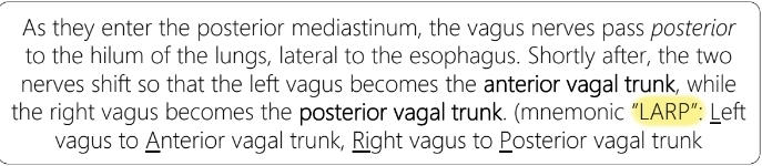
How's does the arrangement of the vagal nerves change around the esophagus
210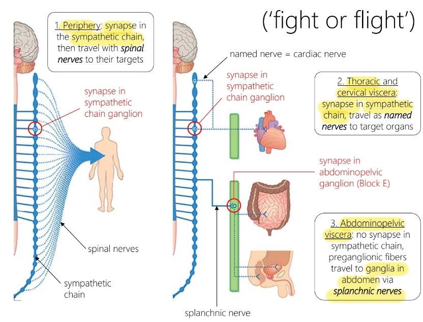
Describe the sympathetic system to the following: periphery, Thoracic and cervical viscera and abdominopelvic viscera
211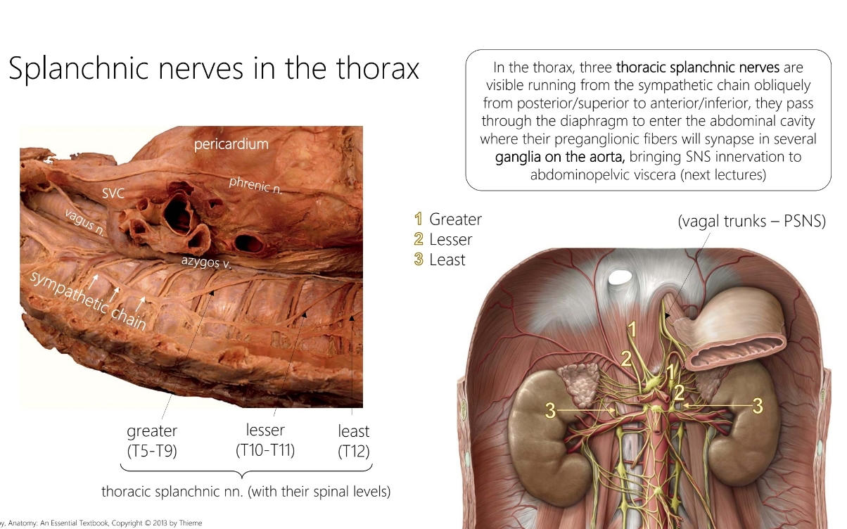
Describe the splanchnic nerves
212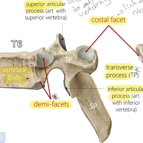
Name these structures 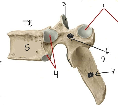
213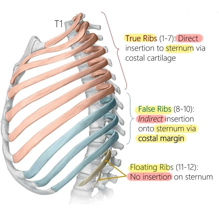
What are the different kinds of ribs
214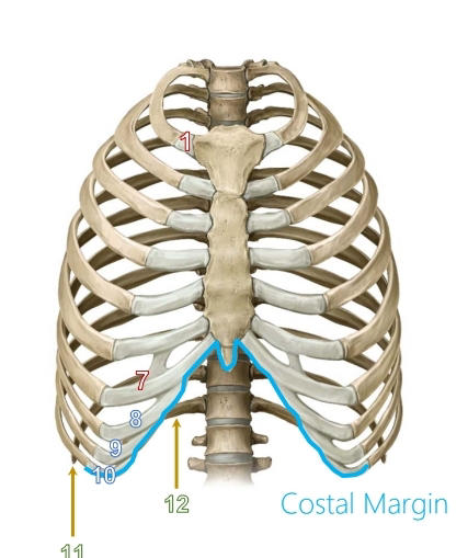
What is the structure in blue called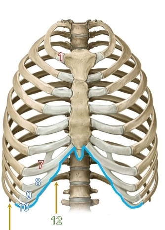
215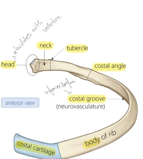
Name these structures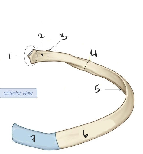
216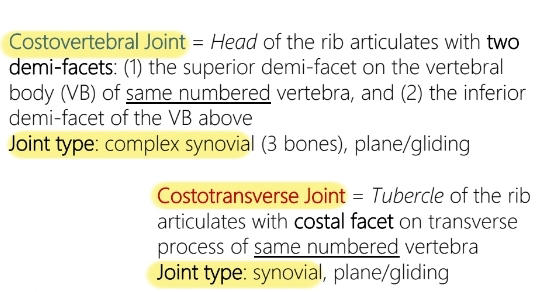
Name the two rib articulations with the vertebre
217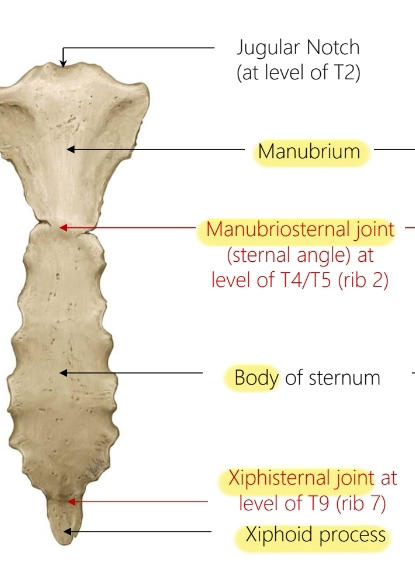
Name these structures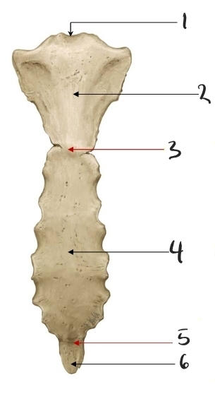
218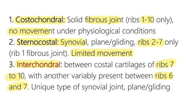
Name the three rib articulations with the sternum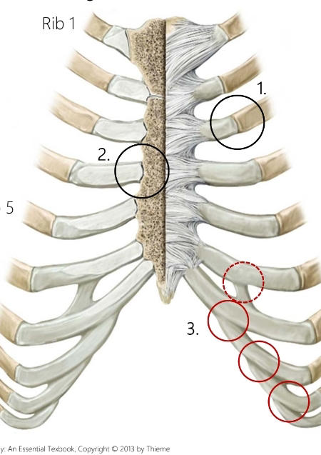
219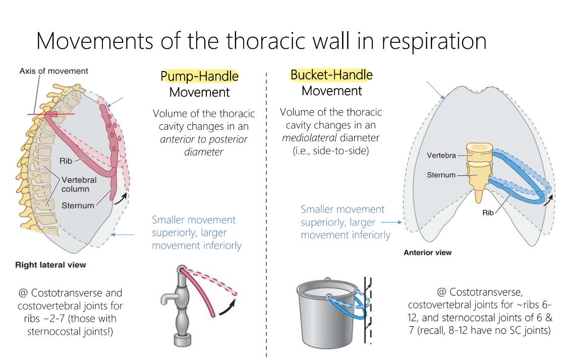
Describe the movements of the thoracic wall during inspiration
220
What are the accessory muscles of the Thoracic Wall and what do they do
External intercostal (hands in pockets, elevation), internal intercostal (opposite), innermost intercostal (opposite) and transverse thoracis (sternum body and xiphoid to costal cartilage of ribs 2-6, depression)
221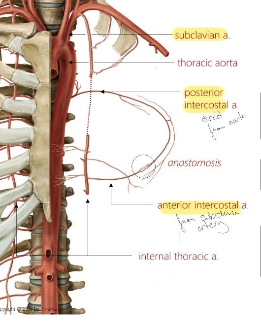
Name the vasculature of the Thoracic wall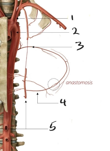
222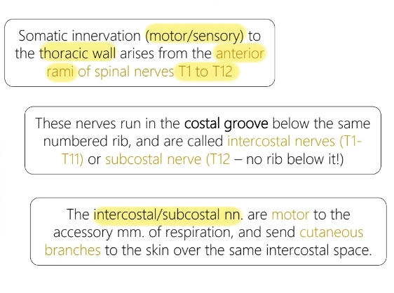
Name the innervation of the Thoracic wall
223
What is the thoracic neurovasculature bundle acronym
VAN
224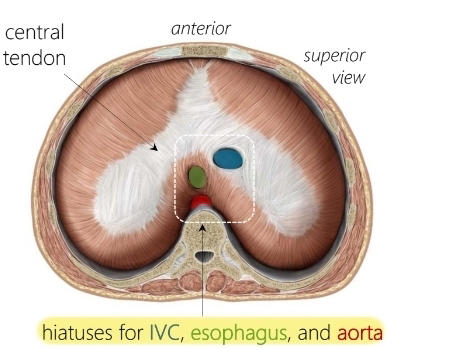
Name these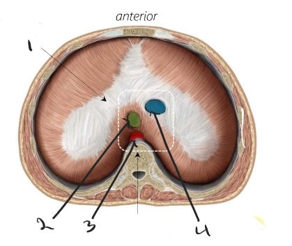
225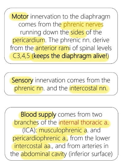
Name the neurovasculature supply to the diaphragm
226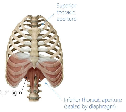
Name these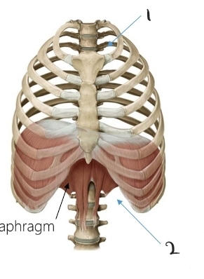
227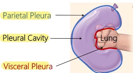
Name the layers of the pleura
228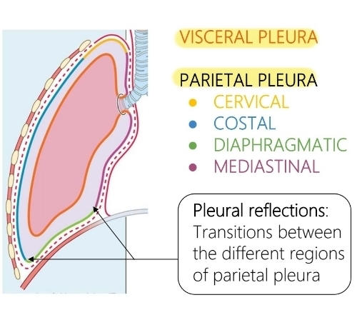
Name the portions of the parietal pleura
229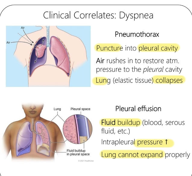
What is pneumothorax and pleural effusion
230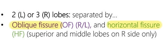
What distinguishes the two lungs and what are the names of the fissures
231
What are the abbreviations for the hilum of each lung
Right: BAV Left: ABV
232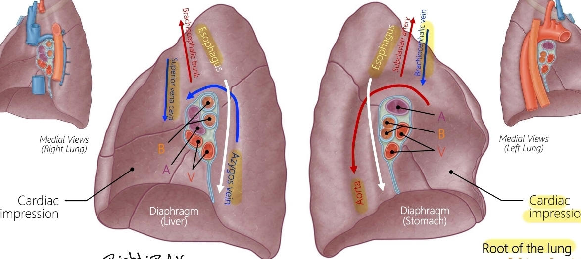
Name these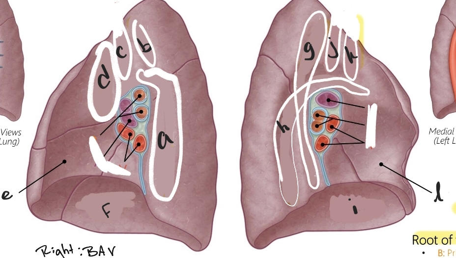
233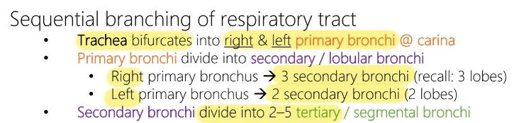
Name the branches of the trachea
234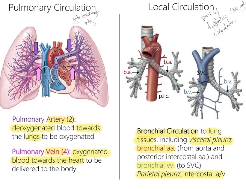
Describe pulmonary circulation vs local circulation