Exam 3: Pelvis and Perineum
Study these flashcardsFree · spaced repetition · iPhone, Android, Mac & web
First 300 of 542 cards
1
Image occlusion: name the hidden labels
2
Image occlusion: name the hidden labels
3
Image occlusion: name the hidden labels
4
Image occlusion: name the hidden labels
5
Image occlusion: name the hidden labels
6
Image occlusion: name the hidden labels
7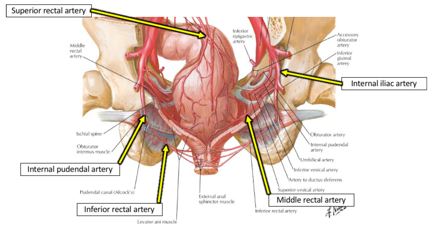
Image occlusion: name the hidden labels
8
Image occlusion: name the hidden labels
9
Image occlusion: name the hidden labels
10
Image occlusion: name the hidden labels
11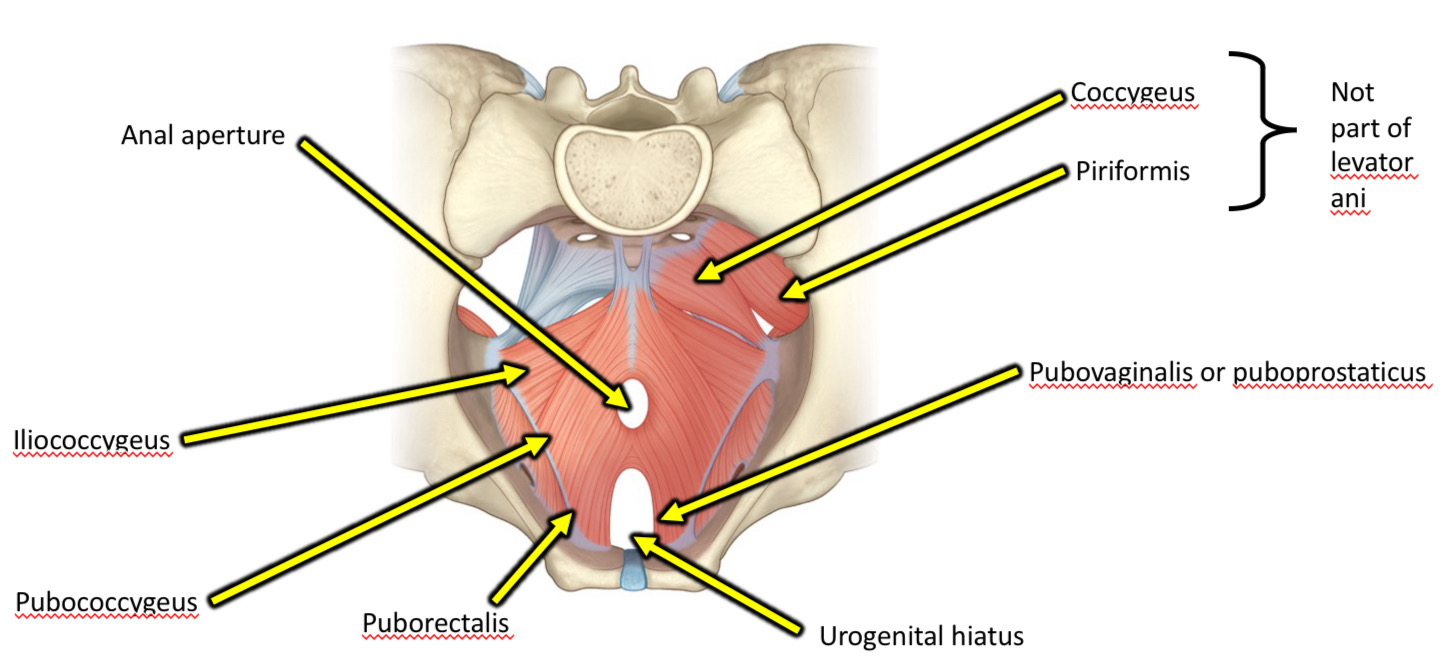
Image occlusion: name the hidden labels
12
Image occlusion: name the hidden labels
13
Image occlusion: name the hidden labels
14
Image occlusion: name the hidden labels
15
Image occlusion: name the hidden labels
16
Image occlusion: name the hidden labels
17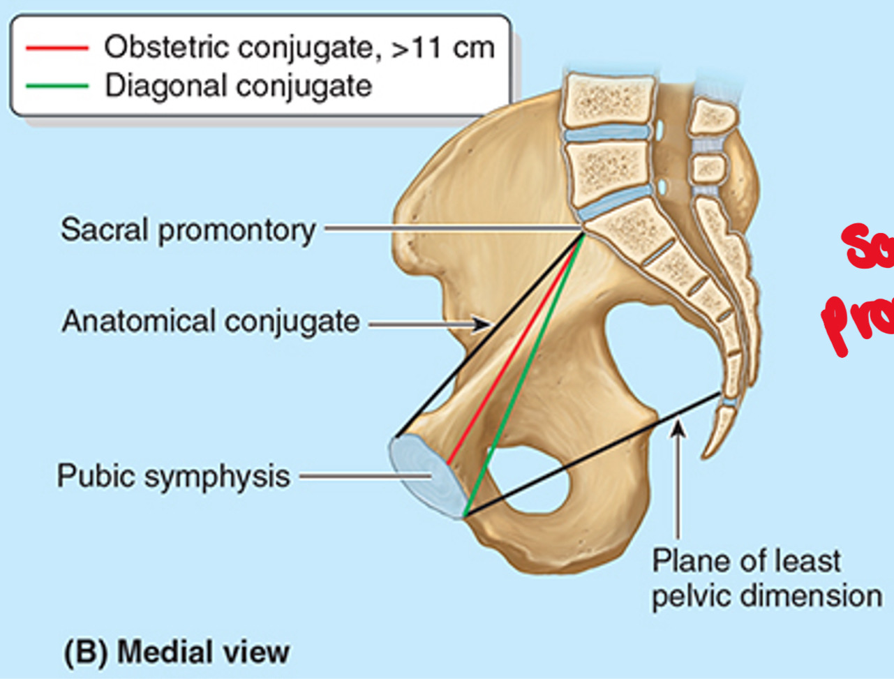
Image occlusion: name the hidden labels
18
Image occlusion: name the hidden labels
19
Image occlusion: name the hidden labels
20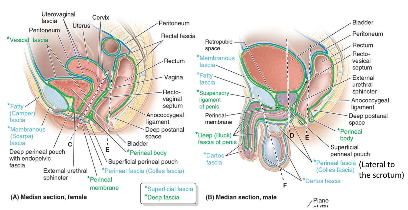
Image occlusion: name the hidden labels
21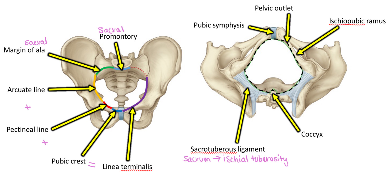
Image occlusion: name the hidden labels
22
Image occlusion: name the hidden labels
23
Image occlusion: name the hidden labels
24
Image occlusion: name the hidden labels
25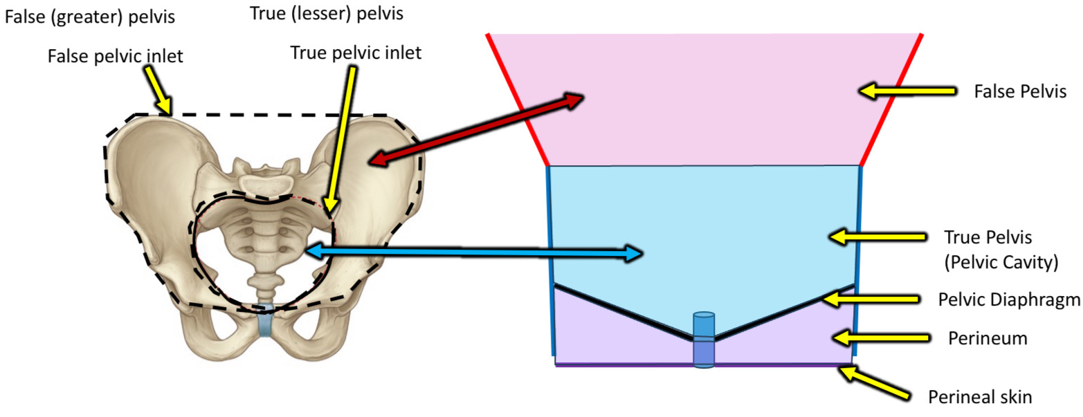
Image occlusion: name the hidden labels
26
Image occlusion: name the hidden labels
27
What does the expression "bridge over troubled water" mean?
Female: Uterine artery/vein pass over urether.
Male: ductus deferens passes over urether.
Male: ductus deferens passes over urether.
28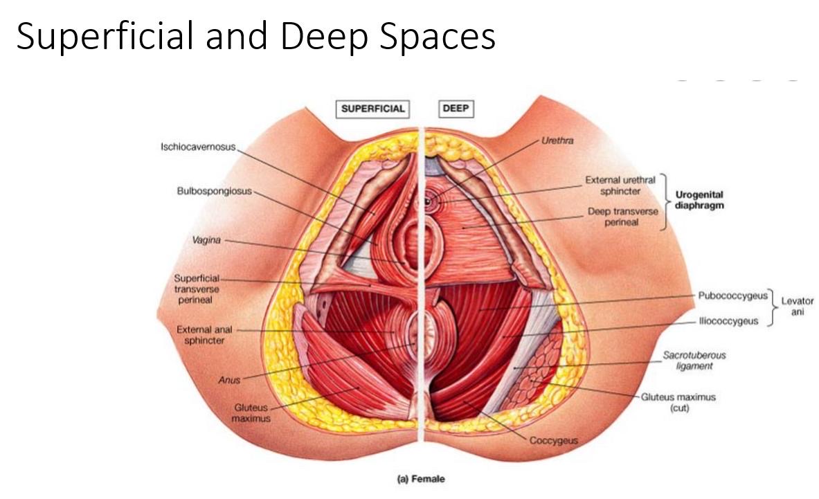
Image occlusion: name the hidden labels
29
Image occlusion: name the hidden labels
30
Image occlusion: name the hidden labels
31
Image occlusion: name the hidden labels
32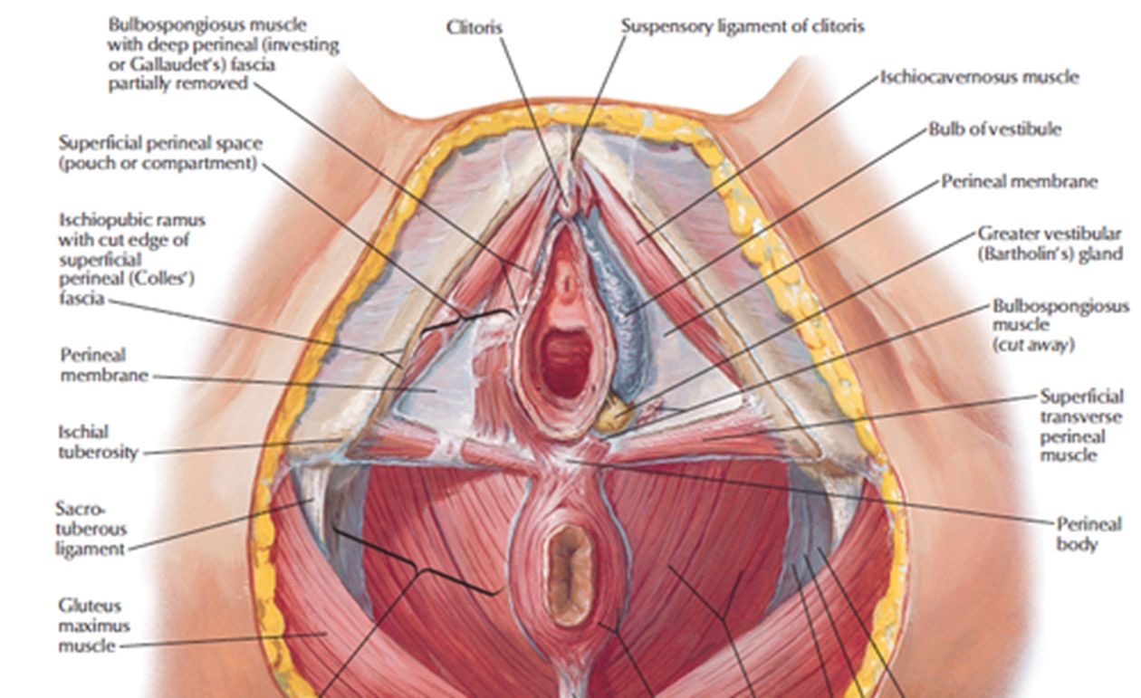
Image occlusion: name the hidden labels
33
Image occlusion: name the hidden labels
34
Image occlusion: name the hidden labels
35
Image occlusion: name the hidden labels
36
Image occlusion: name the hidden labels
37
Image occlusion: name the hidden labels
38
Image occlusion: name the hidden labels
39
Image occlusion: name the hidden labels
40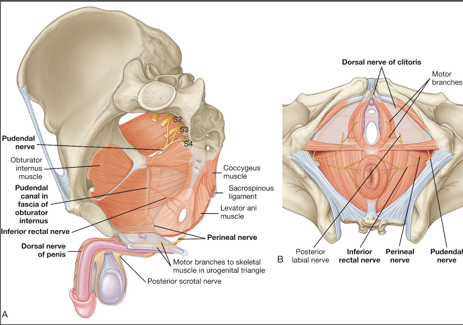
Image occlusion: name the hidden labels
41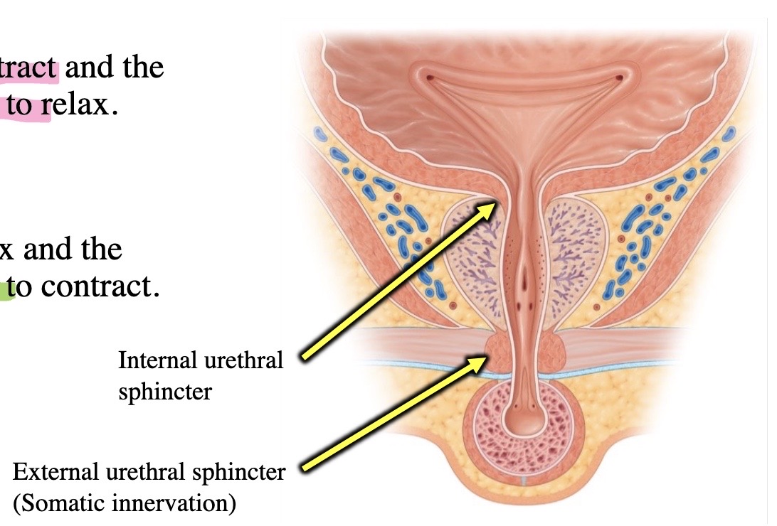
Image occlusion: name the hidden labels
42
Image occlusion: name the hidden labels
43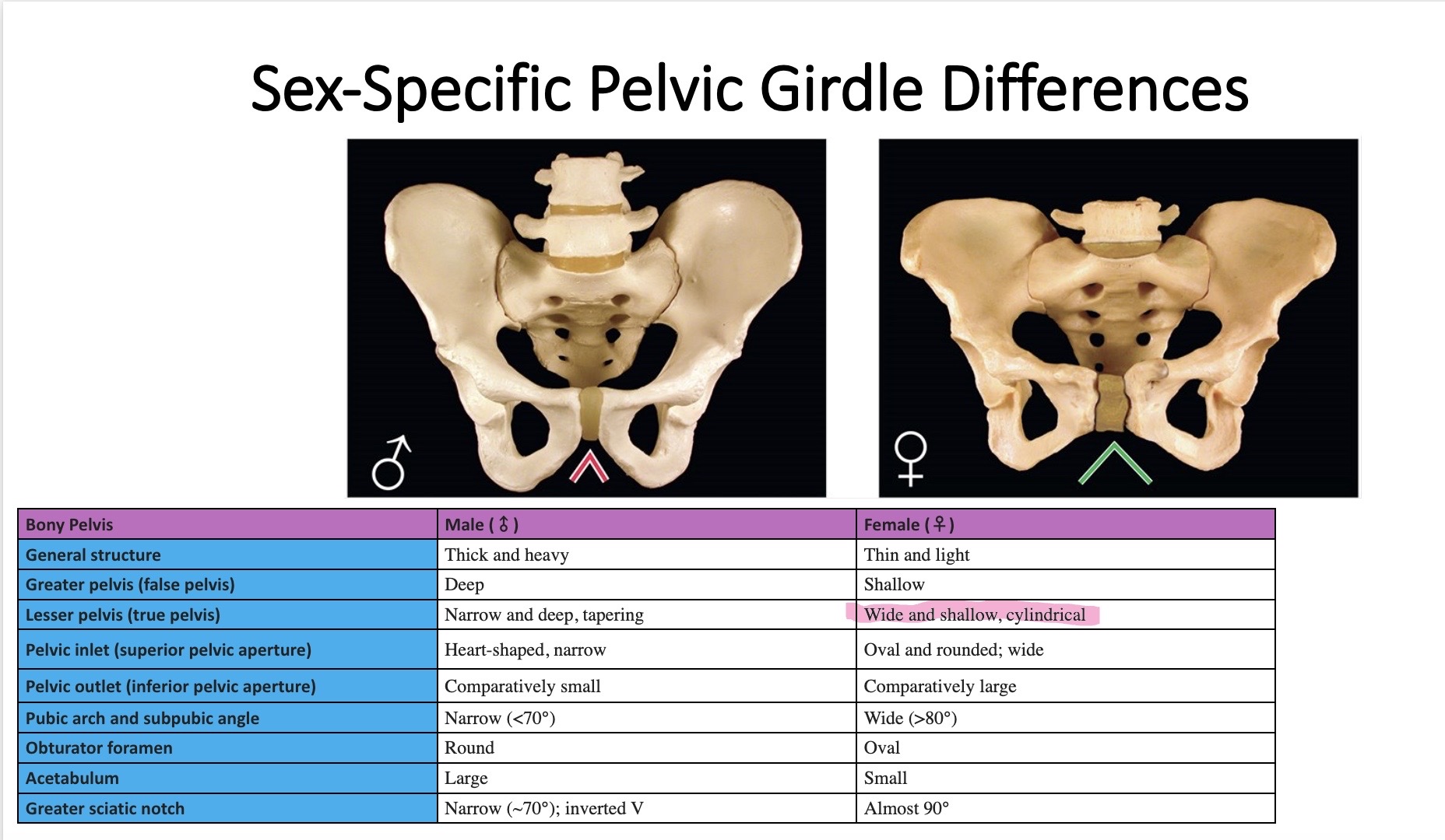
Image occlusion: name the hidden labels
44
Image occlusion: name the hidden labels
45
Image occlusion: name the hidden labels
46
Image occlusion: name the hidden labels
47
Image occlusion: name the hidden labels
48
Image occlusion: name the hidden labels
49
Image occlusion: name the hidden labels
50
Image occlusion: name the hidden labels
51
Image occlusion: name the hidden labels
52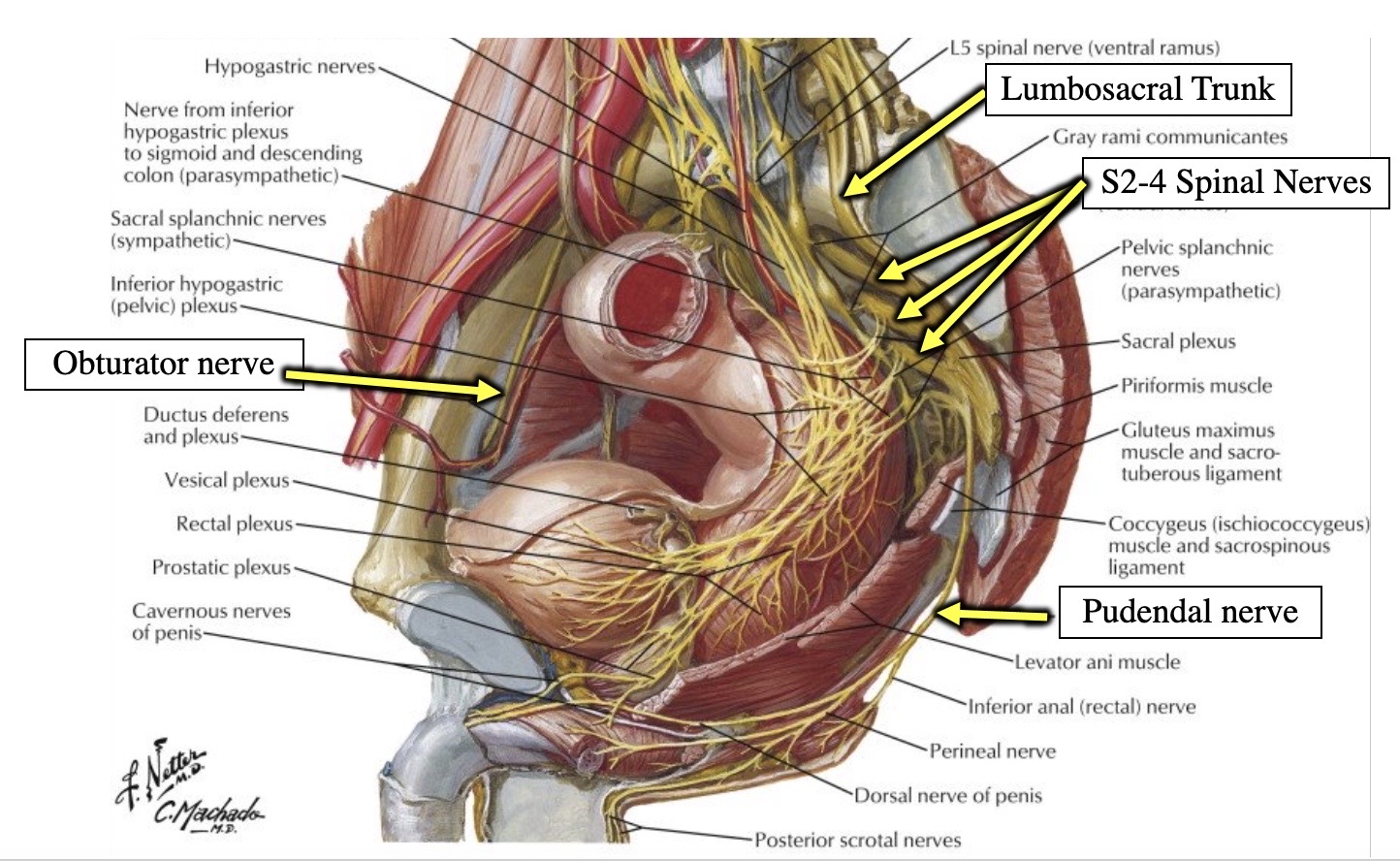
Image occlusion: name the hidden labels
53
Image occlusion: name the hidden labels
54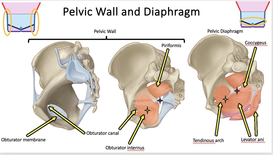
Image occlusion: name the hidden labels
55
Image occlusion: name the hidden labels
56
Functions of the levator ani:
Increase in intra-abdominal pressure.
Sphincter of the anal canal.
Sphincter of vagina.
It is important in the initiation and cessation of micturition.
Increase in intra-abdominal pressure.
Sphincter of the anal canal.
Sphincter of vagina.
It is important in the initiation and cessation of micturition.
Functions of the levator ani:
Support of the pelvic viscera.
Increase in intra-abdominal pressure.
Sphincter of the anal canal.
Sphincter of vagina.
It is important in the initiation and cessation of micturition.
Support of the pelvic viscera.
Increase in intra-abdominal pressure.
Sphincter of the anal canal.
Sphincter of vagina.
It is important in the initiation and cessation of micturition.
57
Functions of the levator ani:
Support of the pelvic viscera.
Sphincter of the anal canal.
Sphincter of vagina.
It is important in the initiation and cessation of micturition.
Support of the pelvic viscera.
Sphincter of the anal canal.
Sphincter of vagina.
It is important in the initiation and cessation of micturition.
Functions of the levator ani:
Support of the pelvic viscera.
Increase in intra-abdominal pressure.
Sphincter of the anal canal.
Sphincter of vagina.
It is important in the initiation and cessation of micturition.
Support of the pelvic viscera.
Increase in intra-abdominal pressure.
Sphincter of the anal canal.
Sphincter of vagina.
It is important in the initiation and cessation of micturition.
58
Functions of the levator ani:
Support of the pelvic viscera.
Increase in intra-abdominal pressure.
Sphincters of the
It is important in the initiation and cessation of micturition.
Support of the pelvic viscera.
Increase in intra-abdominal pressure.
Sphincters of the
It is important in the initiation and cessation of micturition.
Functions of the levator ani:
Support of the pelvic viscera.
Increase in intra-abdominal pressure.
Sphincters of the anal canal and vagina.
It is important in the initiation and cessation of micturition.
Support of the pelvic viscera.
Increase in intra-abdominal pressure.
Sphincters of the anal canal and vagina.
It is important in the initiation and cessation of micturition.
59
Functions of the levator ani:
Support of the pelvic viscera.
Increase in intra-abdominal pressure.
Sphincter of the anal canal.
Sphincter of vagina.
It is important in the initiation and cessation of
Support of the pelvic viscera.
Increase in intra-abdominal pressure.
Sphincter of the anal canal.
Sphincter of vagina.
It is important in the initiation and cessation of
Functions of the levator ani:
Support of the pelvic viscera.
Increase in intra-abdominal pressure.
Sphincter of the anal canal.
Sphincter of vagina.
It is important in the initiation and cessation of micturition.
Support of the pelvic viscera.
Increase in intra-abdominal pressure.
Sphincter of the anal canal.
Sphincter of vagina.
It is important in the initiation and cessation of micturition.
60
The is a wide but thin muscular layer of tissue that forms the inferior border of the abdominopelvic cavity. Composed of a broad, funnel-shaped sling of fascia and muscle, it extends from the symphysis pubis to the coccyx and from one lateral sidewall to the other.
The pelvic diaphragm is a wide but thin muscular layer of tissue that forms the inferior border of the abdominopelvic cavity. Composed of a broad, funnel-shaped sling of fascia and muscle, it extends from the symphysis pubis to the coccyx and from one lateral sidewall to the other. 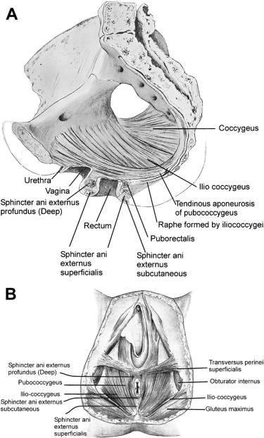
61
The pelvic diaphragm is a wide but thin muscular layer of tissue that forms the inferior border of the abdominopelvic cavity. Composed of a broad, funnel-shaped sling of fascia and muscle, it extends from the and from one lateral sidewall to the other.
The pelvic diaphragm is a wide but thin muscular layer of tissue that forms the inferior border of the abdominopelvic cavity. Composed of a broad, funnel-shaped sling of fascia and muscle, it extends from the symphysis pubis to the coccyx and from one lateral sidewall to the other. 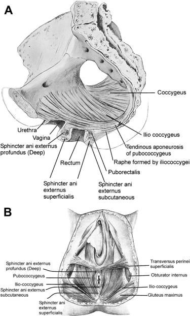
62
Image occlusion: name the hidden labels
63
Image occlusion: name the hidden labels
64
Image occlusion: name the hidden labels
65
Sciatic nerve is a combination of which spinal levers of anterior rami?
L4-S3. Motor innervation from the dorsal divisions of spinal segments L5-S2.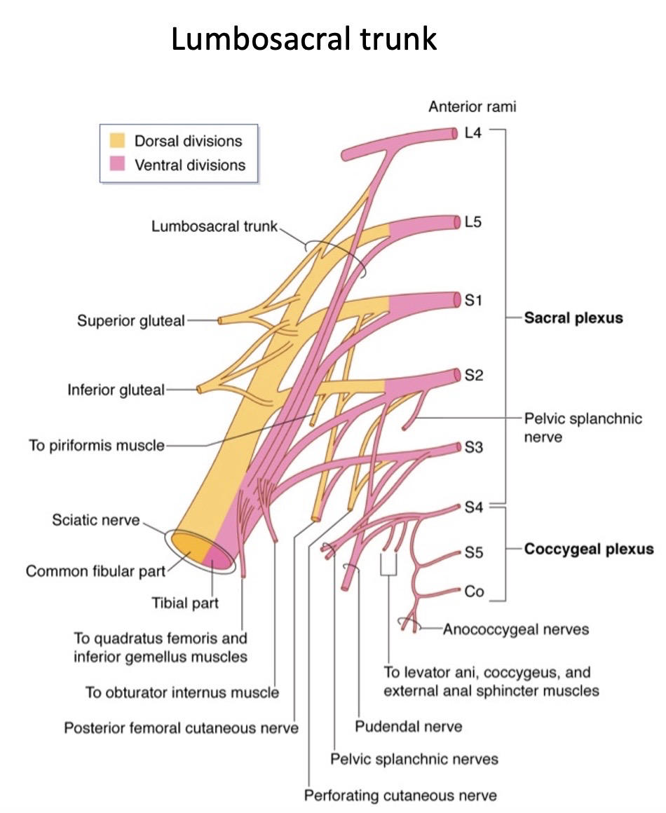
66
What muscles are innervated by this nerve? Sensory, motor, or mixed innervation?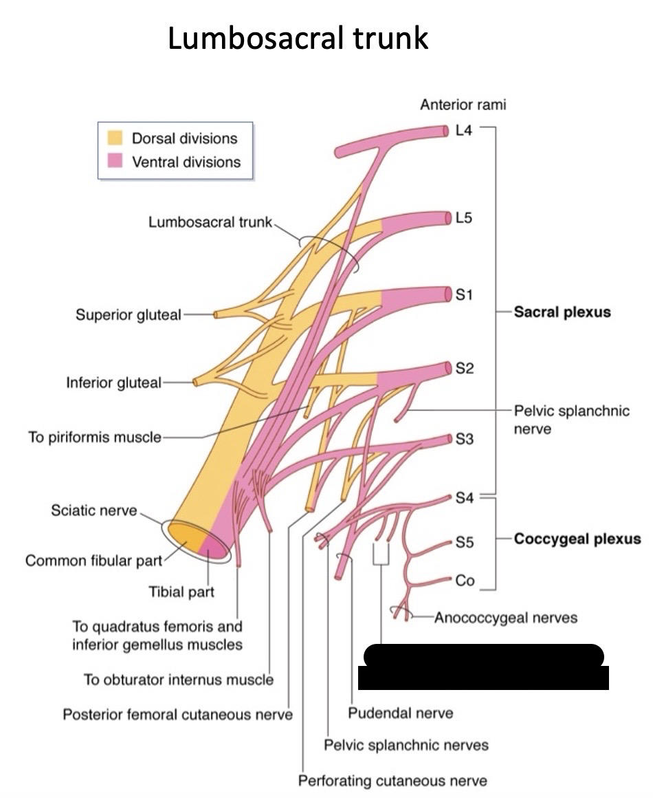
Levator ani, coccygeus, external anal sphincter. Sensory innervation from the ventral divisions of the coccygeal plexus (S4, S5, Co)
67
S1-3 anterio rami nerves sit superior to which muscle?
S1-3 are part of the sacral plexus (L4-S4) and sit on top of piriformis muscle on the posterior wall of pelvic cavity.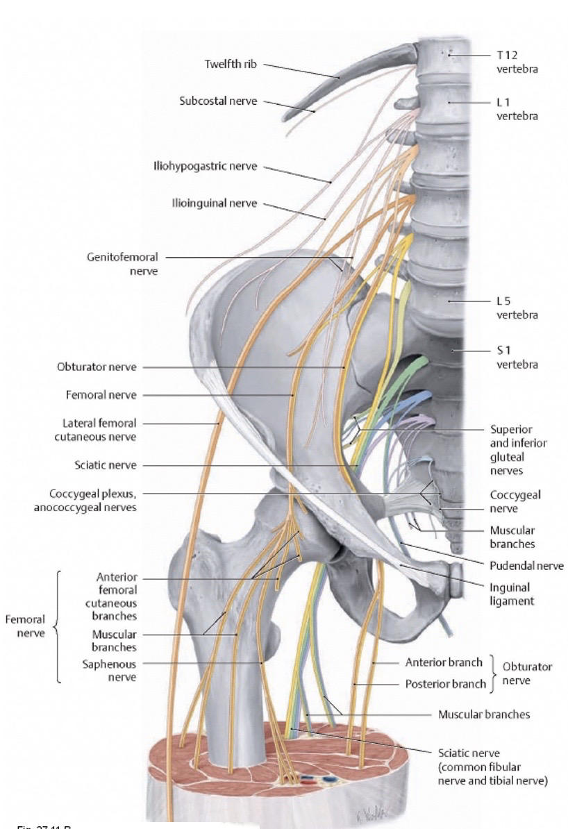
68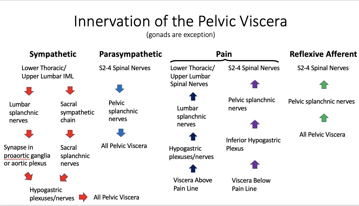
Image occlusion: name the hidden labels
69
Image occlusion: name the hidden labels
70
Image occlusion: name the hidden labels
71
Image occlusion: name the hidden labels
72
Image occlusion: name the hidden labels
73
Image occlusion: name the hidden labels
74
Image occlusion: name the hidden labels
75
Image occlusion: name the hidden labels
76
Image occlusion: name the hidden labels
77
Image occlusion: name the hidden labels
78
Image occlusion: name the hidden labels
79
Image occlusion: name the hidden labels
80
Image occlusion: name the hidden labels
81
Image occlusion: name the hidden labels
82
Image occlusion: name the hidden labels
83
Image occlusion: name the hidden labels
84
Image occlusion: name the hidden labels
85
Image occlusion: name the hidden labels
86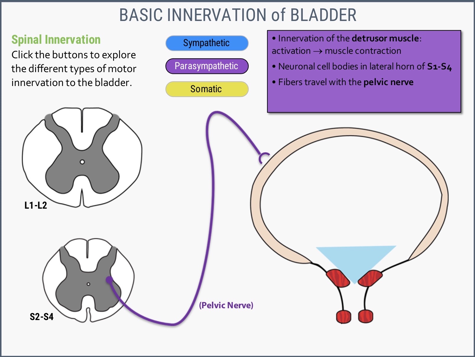
Image occlusion: name the hidden labels
87
Image occlusion: name the hidden labels
88
Image occlusion: name the hidden labels
89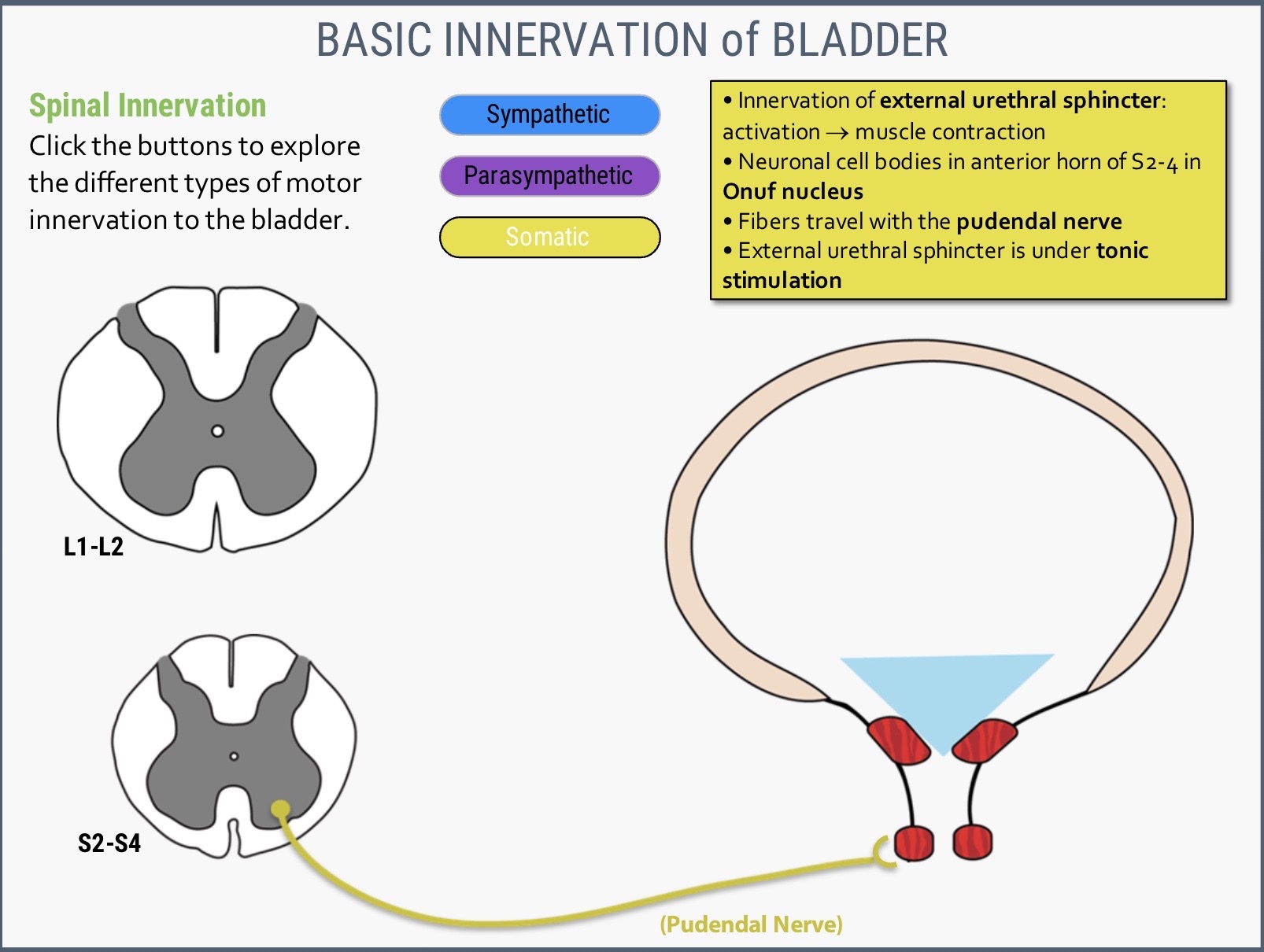
Image occlusion: name the hidden labels
90
Image occlusion: name the hidden labels
91
Image occlusion: name the hidden labels
92
Spastic reflex bladder/ bowel: spinal cord injury
UMN injury: spinal cord reflex is intact and internal sphincter relaxes.
Cerebral cortex control is lost, can't send somatic efferent signal to control external sphincter to contract and hold pee/poo. Results in urine/feces leakage.
UMN injury: spinal cord reflex is intact and internal sphincter relaxes.
Cerebral cortex control is lost, can't send somatic efferent signal to control external sphincter to contract and hold pee/poo. Results in urine/feces leakage.
Spastic reflex bladder/ bowel: spinal cord injury above spinal level S2.
UMN injury: spinal cord reflex is intact and internal sphincter relaxes.
Cerebral cortex control is lost, can't send somatic efferent signal to control external sphincter to contract and hold pee/poo. Results in urine/feces leakage.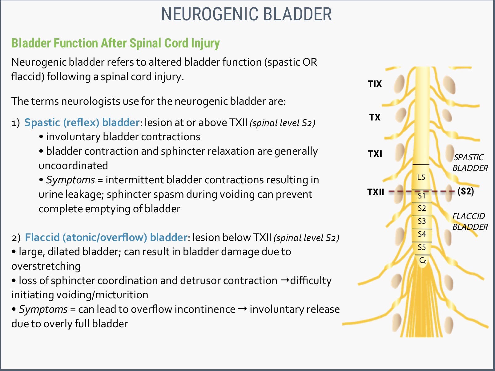
UMN injury: spinal cord reflex is intact and internal sphincter relaxes.
Cerebral cortex control is lost, can't send somatic efferent signal to control external sphincter to contract and hold pee/poo. Results in urine/feces leakage.
93
Flaccid bladder/ bowel: spinal cord injury
LMN injury: spinal cord reflex and cortical control are lost. Internal anal sphincter remains tonically active and person can't void.
Bladder and rectum fill with contents until overflow causes automatic release.
LMN injury: spinal cord reflex and cortical control are lost. Internal anal sphincter remains tonically active and person can't void.
Bladder and rectum fill with contents until overflow causes automatic release.
Flaccid bladder/ bowel: spinal cord injury below spinal level S2.
LMN injury: spinal cord reflex and cortical control are lost. Internal anal sphincter remains tonically active and person can't void.
Bladder and rectum fill with contents until overflow causes automatic release.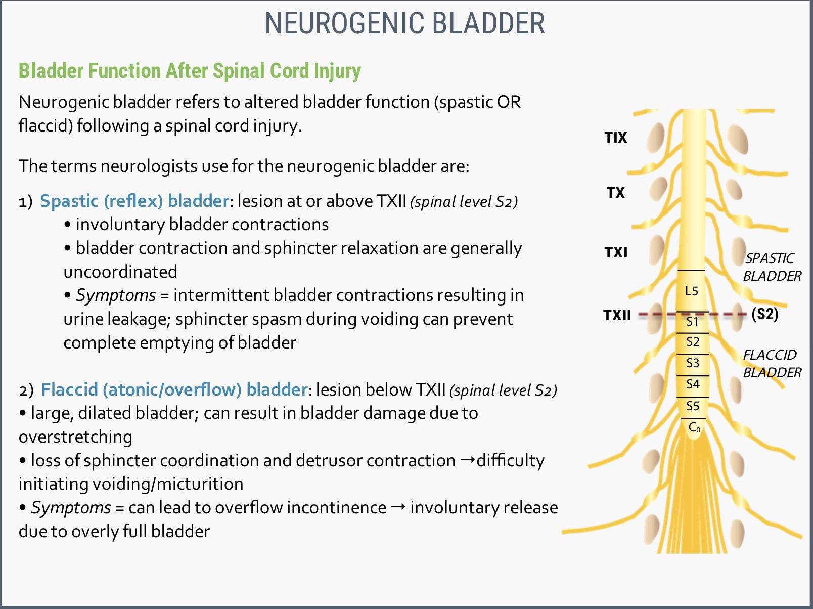
LMN injury: spinal cord reflex and cortical control are lost. Internal anal sphincter remains tonically active and person can't void.
Bladder and rectum fill with contents until overflow causes automatic release.
94
The division of gives off and parietal branches.
The division only gives parietal branches.
The division only gives parietal branches.
The anterior division of internal iliac artery gives off visceral and parietal branches.
The posterior division only gives parietal branches.
The posterior division only gives parietal branches.
95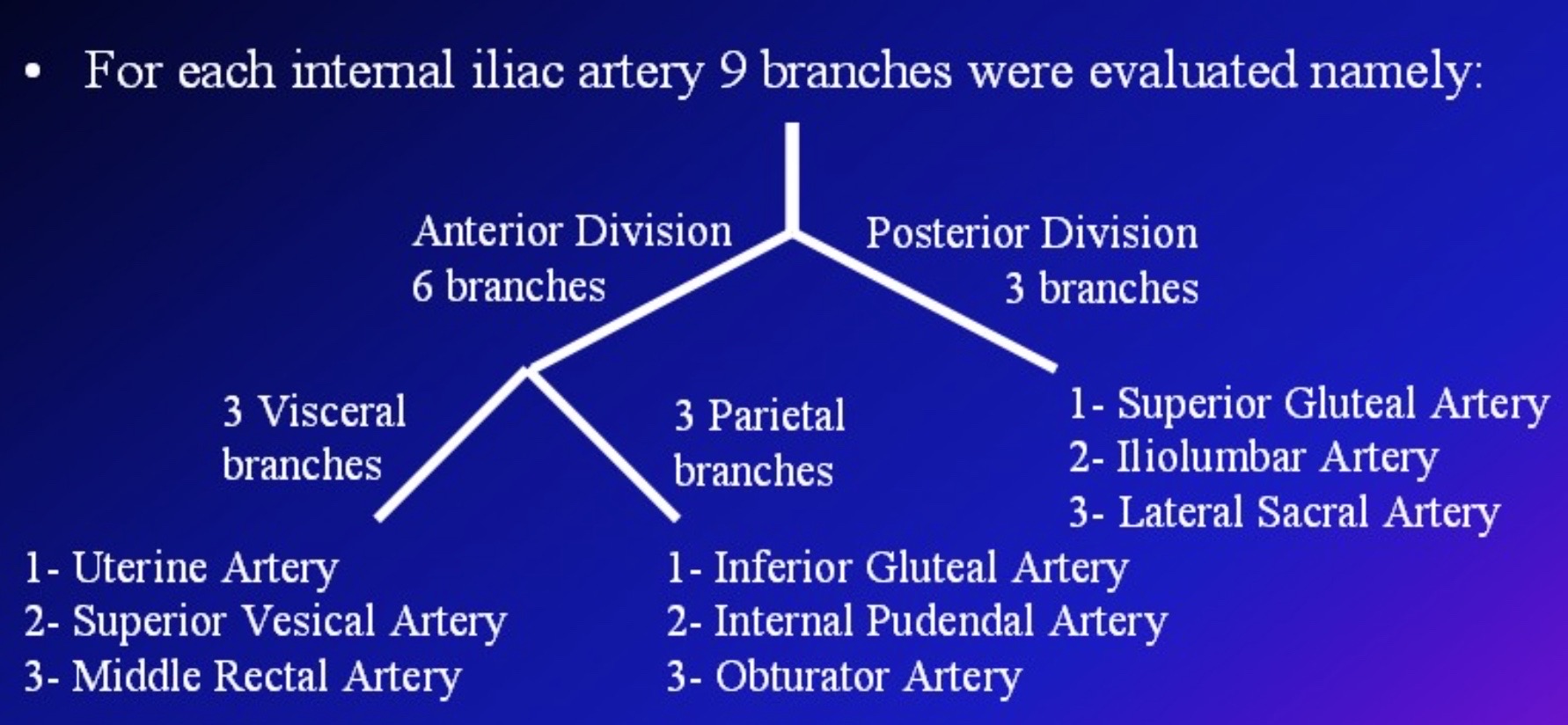
Image occlusion: name the hidden labels
96
Image occlusion: name the hidden labels
97
Image occlusion: name the hidden labels
98
Image occlusion: name the hidden labels
99
Image occlusion: name the hidden labels
100
Image occlusion: name the hidden labels
101
Obturator canal is formed between the and
Obturator canal is formed between the obturator membrane and pelvis (pubic rami).
102
Corona mortis refers to an anatomical variant anastamosis between the obturator artery and vein and the OR .
Corona mortis refers to an anatomical variant anastamosis between the obturator artery and vein and the external iliac artery OR inferior epigastric branch of the external iliac artery. 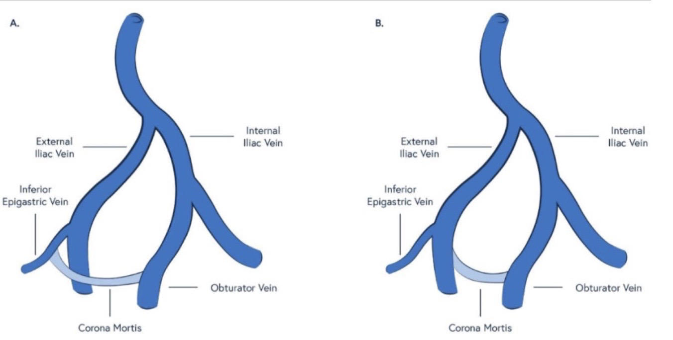
103
Pelvic girdle fractures usually involve due to the pelvis' round shape.
Pelvic girdle fractures usually involve 2 break points due to the pelvis' round shape.
104
The female pelvic outlet has greater . The head of the infant turns to face the posterior wall of the pelvic cavity to better accommodate passage during delivery.
The female pelvic outlet has greater anteroposterior (AP) diameter. The head of the infant turns to face the posterior wall of the pelvic cavity to better accommodate passage during delivery. 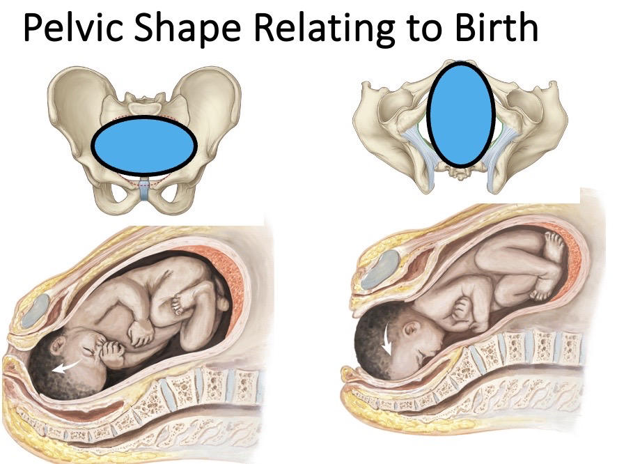
105
ANTERIOR-POSTERIOR RELATIONSHIPS OF THE PELVIC VISCERA
List the main pelvic visceral structures front to back.
List the main pelvic visceral structures front to back.
Female: bladder, uterus and vagina (most commonly anteroflexed and anteroverted), sigmoid colon and rectum.
Male: bladder, seminal vesicles, prostate, vas deferens, sigmoid colon and rectum.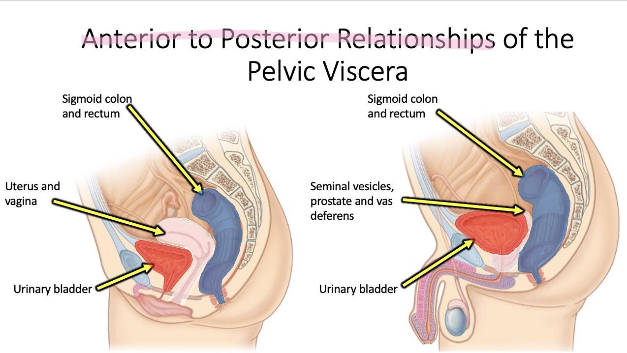
Male: bladder, seminal vesicles, prostate, vas deferens, sigmoid colon and rectum.
106
The is nervous tissue that connects the lumbar plexus with the sacral plexus. anterior rami combine and descend to join the sacral plexus.
The lumbosacral trunk is nervous tissue that connects the lumbar plexus with the sacral plexus. L4-5 anterior rami combine and descend to join the sacral plexus. 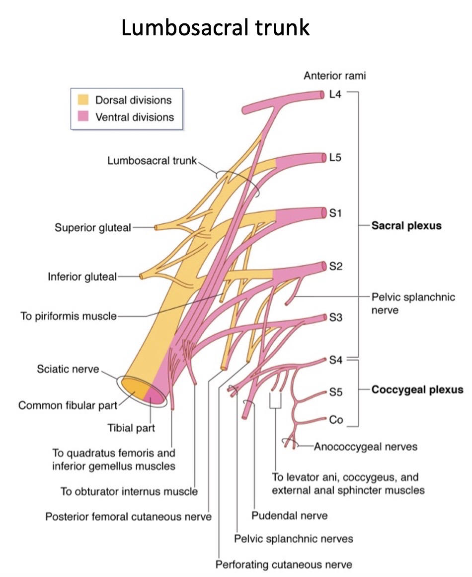
107
The and arise from L1 spinal nerve and provide motor supply to some muscles of and to the skin of the groin area. 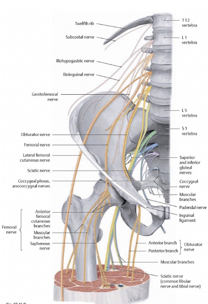
The ilioinguinal and iliohypogastric nerves arise from L1 spinal nerve and provide motor supply to some muscles of the abdominal wall and sensory inputs to the skin of the groin area.
108
The arises from L1 and L2 spinal nerves and then divides into . In males, supplies sensory innervation to skin of scrotum and cremaster muscle.
In females, it supplies the skin around the mons pubis and labia majora.
The femoral nerve supplies cutaneous innervation to .
In females, it supplies the skin around the mons pubis and labia majora.
The femoral nerve supplies cutaneous innervation to .
The genitofemoral nerve arises from L1 and L2 spinal nerves and then divides into genital and femoral branches. In males, genital nerve supplies sensory innervation to skin of scrotum and cremaster muscle.
In females, it supplies the skin around the mons pubis and labia majora.
The femoral nerve supplies cutaneous innervation to upper anteromedial thigh.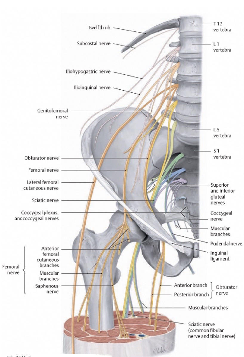
In females, it supplies the skin around the mons pubis and labia majora.
The femoral nerve supplies cutaneous innervation to upper anteromedial thigh.
109
The arises from S2-4 and provides sensory inputs to the and muscular input to .
The pudendal nerve arises from S2-4 and provides sensory inputs to the external genitalia and muscular input to perineal muscles. 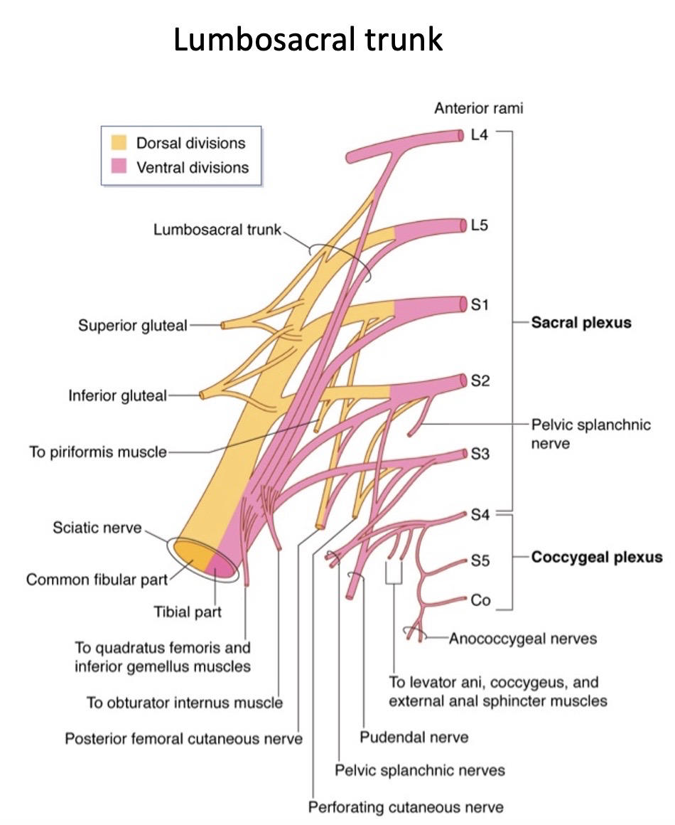
110
The lumbosacral plexus gives off nerves named by the muscle they innervate:
1) (L4-S1)
2) (L5-S2)
3) (S1-S2)
4) (S3-4)
5) (sacral plexus), which supply the skin of the buttocks and posteromedial thigh.
1) (L4-S1)
2) (L5-S2)
3) (S1-S2)
4) (S3-4)
5) (sacral plexus), which supply the skin of the buttocks and posteromedial thigh.
The lumbosacral plexus gives off nerves named by the muscle they innervate:
1) quadratus femoris nerve (L4-S1)
2) obturator internus nerve (L5-S2)
3) piriformis nerve (S1-S2)
4) nerves to the levator ani and coccygeus (S3-4)
5) posterior cutaneous nerve and perforating cutaneous branches (sacral plexus), which supply the skin of the buttocks and posteromedial thigh.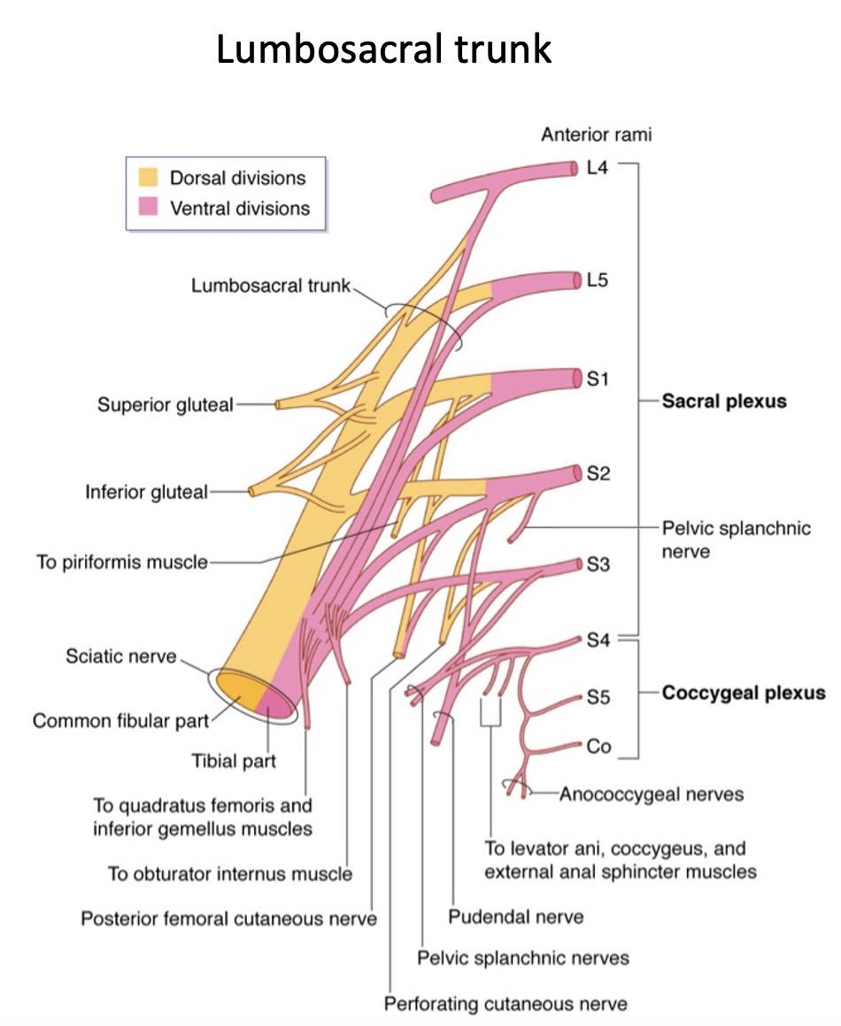
1) quadratus femoris nerve (L4-S1)
2) obturator internus nerve (L5-S2)
3) piriformis nerve (S1-S2)
4) nerves to the levator ani and coccygeus (S3-4)
5) posterior cutaneous nerve and perforating cutaneous branches (sacral plexus), which supply the skin of the buttocks and posteromedial thigh.
111
Most common cause of lumbosacral nerve injury is
Described as that allows the to bulge out posterior and laterally, causing compression of the local spinal nerves. Symptoms are shooting pain down the leg, numbness and muscle weakness in the areas innervated by the affected nerves.
Described as that allows the to bulge out posterior and laterally, causing compression of the local spinal nerves. Symptoms are shooting pain down the leg, numbness and muscle weakness in the areas innervated by the affected nerves.
Most common cause of lumbosacral nerve injury is spinal disc herniation.
Described as a tear in the outer part of the intervertebral disc that allows the nucleus pulposus to bulge out posterior and laterally, causing compression of the local spinal nerves. Symptoms are shooting pain down the leg, numbness and muscle weakness in the areas innervated by the affected nerves.
Described as a tear in the outer part of the intervertebral disc that allows the nucleus pulposus to bulge out posterior and laterally, causing compression of the local spinal nerves. Symptoms are shooting pain down the leg, numbness and muscle weakness in the areas innervated by the affected nerves.
112
Sacral splanchnic nerves contain:
neurons: These neurons come from the T10 to L2 lateral horns of the spinal cord.
neurons: These neurons follow the sympathetic fibers and travel to the spinal cord.
Sacral splanchnic nerves supply the pelvic , and their fibers ascend to the aortic and inferior mesenteric plexuses. Sacral splanchnic nerves connect the sacral part of the sympathetic trunk to the in the pelvis.
neurons: These neurons come from the T10 to L2 lateral horns of the spinal cord.
neurons: These neurons follow the sympathetic fibers and travel to the spinal cord.
Sacral splanchnic nerves supply the pelvic , and their fibers ascend to the aortic and inferior mesenteric plexuses. Sacral splanchnic nerves connect the sacral part of the sympathetic trunk to the in the pelvis.
Sacral splanchnic nerves contain:
Pre-ganglionic sympathetic neurons: These neurons come from the T10 to L2 lateral horns of the spinal cord.
Sensory visceral afferent neurons: These neurons follow the sympathetic fibers and travel to the spinal cord.
Sacral splanchnic nerves supply the pelvic vessels and organs, and their preganglionic sympathetic fibers ascend to the aortic and inferior mesenteric plexuses. Sacral splanchnic nerves connect the sacral part of the sympathetic trunk to the inferior hypogastric plexus in the pelvis.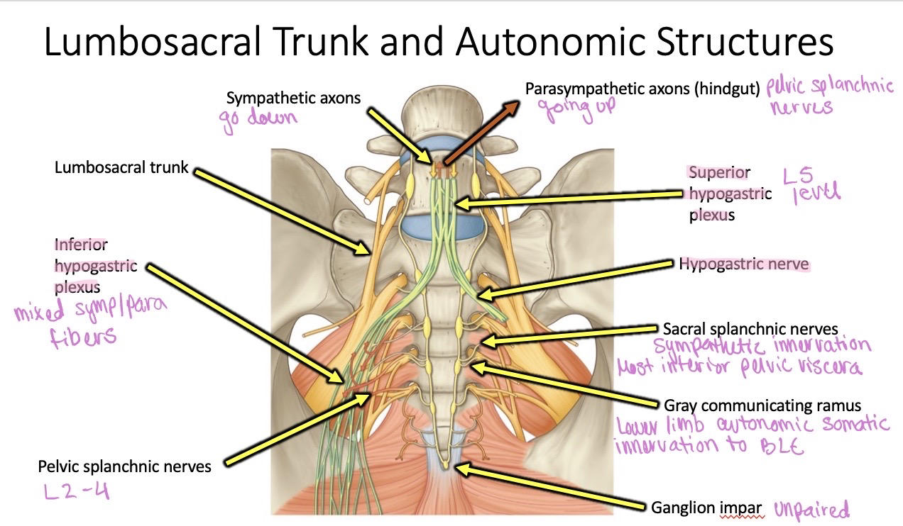
Pre-ganglionic sympathetic neurons: These neurons come from the T10 to L2 lateral horns of the spinal cord.
Sensory visceral afferent neurons: These neurons follow the sympathetic fibers and travel to the spinal cord.
Sacral splanchnic nerves supply the pelvic vessels and organs, and their preganglionic sympathetic fibers ascend to the aortic and inferior mesenteric plexuses. Sacral splanchnic nerves connect the sacral part of the sympathetic trunk to the inferior hypogastric plexus in the pelvis.
113
are parasympathetic and come from anterior rami of segments of the spinal cord and supply the pelvic organs and hindgut:
The nerves travel along the posterior pelvic and abdominal wall or indirectly via the .
Functions:
Colon: Emptying of the colon
Bladder: Emptying of the bladder
Male genitalia: Penile erection and ejaculation
Female genitalia: Increased vaginal secretions, clitoral erection, and labia minora engorgement.
can increase the risk of injuring the pelvic splanchnic nerves.
The nerves travel along the posterior pelvic and abdominal wall or indirectly via the .
Functions:
Colon: Emptying of the colon
Bladder: Emptying of the bladder
Male genitalia: Penile erection and ejaculation
Female genitalia: Increased vaginal secretions, clitoral erection, and labia minora engorgement.
can increase the risk of injuring the pelvic splanchnic nerves.
Pelvic splanchnic nerves are parasympathetic and come from anterior rami of S2 to S4 segments of the spinal cord and supply the pelvic organs and hindgut:
The nerves travel along the posterior pelvic and abdominal wall or indirectly via the hypogastric nerve.
Functions:
Colon: Emptying of the colon
Bladder: Emptying of the bladder
Male genitalia: Penile erection and ejaculation
Female genitalia: Increased vaginal secretions, clitoral erection, and labia minora engorgement.
Pelvic surgery can increase the risk of injuring the pelvic splanchnic nerves.
The nerves travel along the posterior pelvic and abdominal wall or indirectly via the hypogastric nerve.
Functions:
Colon: Emptying of the colon
Bladder: Emptying of the bladder
Male genitalia: Penile erection and ejaculation
Female genitalia: Increased vaginal secretions, clitoral erection, and labia minora engorgement.
Pelvic surgery can increase the risk of injuring the pelvic splanchnic nerves.
114
The artery anastamoses with the and the inferior rectal artery (from internal pudendal).
It's vascular contribution to the pelvic viscera is minor.
It's vascular contribution to the pelvic viscera is minor.
The middle rectal artery anastamoses with the superior rectal artery (from inferior mesenteric artery) and the inferior rectal artery (from internal pudendal).
It's vascular contribution to the pelvic viscera is minor.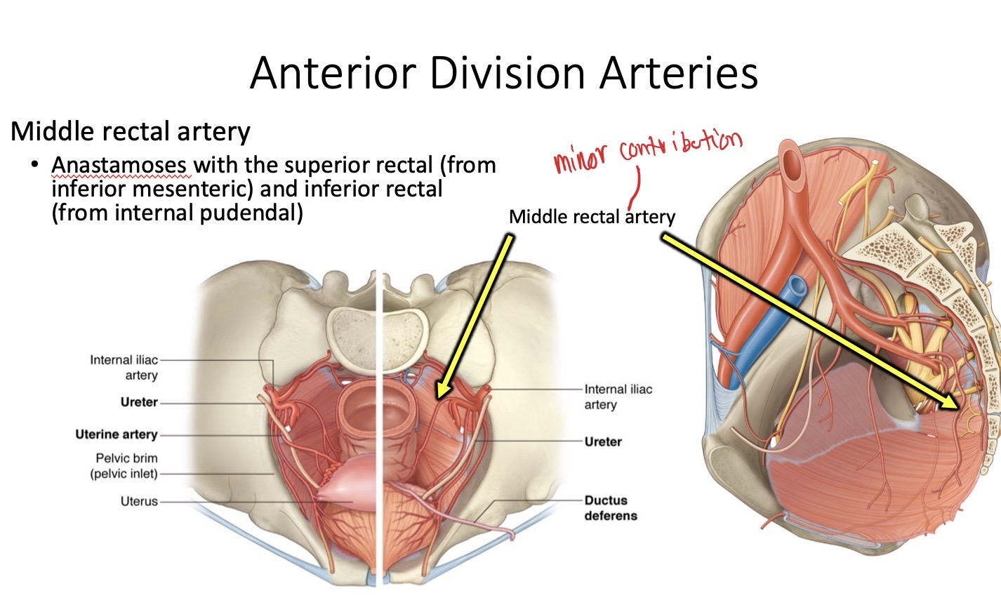
It's vascular contribution to the pelvic viscera is minor.
115
Name the borders of the perineum.
*Hint: perineum is the most inferior part of the trunk where some viscera exit and external genitalia attach.
*Hint: perineum is the most inferior part of the trunk where some viscera exit and external genitalia attach.
Superior- pelvic diaphragm
anterior- pubic symphysis
posterior- coccyx
lateral- ischial tuberosities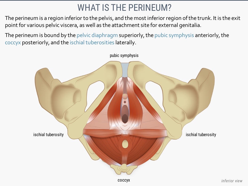
anterior- pubic symphysis
posterior- coccyx
lateral- ischial tuberosities
116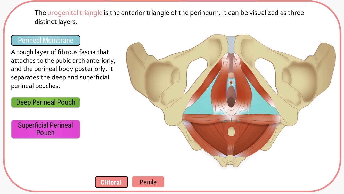
Image occlusion: name the hidden labels
117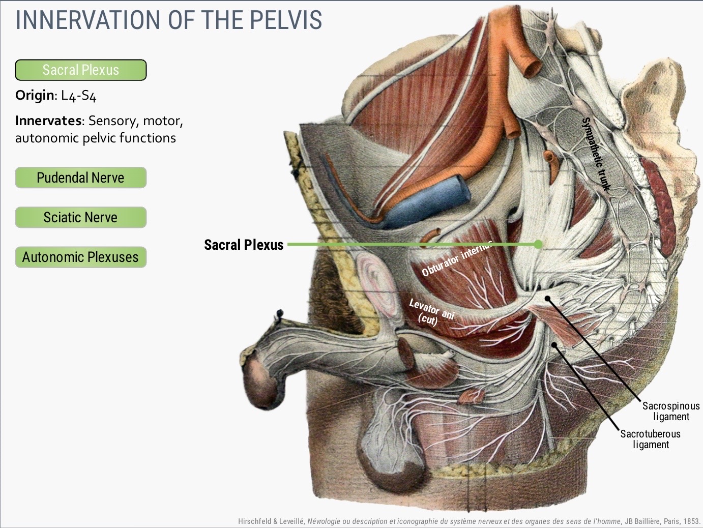
Image occlusion: name the hidden labels
118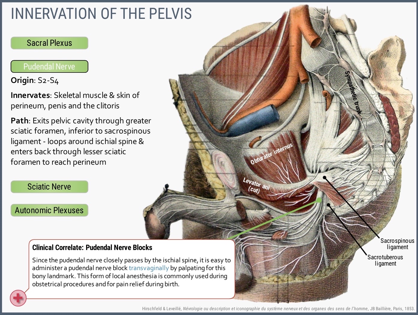
Image occlusion: name the hidden labels
119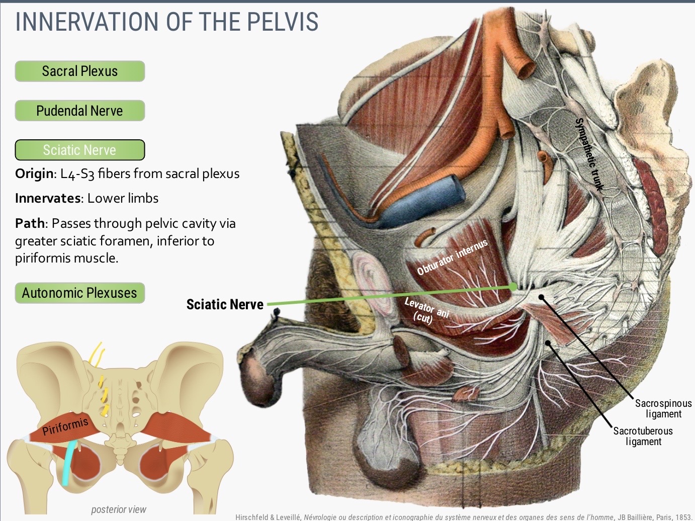
Image occlusion: name the hidden labels
120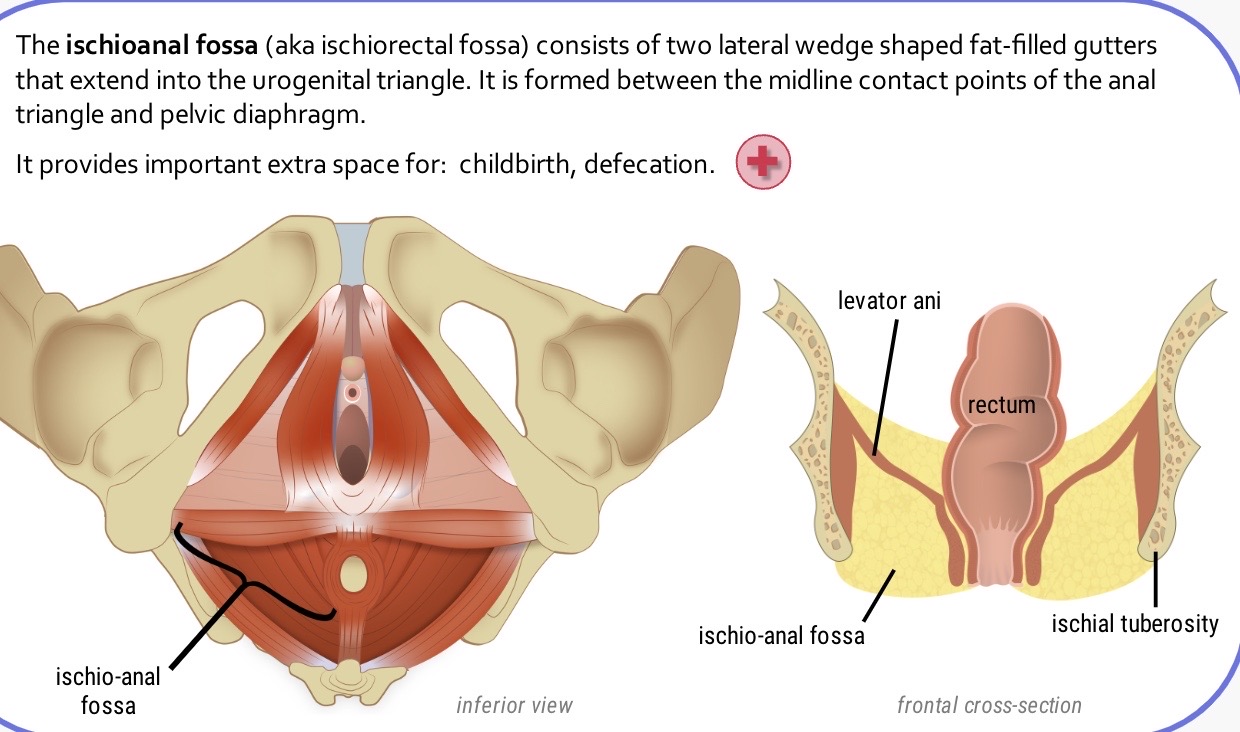
Image occlusion: name the hidden labels
121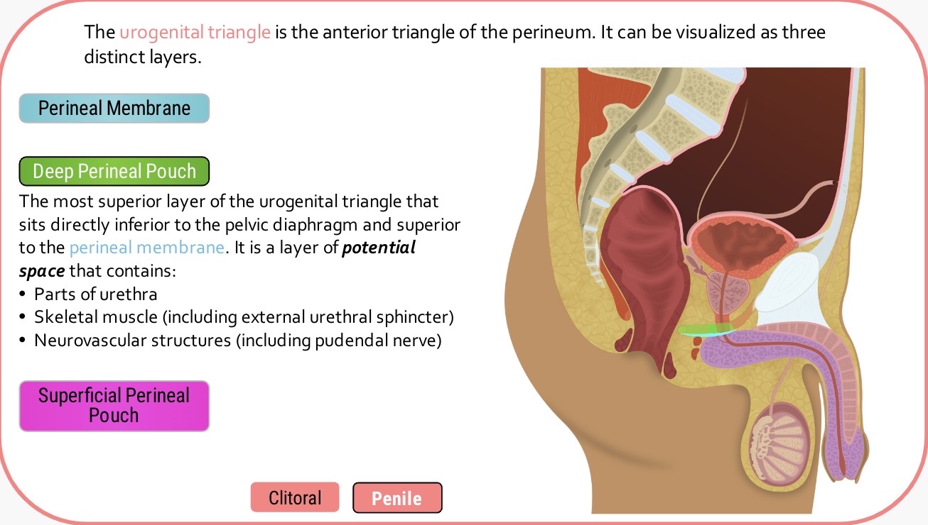
Image occlusion: name the hidden labels
122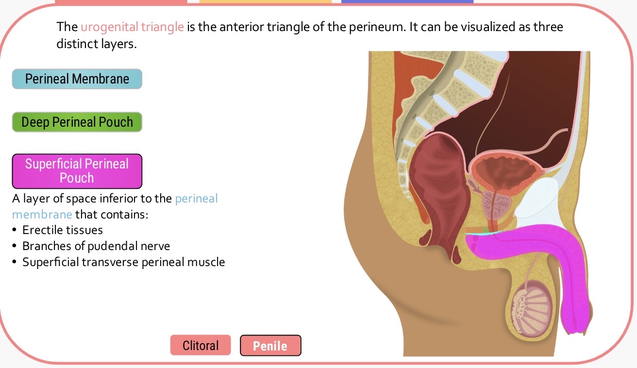
Image occlusion: name the hidden labels
123
Image occlusion: name the hidden labels
124
Image occlusion: name the hidden labels
125
Image occlusion: name the hidden labels
126
Image occlusion: name the hidden labels
127
Image occlusion: name the hidden labels
128
Image occlusion: name the hidden labels
129
Image occlusion: name the hidden labels
130
Image occlusion: name the hidden labels
131
Image occlusion: name the hidden labels
132
Image occlusion: name the hidden labels
133
Image occlusion: name the hidden labels
134
Image occlusion: name the hidden labels
135
Image occlusion: name the hidden labels
136
Image occlusion: name the hidden labels
137
Image occlusion: name the hidden labels
138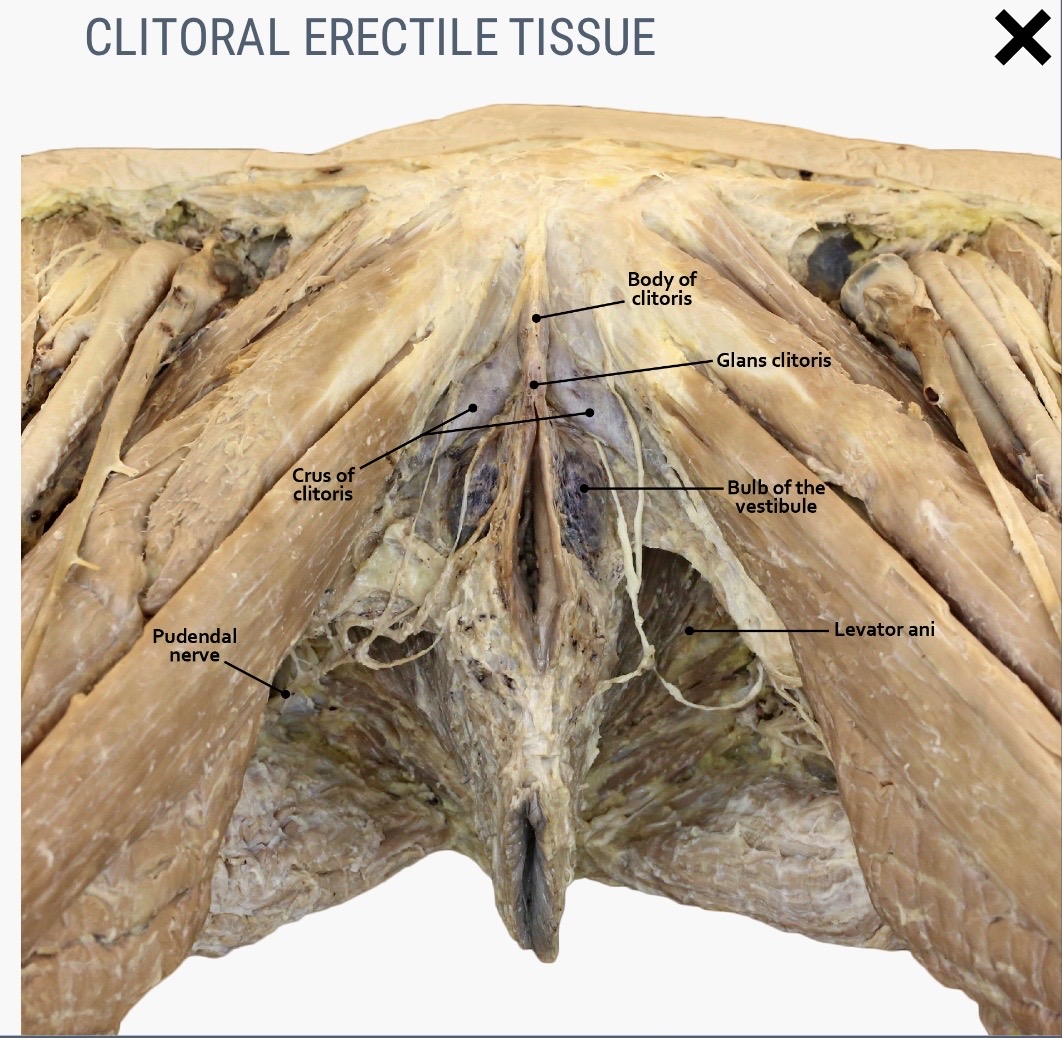
Image occlusion: name the hidden labels
139
Image occlusion: name the hidden labels
140
Image occlusion: name the hidden labels
141
Image occlusion: name the hidden labels
142
Image occlusion: name the hidden labels
143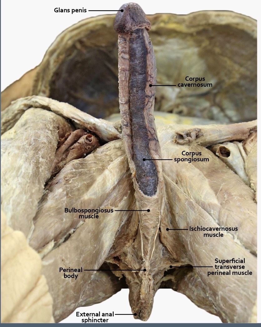
Image occlusion: name the hidden labels
144
Image occlusion: name the hidden labels
145
Image occlusion: name the hidden labels
146
Image occlusion: name the hidden labels
147
Image occlusion: name the hidden labels
148
Image occlusion: name the hidden labels
149
Image occlusion: name the hidden labels
150
Image occlusion: name the hidden labels
151
List the muscles of the perineum in the superficial perineal pouch.
Ischiocavernosus: anchors erectile tissues to perineal membrane. Contracts to push blood from crura of clitoris/penis to distal parts.
Bulbospongiosus: Contracts to push blood to glans clitoris/penis. Anchors erectile tissues to perineal membrane. In males, contracts to assist with emptying urine and ejaculation.
Superficial transverse perineal for stability.
External anal sphincter: constricts anal canal, supports pelvic floor. Under voluntary control by somatic nervous system and stays contracted until relaxed to poop.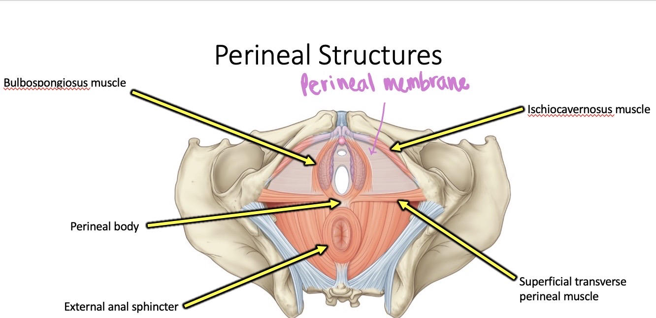
Bulbospongiosus: Contracts to push blood to glans clitoris/penis. Anchors erectile tissues to perineal membrane. In males, contracts to assist with emptying urine and ejaculation.
Superficial transverse perineal for stability.
External anal sphincter: constricts anal canal, supports pelvic floor. Under voluntary control by somatic nervous system and stays contracted until relaxed to poop.
152
List the clitoral erectile tissues, which lie just below the skin of the vulva and labia.
Glans clitoris, bulb of vestibule (come together to form glans clitoris), corpus cavernosum (crura and body come together).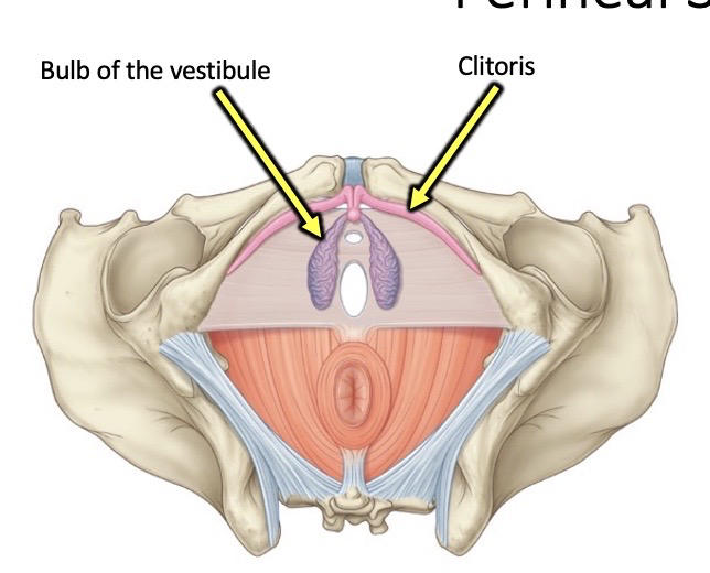
153
List the penile erectile tissues.
Corpus cavernosum forms the crura.
Corpus spongiosum forms the glans penis.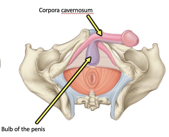
Corpus spongiosum forms the glans penis.
154
List the structures in the deep perineal pouch.
The deep transverse perineus muscle, sphincter urethrae, and perineal membrane form the urogenital diaphragm.
Sphincter urethrae [external urethral sphincter], deep transverse perineal, and compressor urethrae [in females]
MALES: membranous urethra, which passes through the deep perineal pouch. Bulbourethral glands secrete mucous fluid that neutralizes urine and lubricates semen.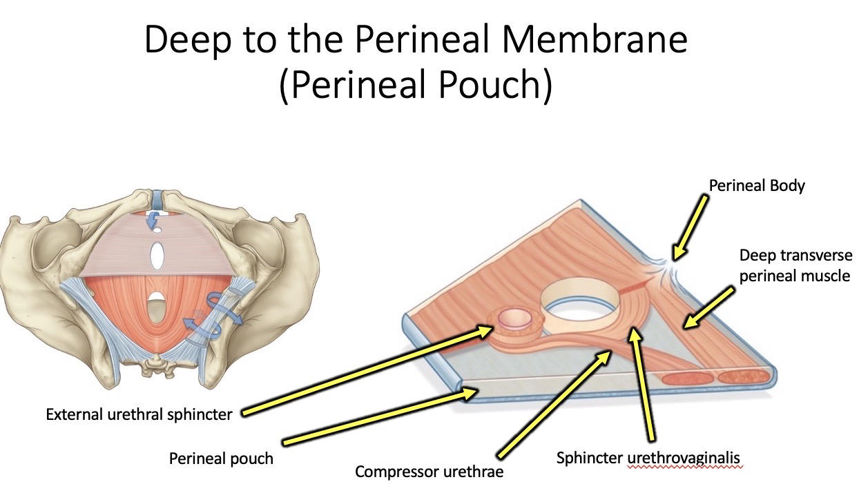
Sphincter urethrae [external urethral sphincter], deep transverse perineal, and compressor urethrae [in females]
MALES: membranous urethra, which passes through the deep perineal pouch. Bulbourethral glands secrete mucous fluid that neutralizes urine and lubricates semen.
155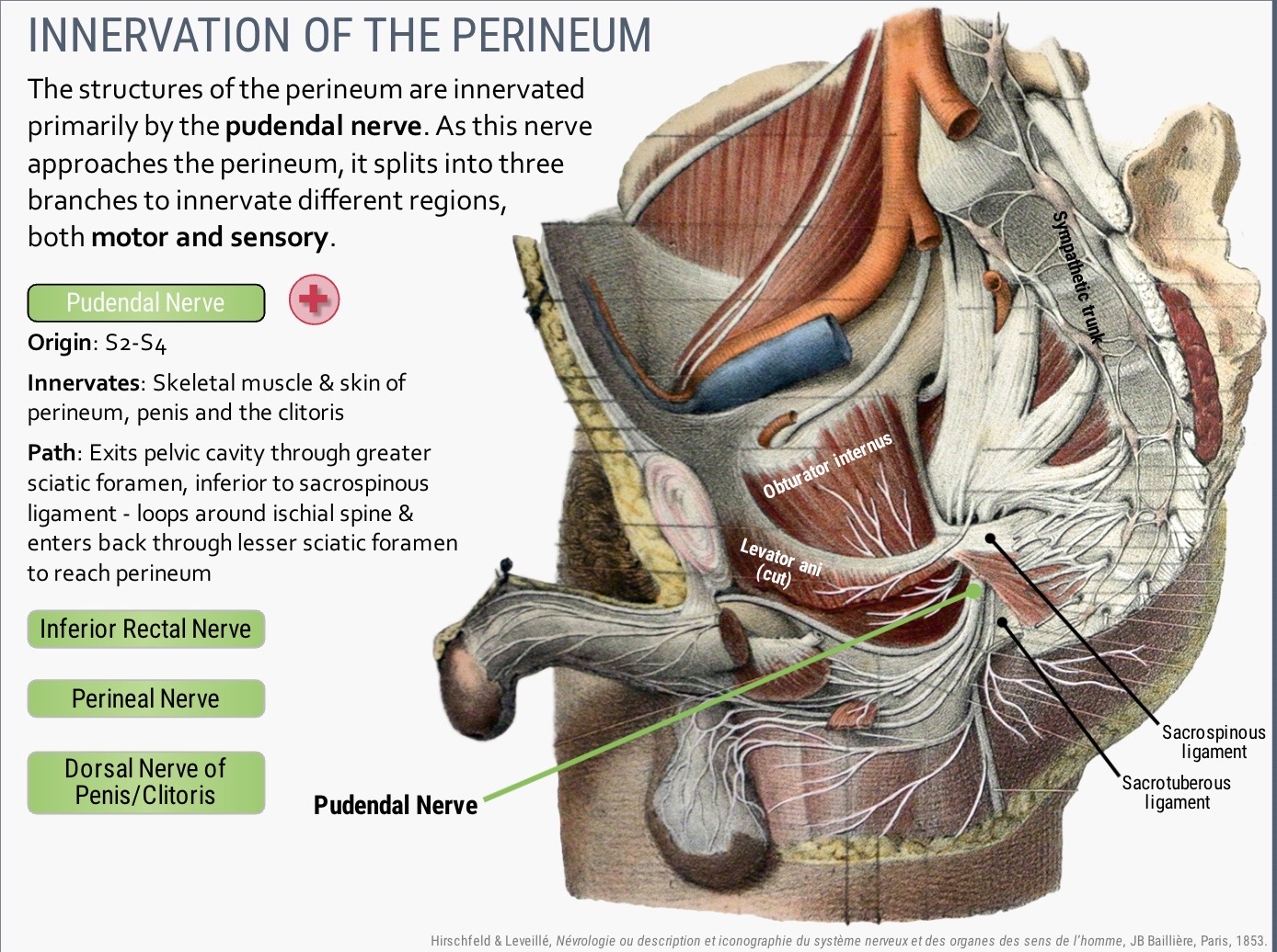
Image occlusion: name the hidden labels
156
Image occlusion: name the hidden labels
157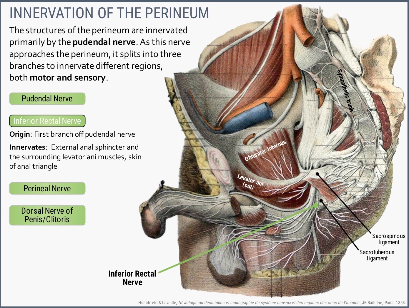
Image occlusion: name the hidden labels
158
Image occlusion: name the hidden labels
159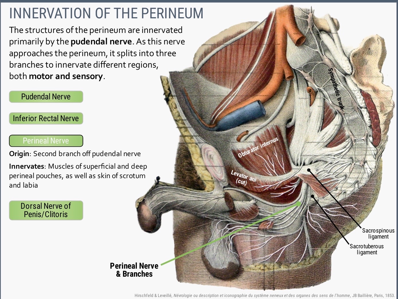
Image occlusion: name the hidden labels
160
Image occlusion: name the hidden labels
161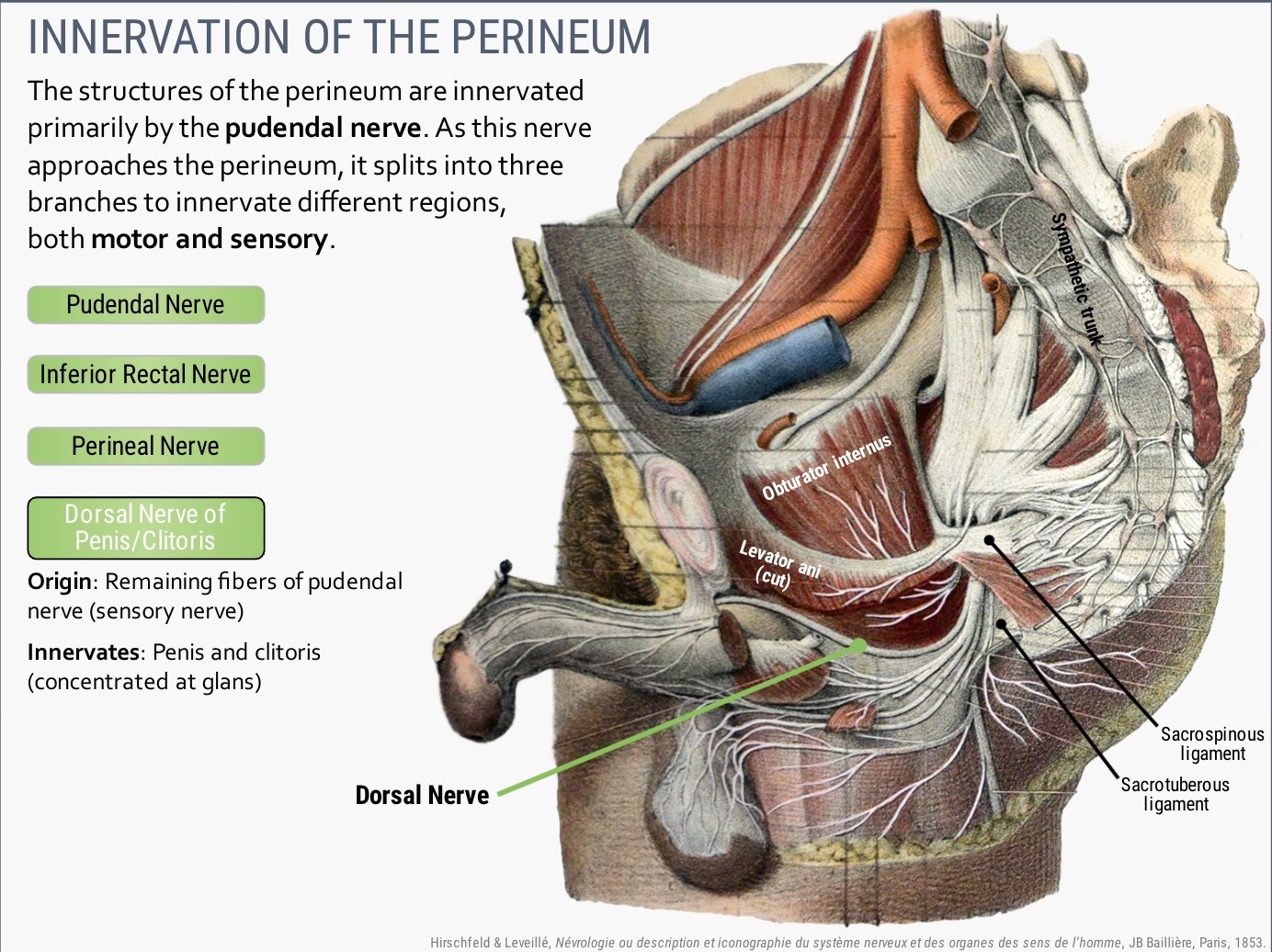
Image occlusion: name the hidden labels
162
Image occlusion: name the hidden labels
163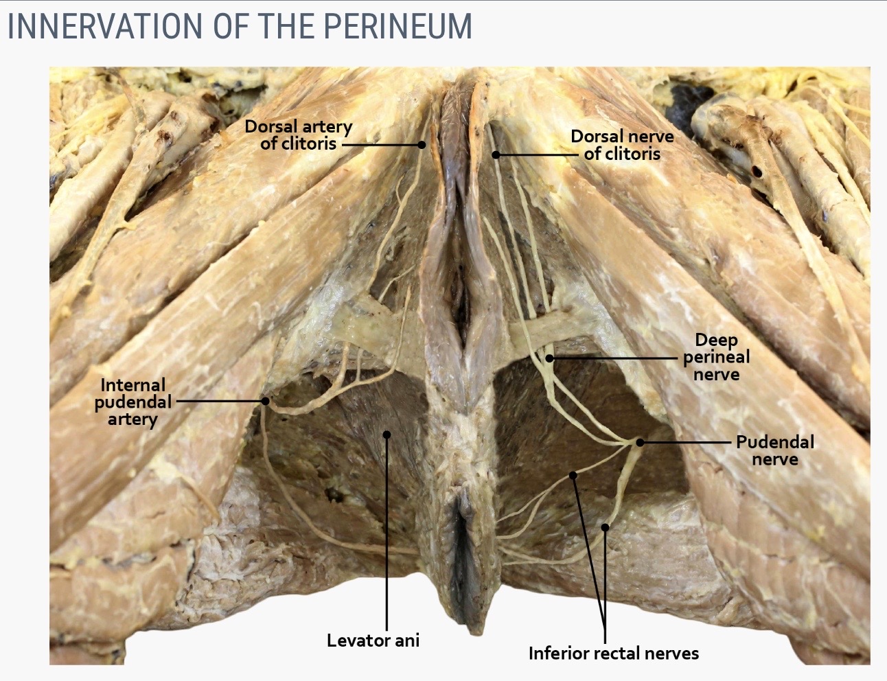
Image occlusion: name the hidden labels
164
Image occlusion: name the hidden labels
165
Image occlusion: name the hidden labels
166
Image occlusion: name the hidden labels
167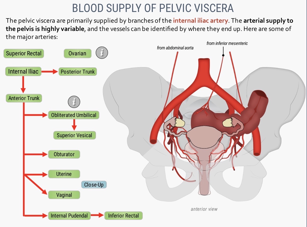
Image occlusion: name the hidden labels
168
Image occlusion: name the hidden labels
169
Image occlusion: name the hidden labels
170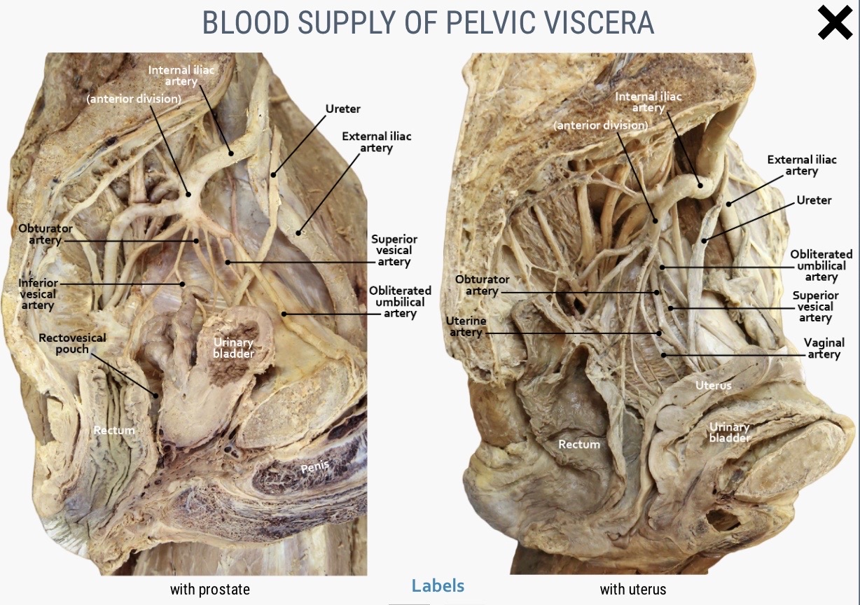
Image occlusion: name the hidden labels
171
Image occlusion: name the hidden labels
172
Image occlusion: name the hidden labels
173
Image occlusion: name the hidden labels
174
Image occlusion: name the hidden labels
175
Image occlusion: name the hidden labels
176
Image occlusion: name the hidden labels
177
Image occlusion: name the hidden labels
178
The vulvar urethra doesn't have a It opens into the vestibule between labia minora and is lubricated by on the anterior wall of vagina.
The vulvar urethra doesn't have a distinct internal urethral sphincter. It opens into the vestibule between labia minora and is lubricated by para-urethral glands on the anterior wall of vagina.
179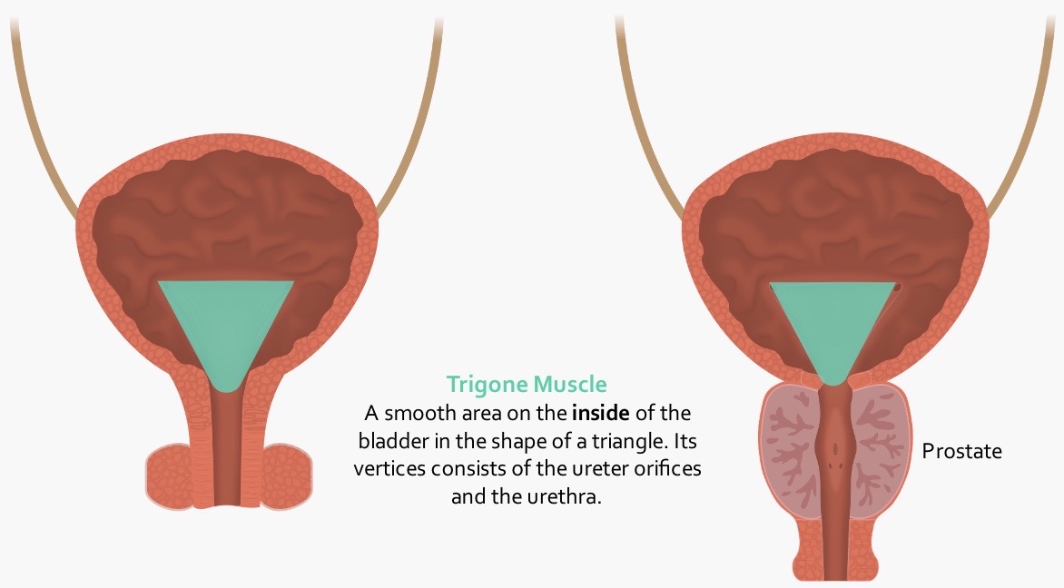
Image occlusion: name the hidden labels
180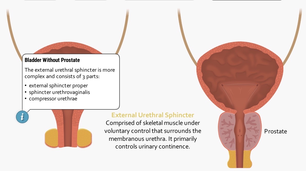
Image occlusion: name the hidden labels
181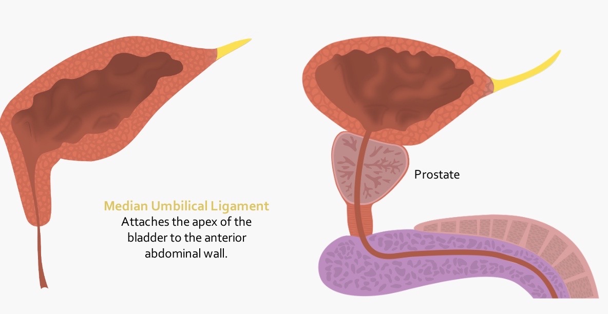
Image occlusion: name the hidden labels
182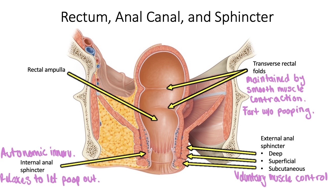
Image occlusion: name the hidden labels
183
Image occlusion: name the hidden labels
184
Image occlusion: name the hidden labels
185
Image occlusion: name the hidden labels
186
Image occlusion: name the hidden labels
187
Image occlusion: name the hidden labels
188
Image occlusion: name the hidden labels
189
Image occlusion: name the hidden labels
190
Image occlusion: name the hidden labels
191
Image occlusion: name the hidden labels
192
Image occlusion: name the hidden labels
193
Image occlusion: name the hidden labels
194
Image occlusion: name the hidden labels
195
Image occlusion: name the hidden labels
196
Image occlusion: name the hidden labels
197
Image occlusion: name the hidden labels
198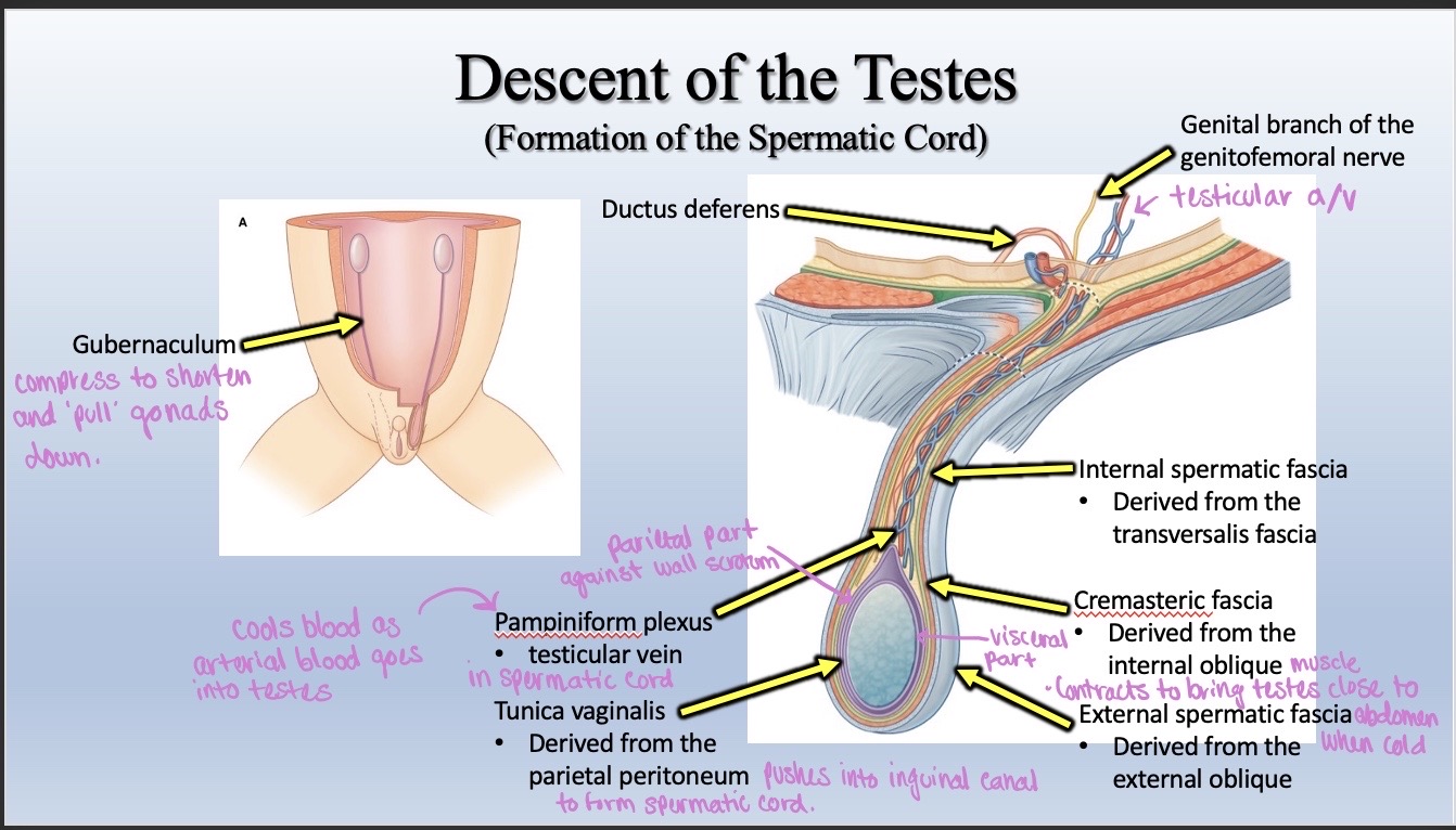
Image occlusion: name the hidden labels
199
Image occlusion: name the hidden labels
200
Image occlusion: name the hidden labels
201
Image occlusion: name the hidden labels
202
Image occlusion: name the hidden labels
203
Image occlusion: name the hidden labels
204
Image occlusion: name the hidden labels
205
Image occlusion: name the hidden labels
206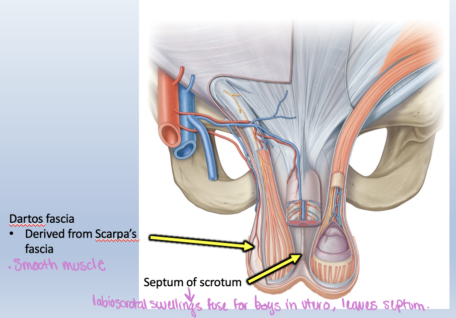
Image occlusion: name the hidden labels
207
Image occlusion: name the hidden labels
208
EXITING ORDER OF IMMATURE AND MATURE SPERM
Spermatozoa are produced in the which drain via the into the Sperm passes through to be stored and mature in the
EJACULATION
>joins to form> duct>urethra
Spermatozoa are produced in the which drain via the into the Sperm passes through to be stored and mature in the
EJACULATION
>joins to form> duct>urethra
EXITING ORDER OF IMMATURE AND MATURE SPERM
Spermatozoa are produced in the seminiferous tubules, which drain via the straight tubules into the rete testis. Sperm passes through efferent ductules to be stored and mature in the epididymis.
EJACULATION
vas deferens>joins seminal vesicle to form>ejaculatory duct>urethra
Spermatozoa are produced in the seminiferous tubules, which drain via the straight tubules into the rete testis. Sperm passes through efferent ductules to be stored and mature in the epididymis.
EJACULATION
vas deferens>joins seminal vesicle to form>ejaculatory duct>urethra
209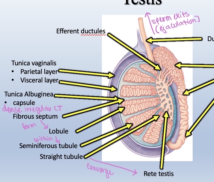
Image occlusion: name the hidden labels
210
Image occlusion: name the hidden labels
211
Image occlusion: name the hidden labels
212
Image occlusion: name the hidden labels
213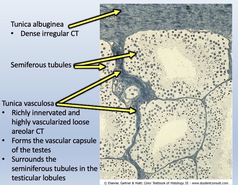
Image occlusion: name the hidden labels
214
Image occlusion: name the hidden labels
215
Image occlusion: name the hidden labels
216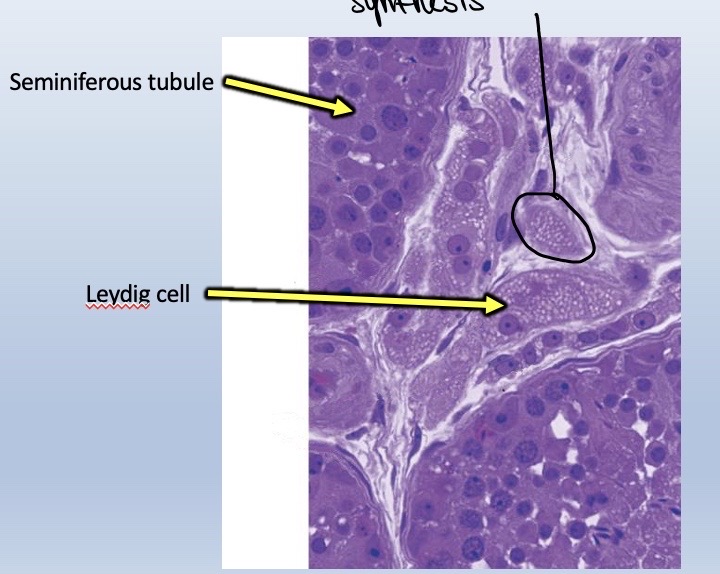
Image occlusion: name the hidden labels
217
Image occlusion: name the hidden labels
218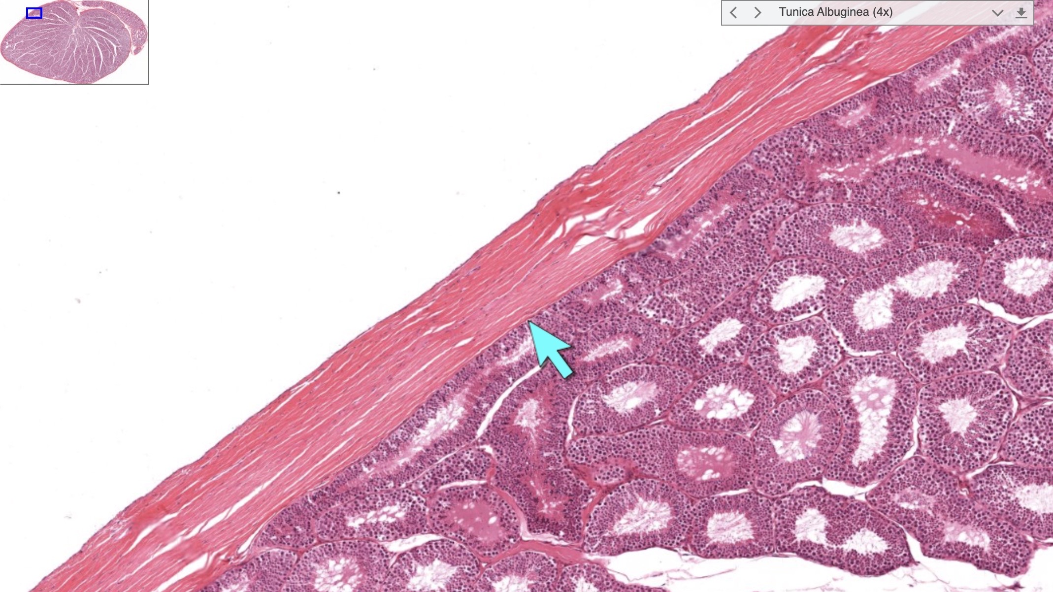
Image occlusion: name the hidden labels
219
Image occlusion: name the hidden labels
220
Identify the structures pointed with the arrow. Describe their function.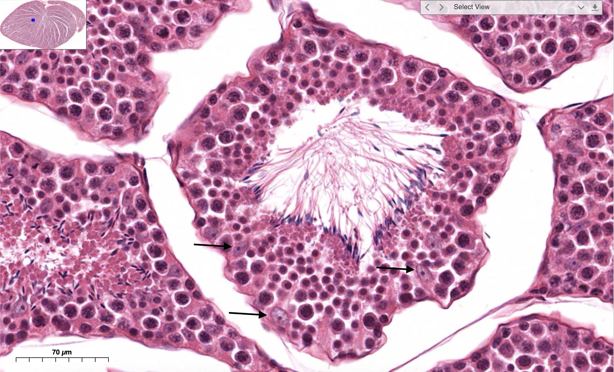
Sertoli cells are avoid or triangular, euchromatic with a prominent and basophilic nucleolus. Usually located close to lumen.
Physical and nutritional support of the developing germ cells
Phagocytosis of cytoplasm eliminated during spermiogenesis.
Synthesis and secretion of:
- inhibin, a hormone that inhibits the release of follicle-stimulating hormone (FSH) by the anterior pituitary.
- androgen-binding protein (ABP), a macromolecule that facilitates an increase in the concentration of testosterone in the seminiferous tubule.
- (during embryogenesis) of antimüllerian hormone, which suppresses the formation of the müllerian duct (precursor of the female reproductive system) and thus establishes the "maleness" of the developing embryo
- fructose-rich medium into the seminiferous tubule lumen that nourishes and facilitates the transport of spermatozoa to the genital ducts
- testicular transferrin, an apoprotein that accepts iron from serum transferrin and conveys it to maturing gametes
Physical and nutritional support of the developing germ cells
Phagocytosis of cytoplasm eliminated during spermiogenesis.
Synthesis and secretion of:
- inhibin, a hormone that inhibits the release of follicle-stimulating hormone (FSH) by the anterior pituitary.
- androgen-binding protein (ABP), a macromolecule that facilitates an increase in the concentration of testosterone in the seminiferous tubule.
- (during embryogenesis) of antimüllerian hormone, which suppresses the formation of the müllerian duct (precursor of the female reproductive system) and thus establishes the "maleness" of the developing embryo
- fructose-rich medium into the seminiferous tubule lumen that nourishes and facilitates the transport of spermatozoa to the genital ducts
- testicular transferrin, an apoprotein that accepts iron from serum transferrin and conveys it to maturing gametes
221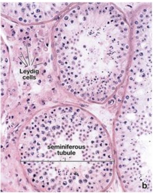
Image occlusion: name the hidden labels
222
Image occlusion: name the hidden labels
223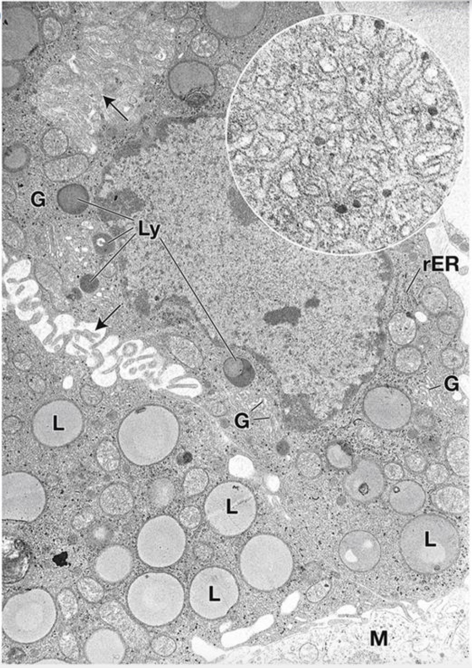
Image occlusion: name the hidden labels
224
what are these?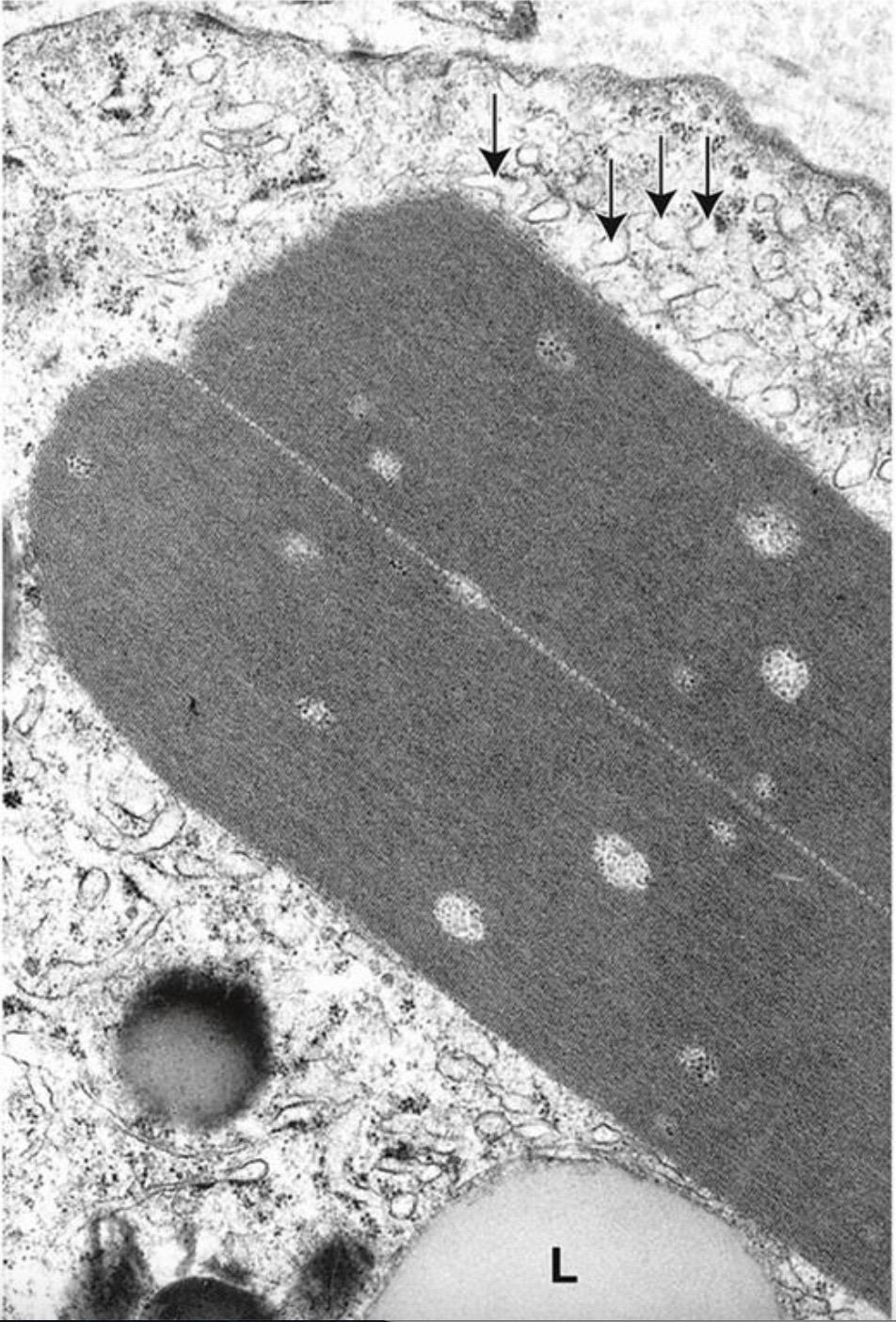
Reinke crystal in the cytoplasm of a human Leydig cell, which are crystallized proteins that have precipitated. Also note the smooth endoplasmic reticulum (sER) (arrows) and a lipid droplet (L) in the cytoplasm. More obvious in Leydig cell tumors.
225
What is this?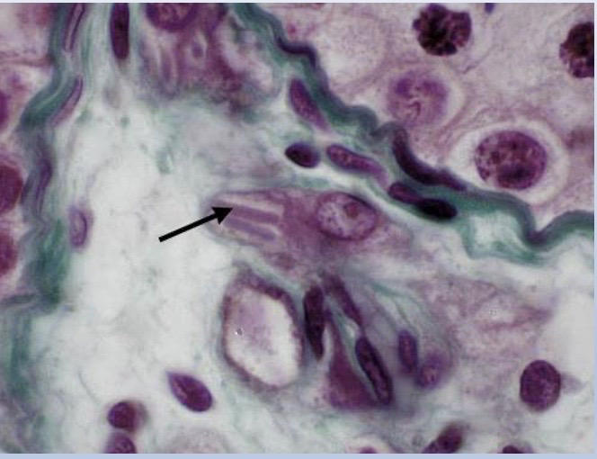
Reinke crystal in Leydig cell.
226
What surrounds seminiferous tubules?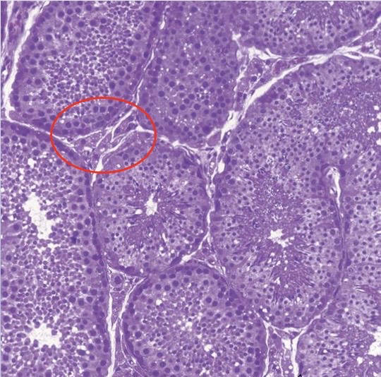
Fibrous connective tissue containing smooth muscle-like myoid cells.
Thick complex germinal epithelium.
Thick complex germinal epithelium.
227
What is the function of the structures pointed by the arrows? What happens if this structure is compromised?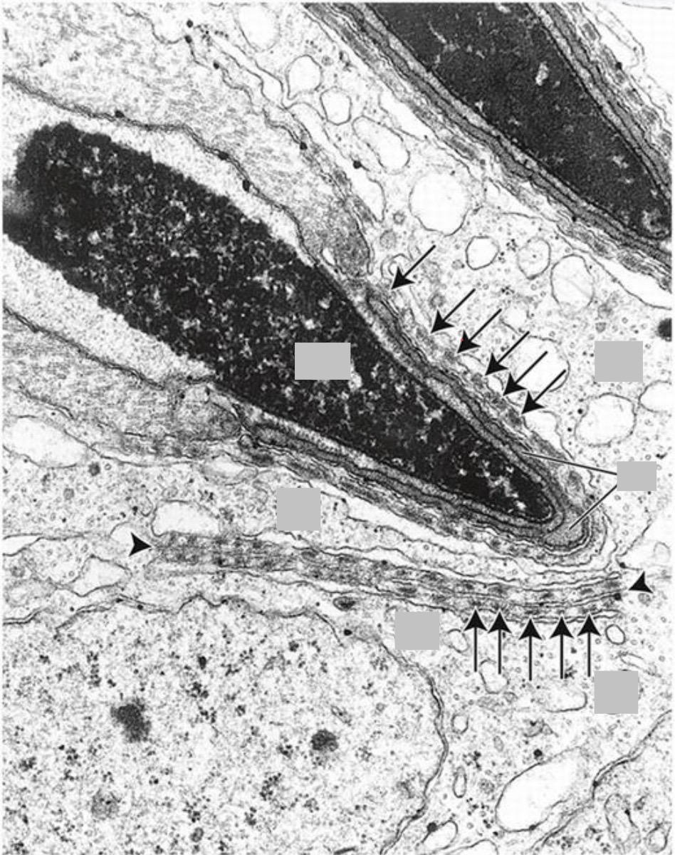
gap/tight junctions formed between sertoli cells and primary spermatocytes in the basal compartment of seminiferous tubules. Form the blood-testis barrier and prevents autoimmune reactions.
Autoimmune attack against seminiferous tubules and developing sperm, leading to infertility.
Autoimmune attack against seminiferous tubules and developing sperm, leading to infertility.
228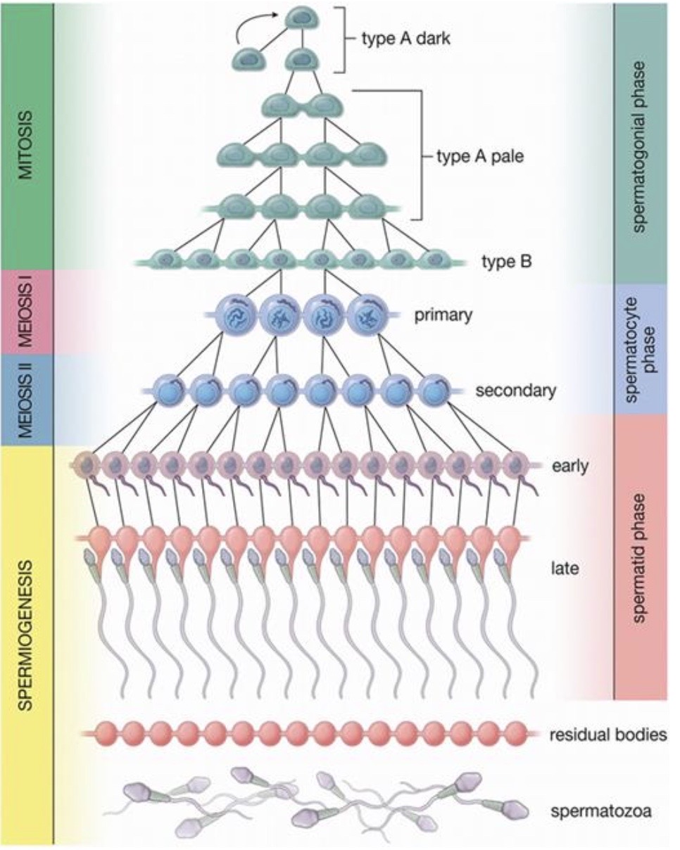
Image occlusion: name the hidden labels
229
Image occlusion: name the hidden labels
230
Image occlusion: name the hidden labels
231
Image occlusion: name the hidden labels
232
Image occlusion: name the hidden labels
233
Image occlusion: name the hidden labels
234
Image occlusion: name the hidden labels
235
Image occlusion: name the hidden labels
236
Image occlusion: name the hidden labels
237
Migrate from the basal to adluminal compartment of seminiferous tubules.
Undergo the first meiotic division
Diploid with 2 chromatids for both members of the homologous chromosomes.
Undergo the first meiotic division
Diploid with 2 chromatids for both members of the homologous chromosomes.
Primary spermatocytes Migrate from the basal to adluminal compartment of seminiferous tubules.
Undergo the first meiotic division
Diploid with 2 chromatids for both members of the homologous chromosomes.
Undergo the first meiotic division
Diploid with 2 chromatids for both members of the homologous chromosomes.
238
Undergo the second meiotic division to produce spermatids
Haploid with 2 chromatids for one member of the homologous chromosomes.
Haploid with 2 chromatids for one member of the homologous chromosomes.
Secondary spermatocytes
Undergo the second meiotic division to produce spermatids
Haploid with 2 chromatids for one member of the homologous chromosomes.
Haploid with 2 chromatids for one member of the homologous chromosomes.
239
are Formed after the completion of meiosis
and are 1n
Spermatids
are Formed after the completion of meiosis
and are 1n
240
are Formed from spermatids that have matured through spermiogenesis
Spermatozoa
are Formed from spermatids that have matured through spermiogenesis
241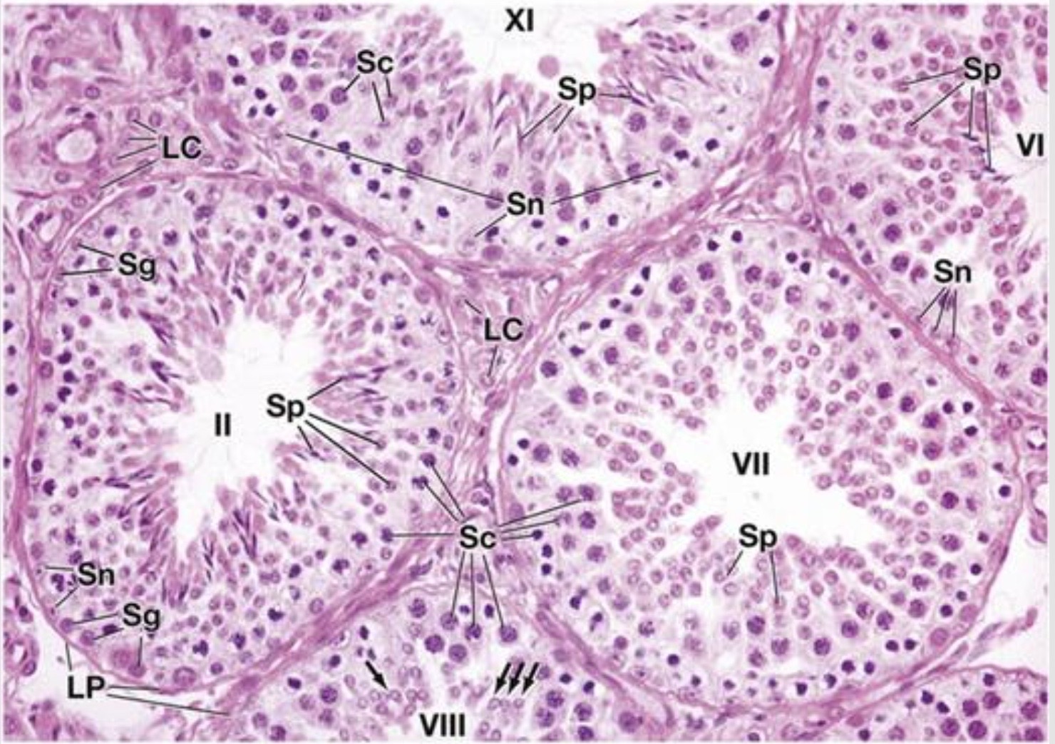
Image occlusion: name the hidden labels
242
Image occlusion: name the hidden labels
243
Image occlusion: name the hidden labels
244
Image occlusion: name the hidden labels
245
Image occlusion: name the hidden labels
246
Image occlusion: name the hidden labels
247
Image occlusion: name the hidden labels
248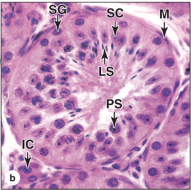
Image occlusion: name the hidden labels
249
Image occlusion: name the hidden labels
250
Image occlusion: name the hidden labels
251
Image occlusion: name the hidden labels
252
Image occlusion: name the hidden labels
253
Which of the labeled structures in this seminiferous tubule histologic slide produces testosterone?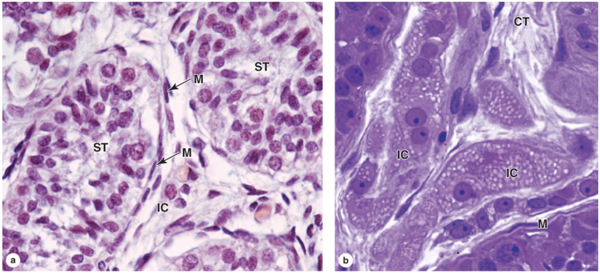
IC = interstitial (Leydig) cells. Characterized by location in the interstitial space between seminiferous tubules (ST) and their association with lipid droplets, better visualized in figure b.
M= myeloid cells
CT= connective tissue
M= myeloid cells
CT= connective tissue
254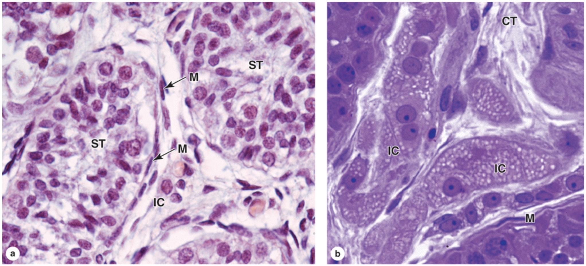
Image occlusion: name the hidden labels
255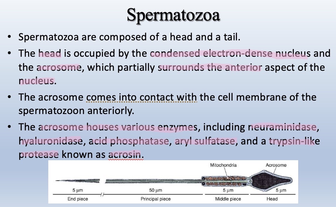
Image occlusion: name the hidden labels
256
Image occlusion: name the hidden labels
257
Image occlusion: name the hidden labels
258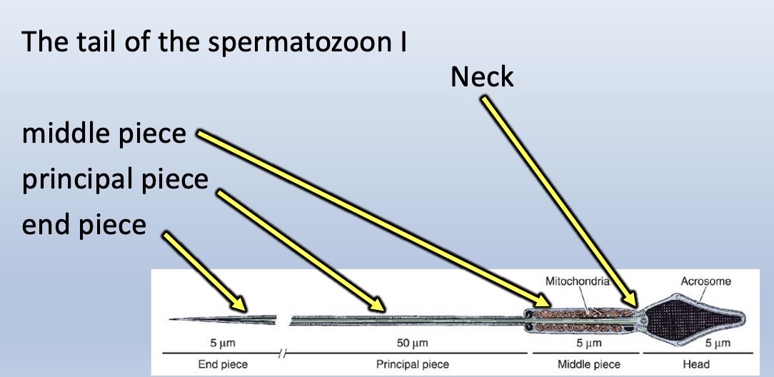
Image occlusion: name the hidden labels
259
Image occlusion: name the hidden labels
260
Image occlusion: name the hidden labels
261
Image occlusion: name the hidden labels
262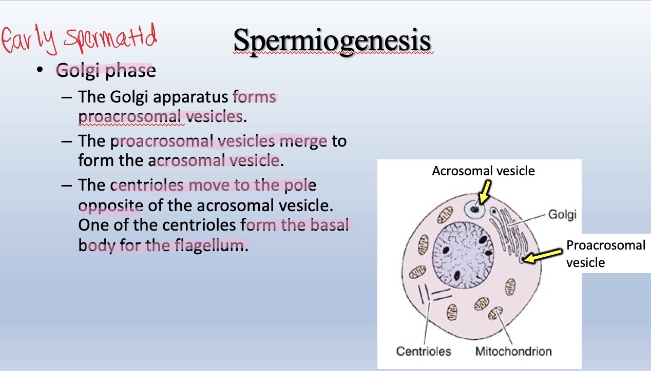
Image occlusion: name the hidden labels
263
Image occlusion: name the hidden labels
264
Image occlusion: name the hidden labels
265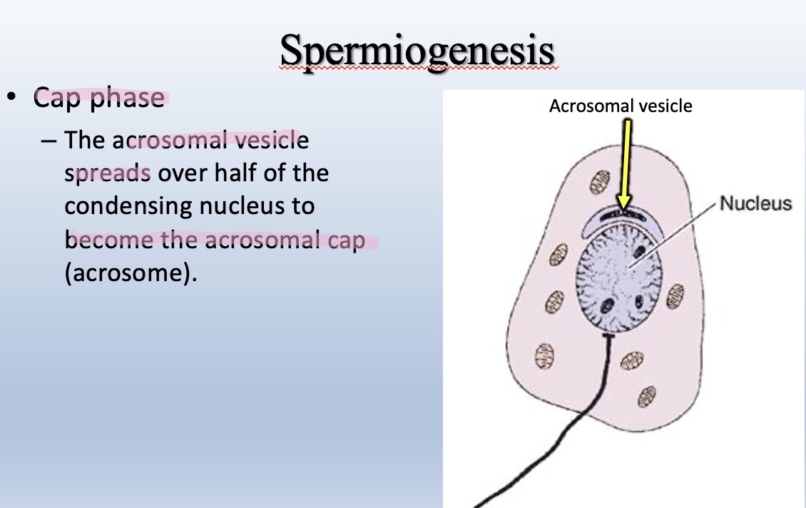
Image occlusion: name the hidden labels
266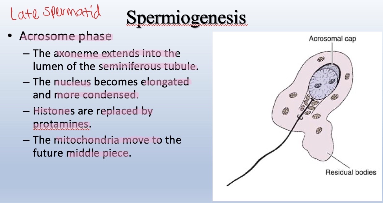
Image occlusion: name the hidden labels
267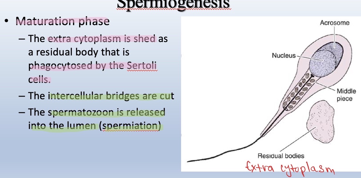
Image occlusion: name the hidden labels
268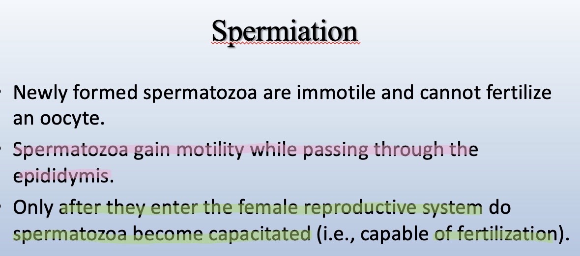
Image occlusion: name the hidden labels
269
What is spermiogenesis? List the basic steps in order,
Process of spermatid maturation into spem.
1) Golgi phase: Golgi forms proacrosomal vesicles, which merge to form the acrosomal vesicle. Centrioles move to the opposite side of acrosomal vesicle and one of them emerge from the basal body of the sperm cell to begin forming the flagellum.
2) Cap phase: acrosomal vesicle spreads over half of the condensing nucleus to form the acrosomal cap (acrosome).
3) Acrosome phase: axoneme (developing flagellum) extends into the lumen of the feminiferous tubule. Sperm cell's nucleus becomes elongated and more condensed. Histomes are replaced by protamines. Mitochondria move to the future middle piece of the sperm.
4) Maturation phase: extra cytoplasm is shed as a residual body and is phagocytosed by Sertoli cells. Intercellular bridges in sperm cell are cut. Differentiated spermatozoon are released into the lumen, this is spermiation.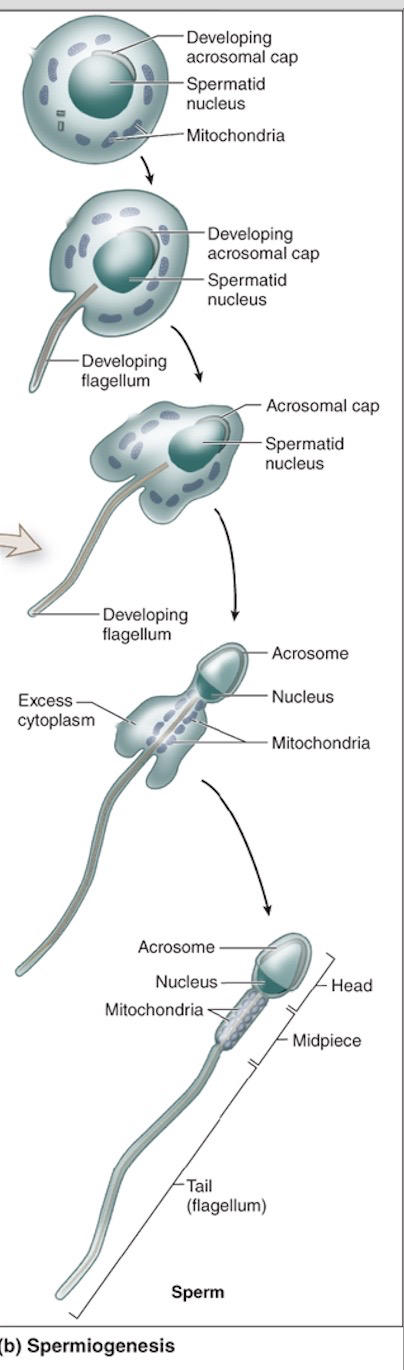
1) Golgi phase: Golgi forms proacrosomal vesicles, which merge to form the acrosomal vesicle. Centrioles move to the opposite side of acrosomal vesicle and one of them emerge from the basal body of the sperm cell to begin forming the flagellum.
2) Cap phase: acrosomal vesicle spreads over half of the condensing nucleus to form the acrosomal cap (acrosome).
3) Acrosome phase: axoneme (developing flagellum) extends into the lumen of the feminiferous tubule. Sperm cell's nucleus becomes elongated and more condensed. Histomes are replaced by protamines. Mitochondria move to the future middle piece of the sperm.
4) Maturation phase: extra cytoplasm is shed as a residual body and is phagocytosed by Sertoli cells. Intercellular bridges in sperm cell are cut. Differentiated spermatozoon are released into the lumen, this is spermiation.
270
What are the labeled structures in these histolical sections of the testes?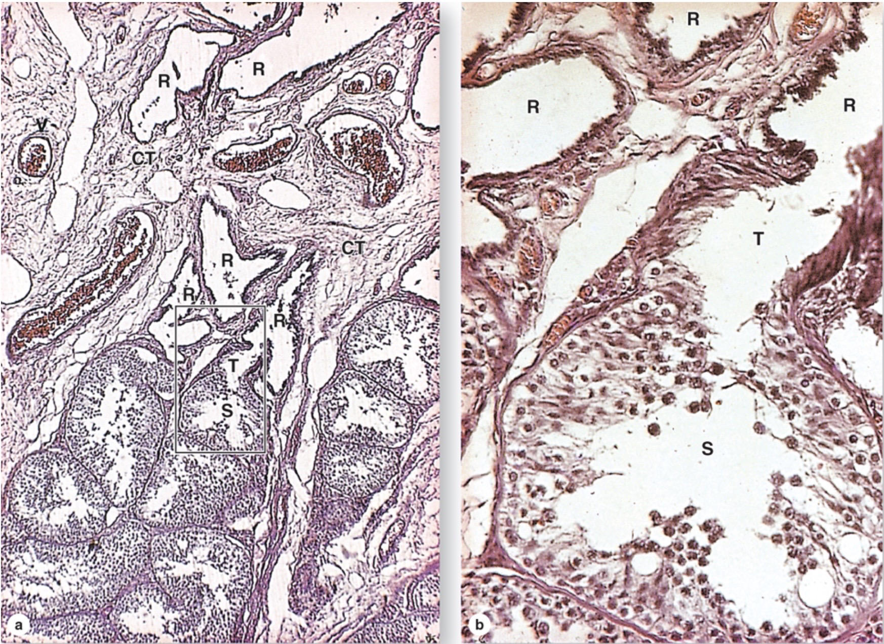
Showing the transition of a seminiferous tubule into a straight tubule, and then into the rete testis.
(a) The seminiferous tubules (S) drain into short, much narrower straight tubules (T), which connect to the rete testis (R), a network of channels embedded along with blood vessels (V) in the connective tissue (CT) of the mediastinum testis.
(b) Transition from wide seminiferous tubule (S) to the straight tubule (T). Initially, the straight tubule wall has only tall Sertoli cells devoid of germ cells. The wall becomes a simple cuboidal epithelium near its connection to the rete testis (R), which is also lined with simple cuboidal epithelium.
(a) The seminiferous tubules (S) drain into short, much narrower straight tubules (T), which connect to the rete testis (R), a network of channels embedded along with blood vessels (V) in the connective tissue (CT) of the mediastinum testis.
(b) Transition from wide seminiferous tubule (S) to the straight tubule (T). Initially, the straight tubule wall has only tall Sertoli cells devoid of germ cells. The wall becomes a simple cuboidal epithelium near its connection to the rete testis (R), which is also lined with simple cuboidal epithelium.
271
lead spermatozoa from the seminiferous tubules into the Lined only by Sertoli cells, transitions into simple cuboidal epithelium with microvilli and a cilum on each cell.
Straight tubules lead spermatozoa from the seminiferous tubules into the rete testis. Lined only by Sertoli cells, transitions into simple cuboidal epithelium with microvilli and a cilum on each cell.
272
The have simple cuboidal epithelium and are usually embedded in tissue. Similar to straight tubules, the have microvilli and a cilium on each cell.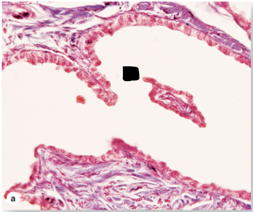
The Rete testis channels have simple cuboidal epithelium and are usually embedded in dense connective tissue. Similar to straight tubules, the rete testis have microvilli and a cilium on each cell.
273
What is this structure and what gives it its scalloped appearance?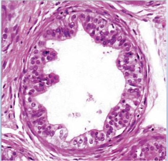
Efferent ductules are lined by alternating clusters of simple non-ciliated cuboidal cells and simple ciliated columnar cells, which gives a scalloped appearance.
The cuboidal cells have many lysosomes and the apical plasmalemmae have numerous invaginations indicative of endocytosis; they're involved in resorption of luminal fluid.
The cilia of the columnar cells probably move the spermatozoa toward the epididymis.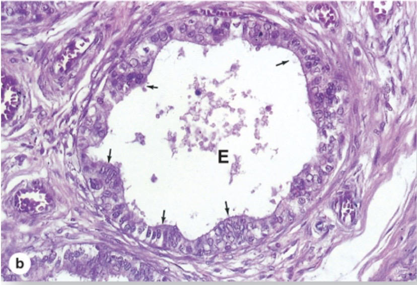
The cuboidal cells have many lysosomes and the apical plasmalemmae have numerous invaginations indicative of endocytosis; they're involved in resorption of luminal fluid.
The cilia of the columnar cells probably move the spermatozoa toward the epididymis.
274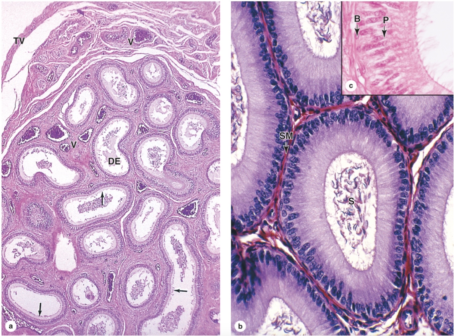
Image occlusion: name the hidden labels
275
Image occlusion: name the hidden labels
276
Identify this structure: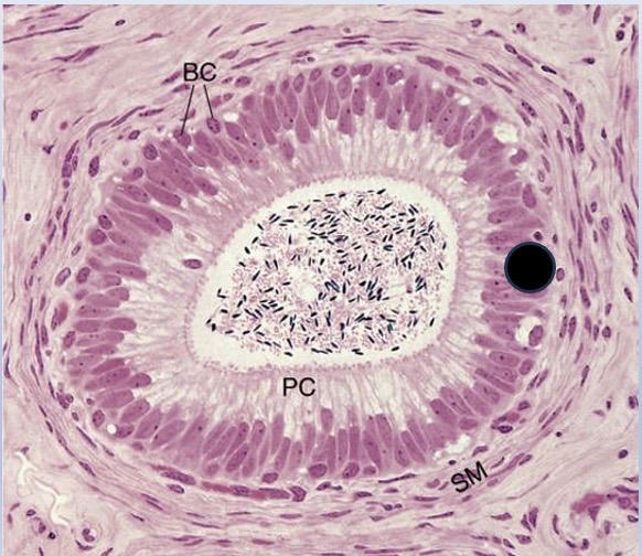
Epididymis characterized by stereociliated pseudostratified columnar epithelium with basal stem cells in the basal domain, which regenerated the principal cells closer to the adluminal domain. Principal cells resorb luminal fluid, phagocytose cytoplasmic remnants, and secret glycerophosphocholine to inhibit spermatozoa capacitacion.
Note the surrounding circular smooth muscle, which conducts the spermatozoa to ductus deferens by peristalsis (contraction).
Note the surrounding circular smooth muscle, which conducts the spermatozoa to ductus deferens by peristalsis (contraction).
277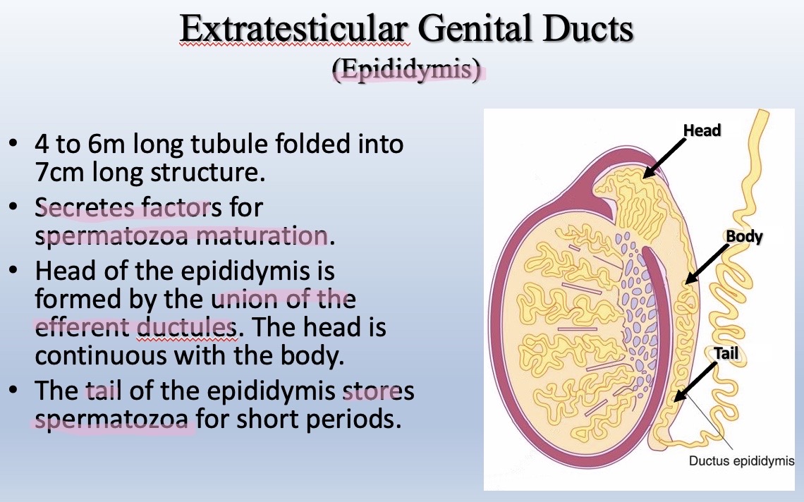
Image occlusion: name the hidden labels
278
Image occlusion: name the hidden labels
279
Image occlusion: name the hidden labels
280
What extratesticular duct is characterized by having 3 surrounding muscular layers and a lamina propria with many elastic fibers?
Ductus deferens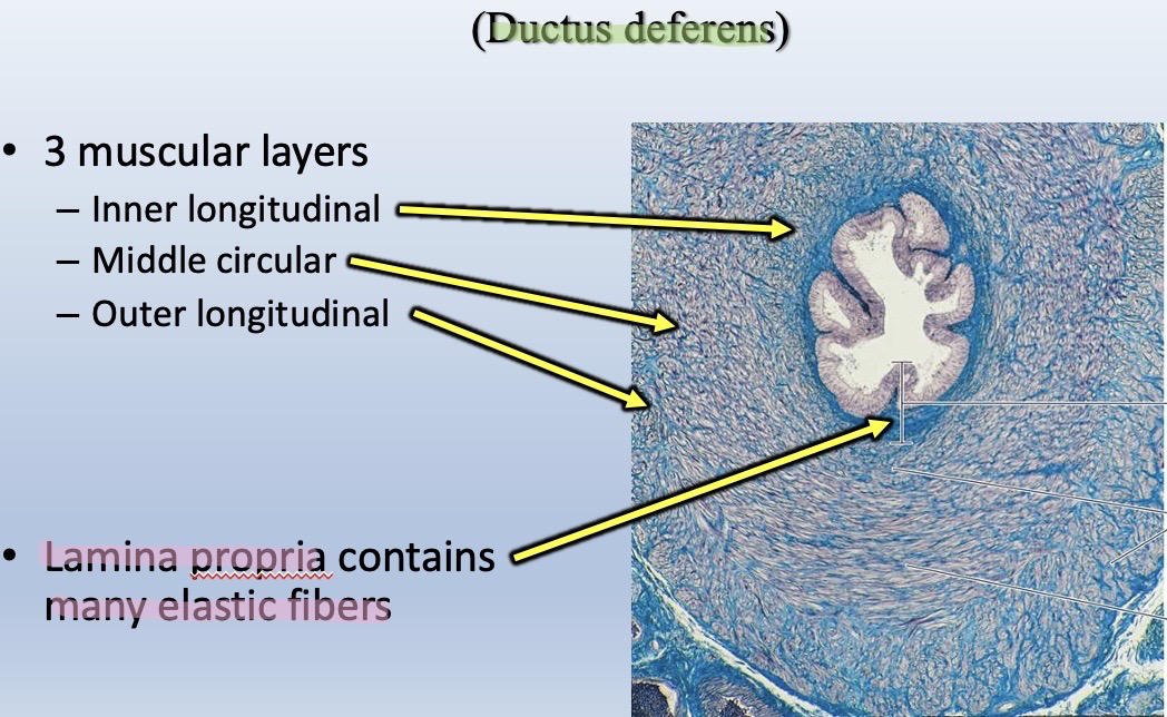
281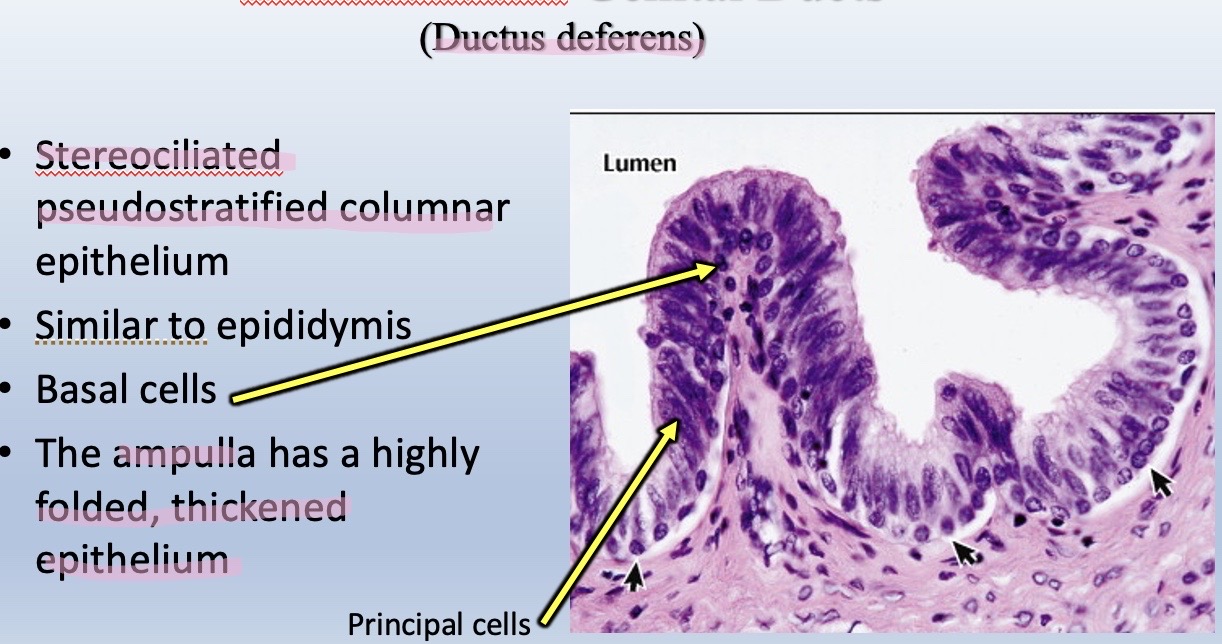
Image occlusion: name the hidden labels
282
Image occlusion: name the hidden labels
283
Identify the structure in these histological slides (H&E and Mallory trichrome stains),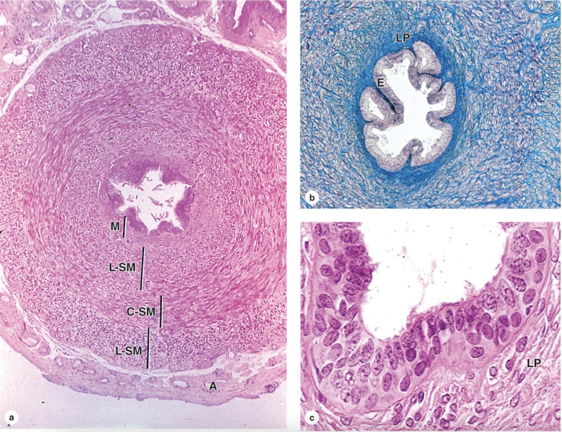
(a) Cross section of the vas deferens shows the mucosa (M), a thick muscularis with inner and outer layers of longitudinal smooth muscle (L-SM) and an intervening layer of circular smooth muscle (C-SM), and an external adventitia (A). The muscularis is specialized for powerful peristaltic movement of sperm at ejaculation.
(b) The lamina propria (LP) is rich in elastic fibers and the thick epithelial lining (E) shows longitudinal folds.
(c) Higher magnification of the mucosa shows that the epithelium is pseudostratified with basal cells and many columnar cells, some with stereocilia.
(b) The lamina propria (LP) is rich in elastic fibers and the thick epithelial lining (E) shows longitudinal folds.
(c) Higher magnification of the mucosa shows that the epithelium is pseudostratified with basal cells and many columnar cells, some with stereocilia.
284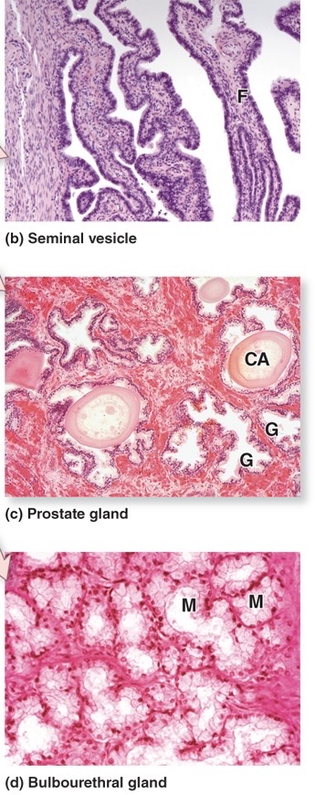
Image occlusion: name the hidden labels
285
Image occlusion: name the hidden labels
286
Image occlusion: name the hidden labels
287
Image occlusion: name the hidden labels
288
Image occlusion: name the hidden labels
289
Image occlusion: name the hidden labels
290
Identify the structure in the histological slides (H&E and PSH).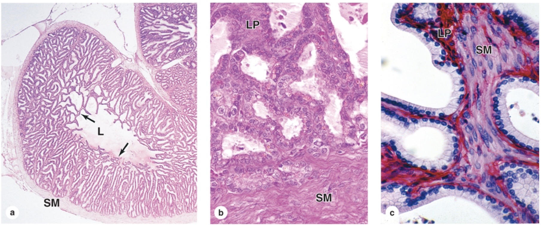
Seminal vesicles characterized by:
2 layers of smooth muscle (SM) surrounding the organ arranged in an inner circular and outer longitudinal layers.
SM are deep to the thin lamina propia (LP).
Mucosal folds with simple columnar epithelium of principal secretory cells:
70% of the ejaculate
Fructose
Prostaglandins
Fibrinogen
Lipochrome
Pigment that gives the pale yellow color of semen
2 layers of smooth muscle (SM) surrounding the organ arranged in an inner circular and outer longitudinal layers.
SM are deep to the thin lamina propia (LP).
Mucosal folds with simple columnar epithelium of principal secretory cells:
70% of the ejaculate
Fructose
Prostaglandins
Fibrinogen
Lipochrome
Pigment that gives the pale yellow color of semen
291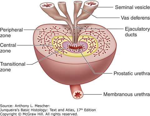
Image occlusion: name the hidden labels
292
Image occlusion: name the hidden labels
293
Image occlusion: name the hidden labels
294
Image occlusion: name the hidden labels
295
Image occlusion: name the hidden labels
296
Image occlusion: name the hidden labels
297
Image occlusion: name the hidden labels
298
Image occlusion: name the hidden labels
299
Identify the structure in these micrographs: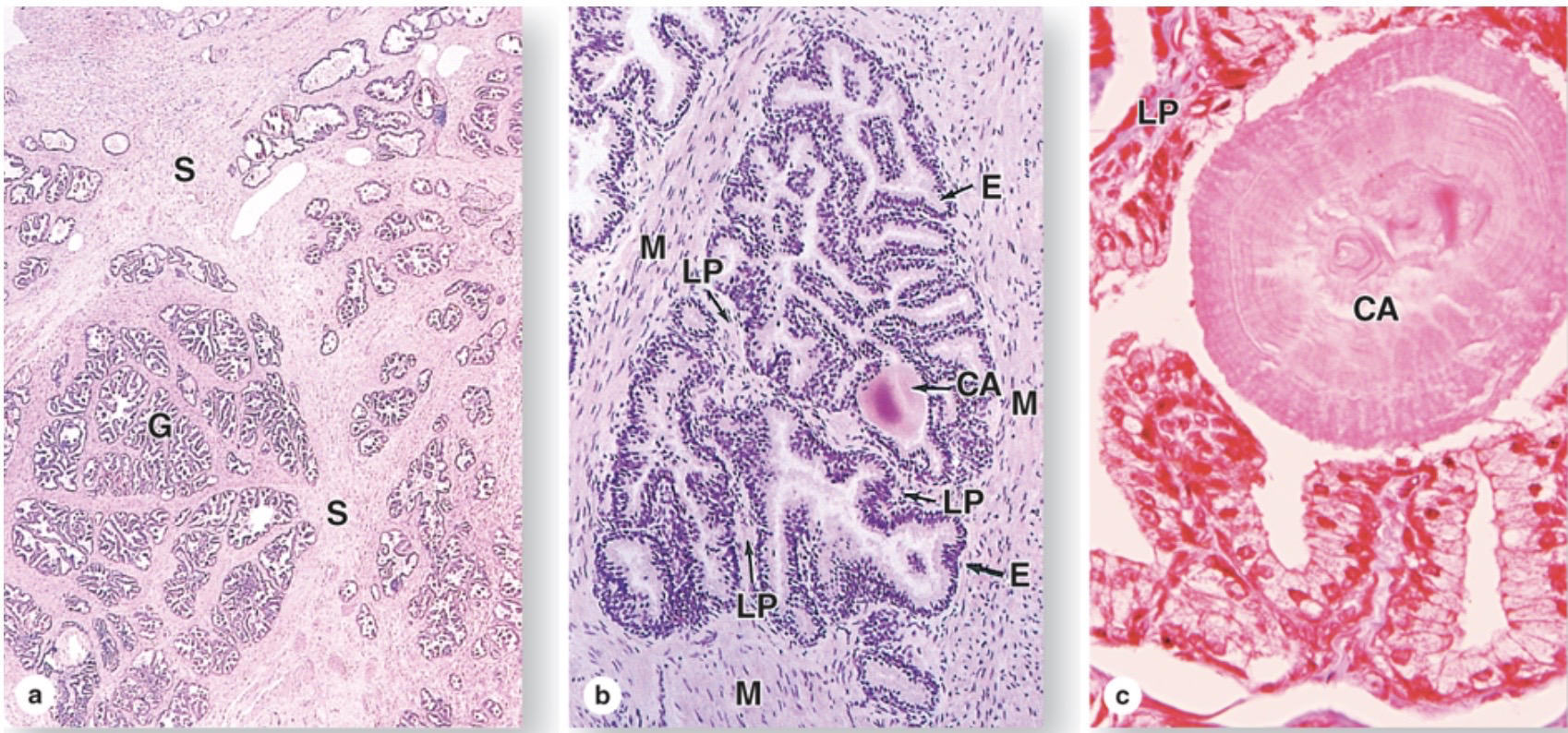
30-50 compound tubuloalveolar glands
Types of glands arranged in the different zones of the prostate
Transitional zone-> mucosal glands with short ducts
Central zone-> submucosal glands with intermediate length ducts
Peripheral zone-> main glands with long ducts
Simple or pseudostratified columnar epithelium
Fibromuscular stroma
Corpora amylacea are Calcified concretions of glycoproteins and keratan sulfate.
Prostate gland.
(a) The prostate has a dense fibromuscular stroma (S) in which are embedded a large number of small tubuloacinar glands (G).
(b) A micrograph of one prostatic gland, showing a corpus amylaceum (CA) concretion and the secretory epithelium (E) surrounded by a thin lamina propria (LP) and thick smooth muscle (M).
(c) Higher magnification shows the lamellar nature of a corpus amylaceum (CA) and the pseudostratified columnar epithelium underlain by lamina propria (LP).
Types of glands arranged in the different zones of the prostate
Transitional zone-> mucosal glands with short ducts
Central zone-> submucosal glands with intermediate length ducts
Peripheral zone-> main glands with long ducts
Simple or pseudostratified columnar epithelium
Fibromuscular stroma
Corpora amylacea are Calcified concretions of glycoproteins and keratan sulfate.
Prostate gland.
(a) The prostate has a dense fibromuscular stroma (S) in which are embedded a large number of small tubuloacinar glands (G).
(b) A micrograph of one prostatic gland, showing a corpus amylaceum (CA) concretion and the secretory epithelium (E) surrounded by a thin lamina propria (LP) and thick smooth muscle (M).
(c) Higher magnification shows the lamellar nature of a corpus amylaceum (CA) and the pseudostratified columnar epithelium underlain by lamina propria (LP).
300
List the major prostatic secretions. What is their main function? Synthesis of these secretions is regulated by what hormone?
1) Acid phosphatase, Fibrinolysin, Prostate-specific antigen (liquifies coagulated semen).
2) Help spermatozoa achieve motility
3) Synthesis of secretions regulated by dihydrotestosterone
2) Help spermatozoa achieve motility
3) Synthesis of secretions regulated by dihydrotestosterone
242 more cards in OutLearn. Open the full deck