Adaptations for transport in animals unit 2
Study these flashcardsFree · spaced repetition · iPhone, Android, Mac & web
All cards
1
What are the 4 types of circulatory systems?
Open
Closed
Single
Double
Closed
Single
Double
2
What is a open circulatory system?
Blood pumped to haemocoel- bathes at organs and returns slowly to heart
Little control over direction of blood flow
Blood not in vessels
Little control over direction of blood flow
Blood not in vessels
3
What is a closed circulatory system?
Blood in vessels
Flow is rapid and direction controlled
Organs bathed by tissue fluid that leaks from capillaries
Flow is rapid and direction controlled
Organs bathed by tissue fluid that leaks from capillaries
4
What is a single circulatory system?
Blood passes thru heart once per circulation
5
What is a double circulatory system?
Blood passes thru heart twice per circulation
Once in pulmonary (lung) and then thru systemic (body) circulation
Once in pulmonary (lung) and then thru systemic (body) circulation
6
What circulatory systems do insects have?
Open circulatory system
Dorsal tube-shaped heart
No respiratory pigment
Dorsal tube-shaped heart
No respiratory pigment
7
What is a respiratory pigment
Coloured protein, enchances bloods capacity to carry O2 and CO2
Eg. haemoglobin
Eg. haemoglobin
8
What circulatory systems do earthworms have?
Closed circulatory system
5 pseudohearts
Respiratory pigment haemoglobin
5 pseudohearts
Respiratory pigment haemoglobin
9
What circulatory systems do fish have?
Closed single circulatory system
Blood pumped and oxygenated in gills
Gills O2 blood->Tissues
Lower pressure and slower flow around body
Blood pumped and oxygenated in gills
Gills O2 blood->Tissues
Lower pressure and slower flow around body
10
What circulatory systems do mammals have?
Closed double circulatory system
High blood pressure delivers O2 quickly
Lower pressure to lungs preventing hydrostatic pressure
High blood pressure delivers O2 quickly
Lower pressure to lungs preventing hydrostatic pressure
11
What does hydrostatic pressure do?
Force tissue fluid into alveoli and reducing its efficiency
12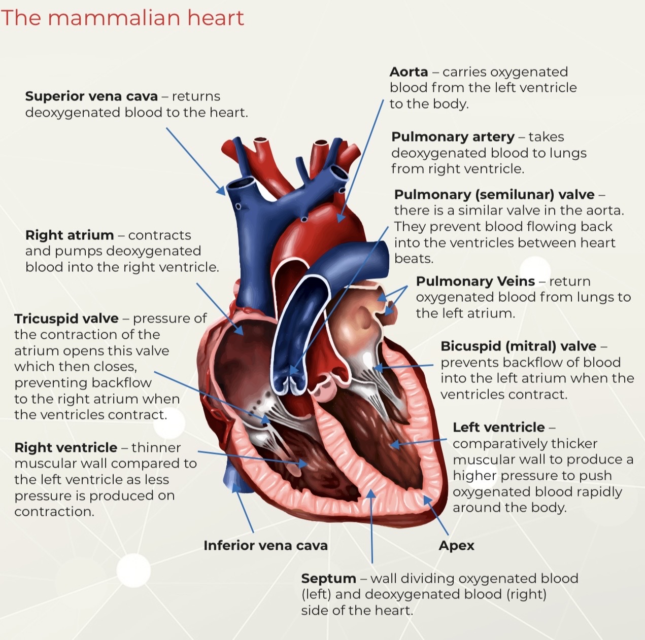
Image occlusion: name the hidden labels
13
Image occlusion: name the hidden labels
14
Image occlusion: name the hidden labels
15
Image occlusion: name the hidden labels
16
Image occlusion: name the hidden labels
17
Image occlusion: name the hidden labels
18
Image occlusion: name the hidden labels
19
Image occlusion: name the hidden labels
20
Image occlusion: name the hidden labels
21
Image occlusion: name the hidden labels
22
Image occlusion: name the hidden labels
23
Image occlusion: name the hidden labels
24
Image occlusion: name the hidden labels
25
What does the superior vena cava do?
Returns deoxygenated blood to heart
26
What does the right atrium do?
Contracts and pumps deoxygenated blood into right ventricle
27
What does the tricuspid valve do?
Pressure of atrium contraction opens valve then closes- preventing backflow to right atrium when ventricles contract
28
What is the right ventricle like?
Thinner muscular wall compared to left bc less pressure produced on contraction
29
What is the septum?
Wall dividing oxygenated blood (left) and deoxygenated blood (right) sides of heart
30
What side of heart is deoxygenated blood on?
Right
31
What side of heart is oxygenated blood on?
Left
32
What does the aorta do?
Carry oxygenated blood from LV to body
33
What does pulmonary artery do?
Take deoxygenated blood to lung from RV
34
What does the pulmonary valve do?
Prevent blood flowing back into ventricles between heartbeats
35
What do the pulmonary veins do?
Return oxygenated blood from lungs to left atrium
36
Whag does the bicuspid mitral valve do
Prevent backflow of blood into left atrium when ventricles contract
37
What is the left ventricle
Comparatively thicker muscular wall to produce higher pressure to push oxygenated blood rapidly around body
38
what is this?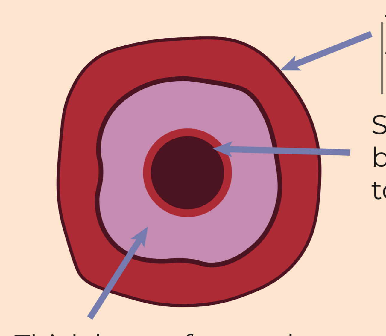
Artery
39
what is this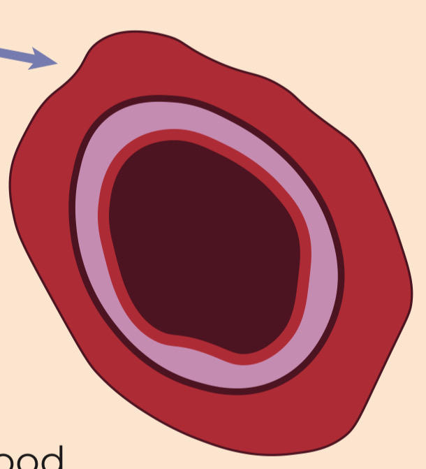
Vein
40
What is this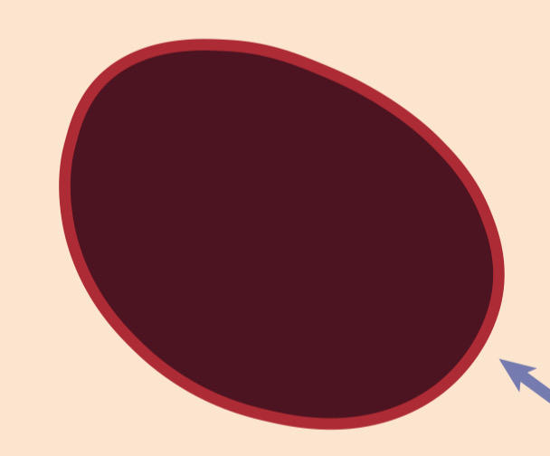
capillary
41
What is the outer layer of the vein and artery
a tough collagen outer coat to prevent overstretching
42
What is the purple part of the artery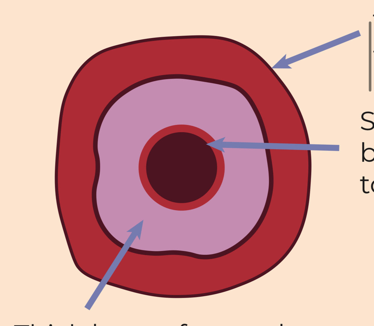
Thick layer of smooth muscle and elastic tissue
muscle Contracts and relaxes to alter blood flow to diff organs
elastic recoils to propel blood forward and even out flow
muscle Contracts and relaxes to alter blood flow to diff organs
elastic recoils to propel blood forward and even out flow
43
What is the lumen like in artery
Small surrounded by smooth endothelium to prevent friction
44
What is the lumen like in a vein
Larger bc blood under lower pressure
Less resistance for blood flow
Less resistance for blood flow
45
What do veins have instead of lots of muscle and elastic fibres?
Semilunar valves, prevent backflow of blood
46
What is the structure of a capillary like?
A single layer of endothelium for short diffusion pathway
47
What is the order of the cardiac cycle
Atrial systole
Ventricular systole
Ventricular diastole
Diastole
Ventricular systole
Ventricular diastole
Diastole
48
What happens in atrial systole
Atrial contract
Pressure open atrio ventricular valves
Blood flow into ventricles
Pressure open atrio ventricular valves
Blood flow into ventricles
49
What happens in ventricular systole
Ventricles contract
Pressure in ventricles higher than in atria
Atrio ventricular valves close
SL valves in aorta and PA open
Blood flow into arteries
Pressure in ventricles higher than in atria
Atrio ventricular valves close
SL valves in aorta and PA open
Blood flow into arteries
50
What happens in ventricular diastole
Ventricle muscle relaxes
SL valve close to prevent backflow of blood
SL valve close to prevent backflow of blood
51
What happens in diastole
Heart muscle relax
atria fills from vena cava and Pulmonary vein
atria fills from vena cava and Pulmonary vein
52
What is the heartbeat? Why?
myogenic. heartbeat produced and intiated by the heart muscle
53
What does the sinoatrial node do when initiating heartbeat
first stage
acts as pacemaker
sends waves of excitation across atria so they contract simultaneously
acts as pacemaker
sends waves of excitation across atria so they contract simultaneously
54
what does the connective tissue in heart do when heartbeat is being initiated?
second stage
Prevents excitation wave from passing down ventricles
Passes to atrio ventricular node
Delay to allow atria to contract completely
Prevents excitation wave from passing down ventricles
Passes to atrio ventricular node
Delay to allow atria to contract completely
55
What does the atrio ventricular node do when heartbeat is being initiated
third stage
transmits impulses down bundle of His to apex of heart
travels up branched Purkinje fibres
makes ventricles contract from bottom upward
all blood pumped out
transmits impulses down bundle of His to apex of heart
travels up branched Purkinje fibres
makes ventricles contract from bottom upward
all blood pumped out
56
What is the dashed line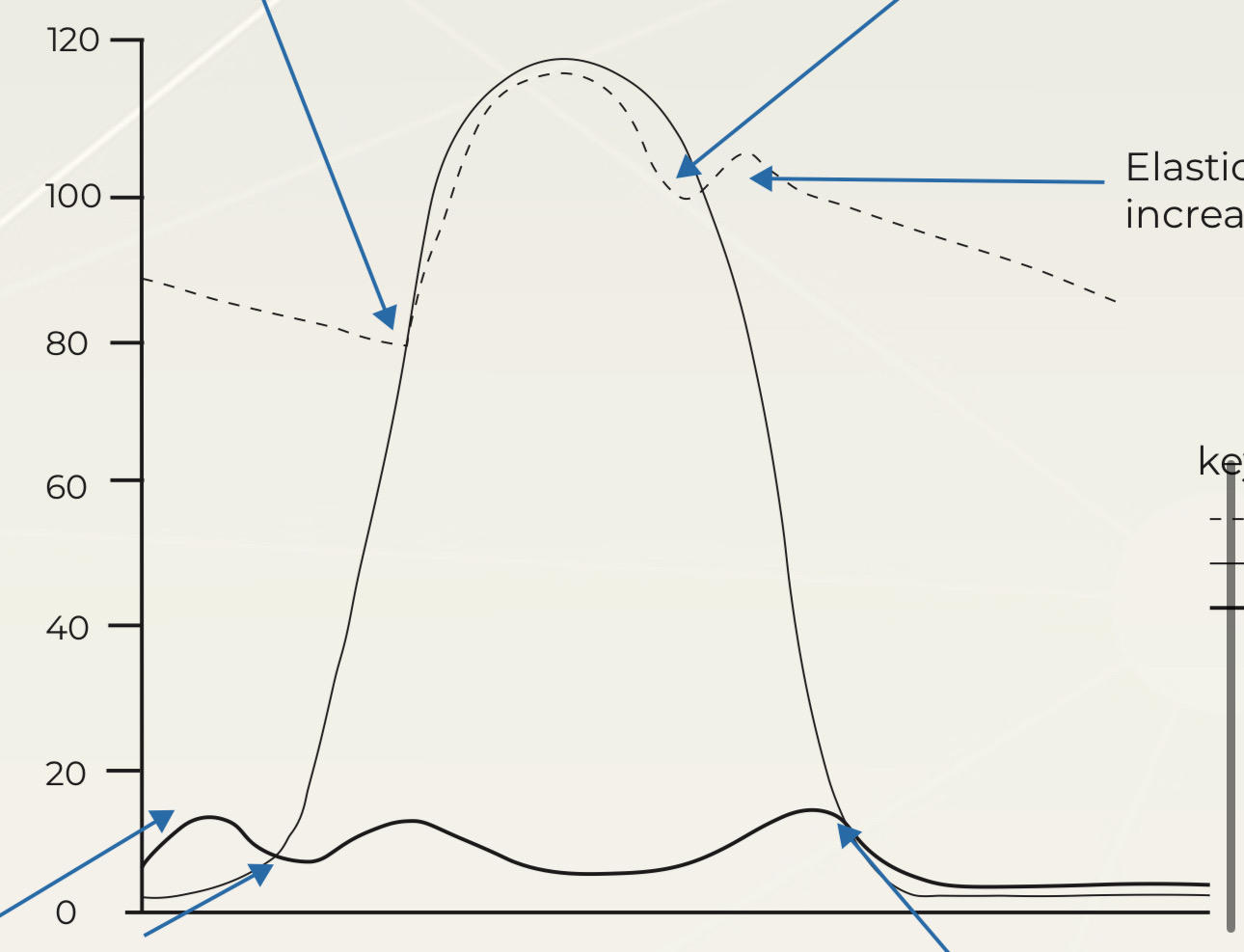
Aorta
57
What is the black thin line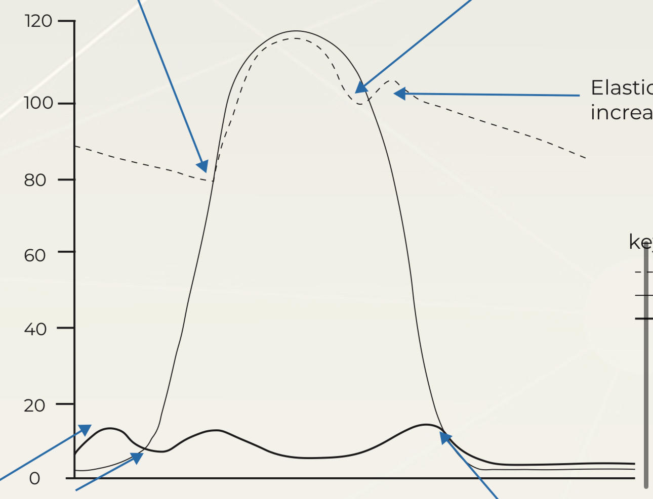
L ventricle
58
What is the black bold line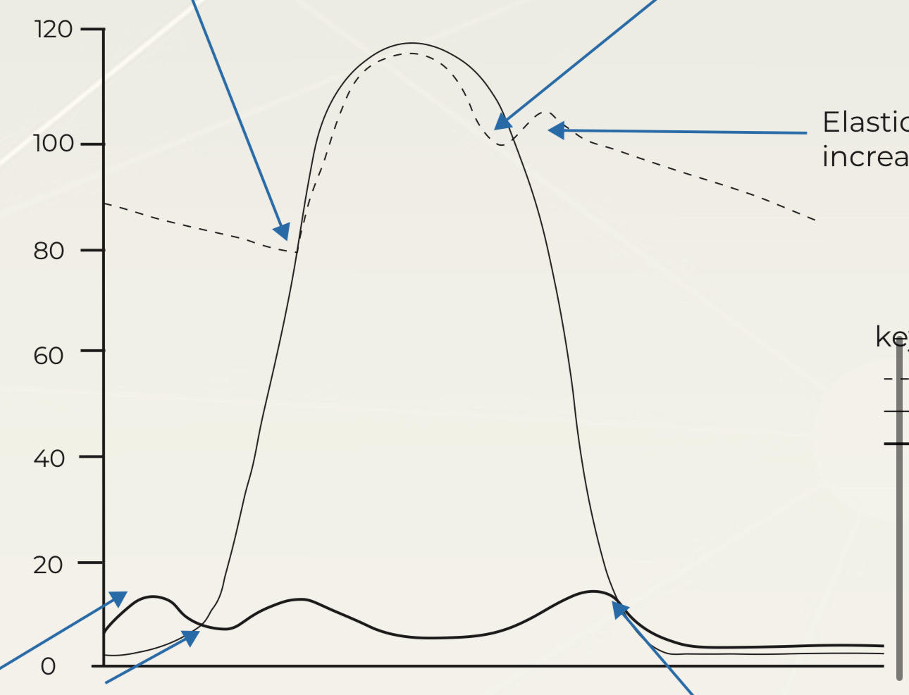
L atrium
59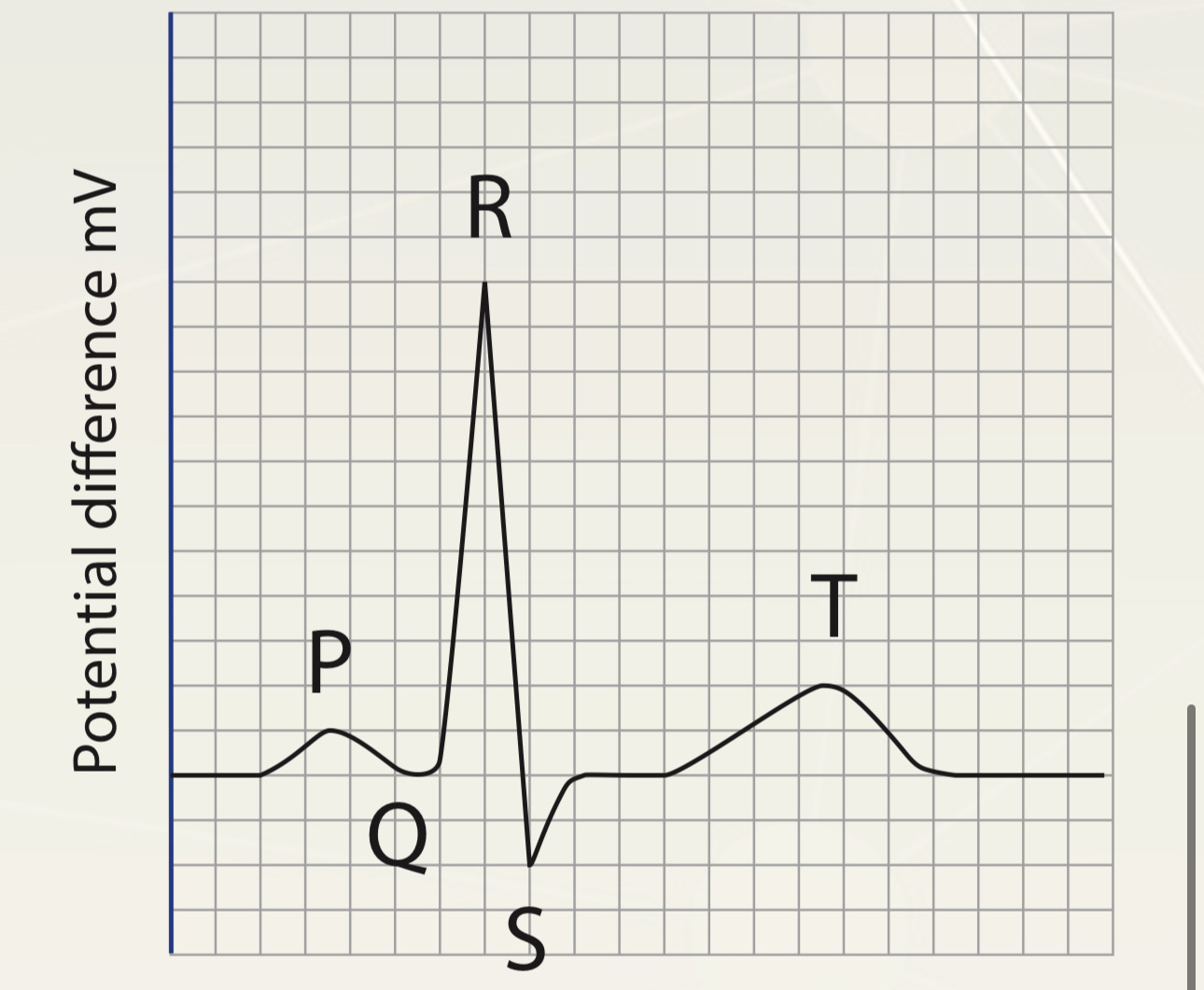
Image occlusion: name the hidden labels
60
Image occlusion: name the hidden labels
61
Image occlusion: name the hidden labels
62
Image occlusion: name the hidden labels
63
Image occlusion: name the hidden labels
64
P wave?
depolarisation of atria
corresponding to atrial systole
corresponding to atrial systole
65
QRS waves?
Spread of depolarisation through ventricles results in ventricular systole
66
T wave?
Repolarisation of ventricles results in ventricular systole
67
How mant O2 can haemoglobin carry? whag does it form
4, oxyhaemoglobin
68
Whats happening in 1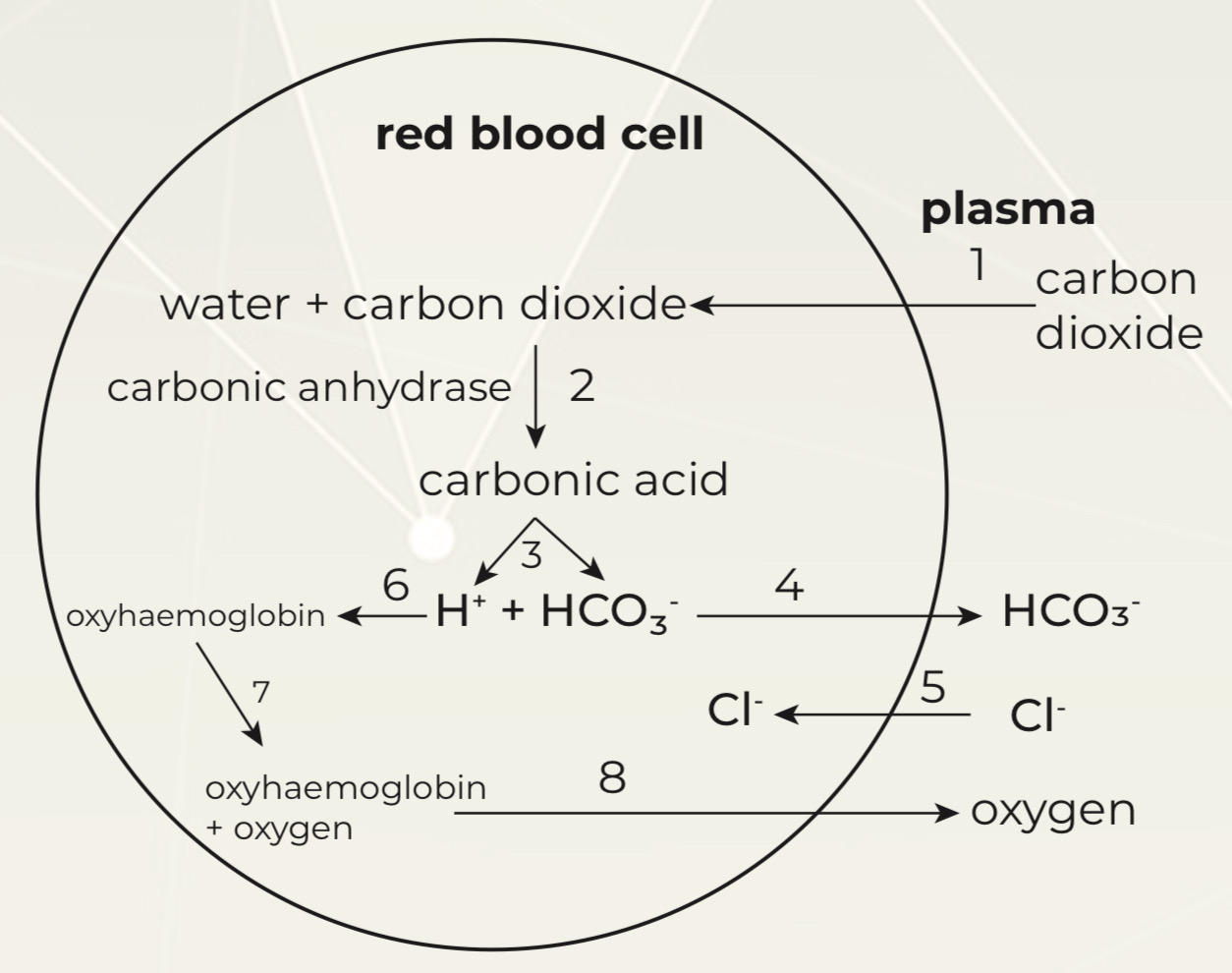
CO2 diffuse into RBC
69
Whats happening in 2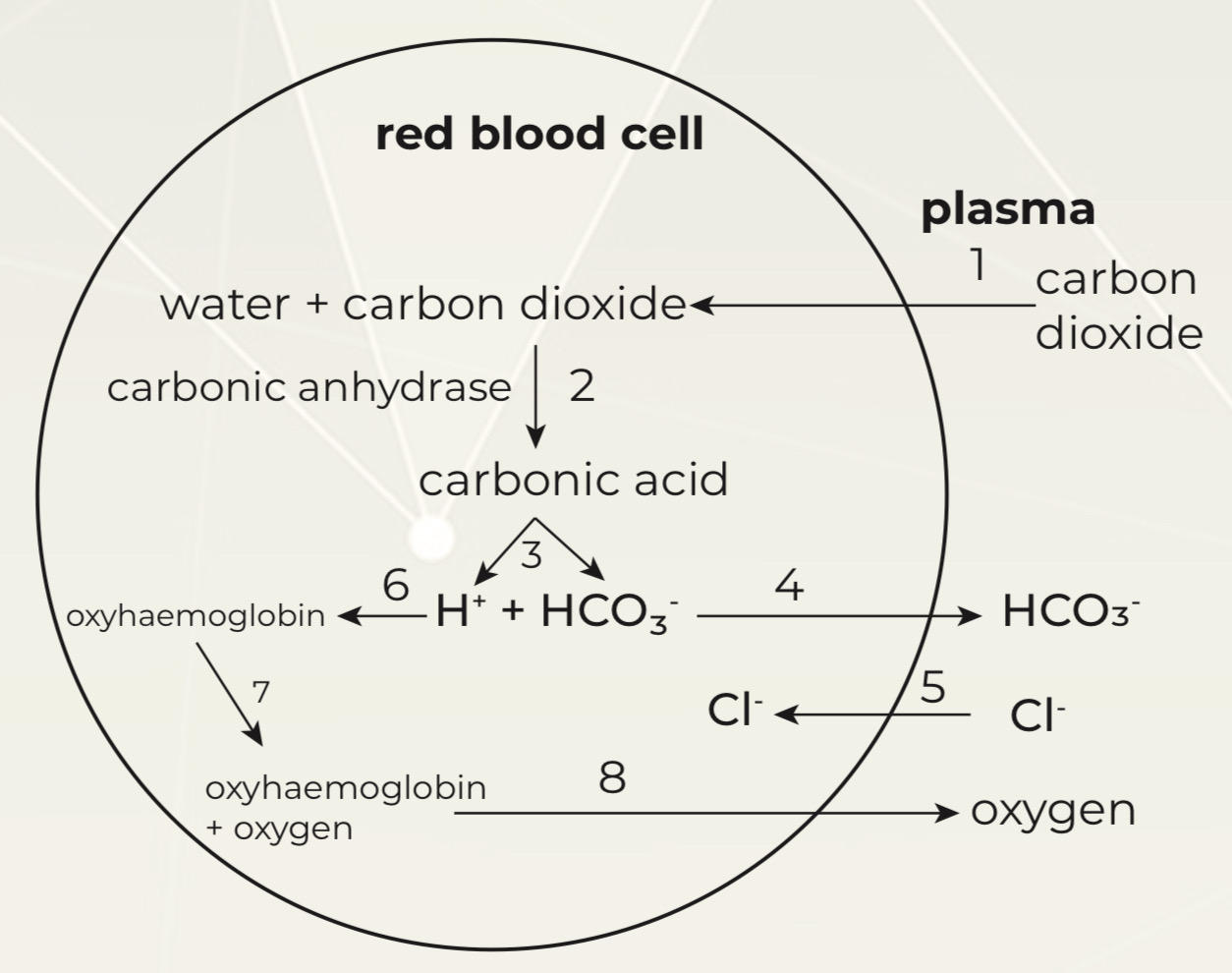
CO2 combine with H2O catalyse by carbonic anhydrase forms carbonic acid
70
Whats happening in 3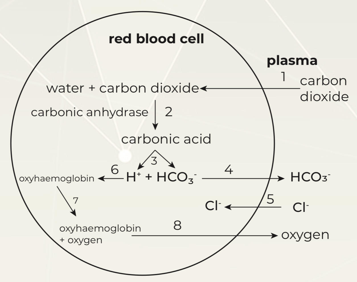
Carbonic acid turn into H ions
H carbonate ions diffuse out cell
H carbonate ions diffuse out cell
71
Whats happening in 4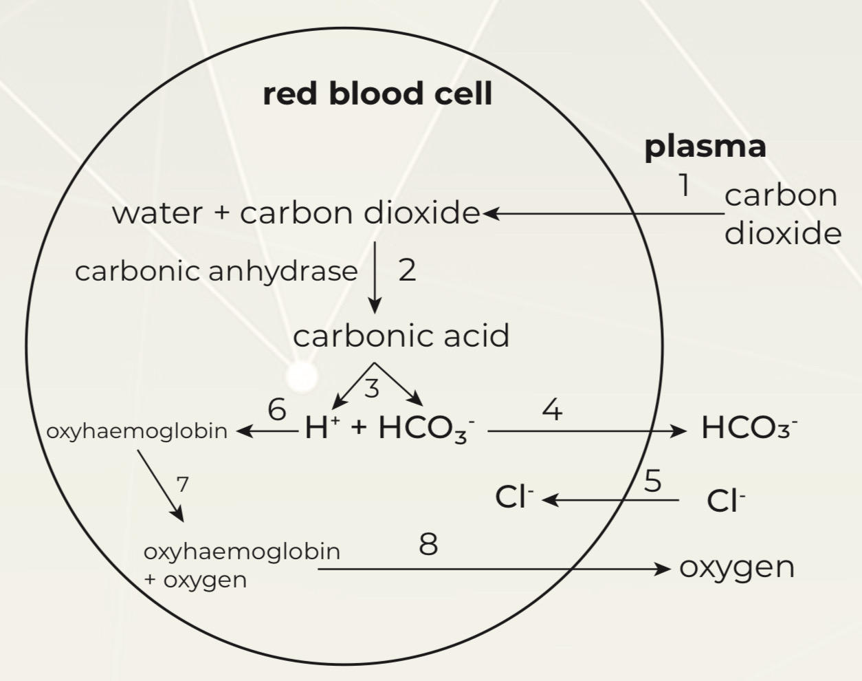
Chloride ions diffuse into rbc to maintain electrochemical neutrality (chloride shift)
72
Whats happening in 5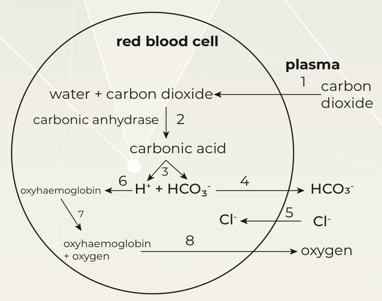
H binds to oxyhaemoglobin to reduce its affinity to O2
The bohr effect
The bohr effect
73
Whats happening in 6 & 7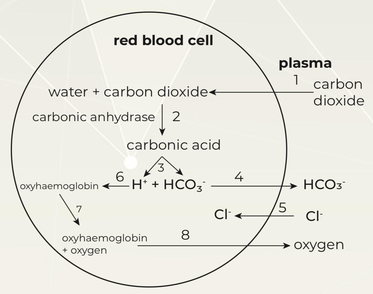
O2 released from haemoglobin (6)
O2 diffuse out of rbc to plasma to body cells (7)
O2 diffuse out of rbc to plasma to body cells (7)
74
What is tissue fluid
Link between blood and cells
surrounds cells and tissues of body
surrounds cells and tissues of body
75
How is tissue fluid formed
arterial end-Hydrostatic pressure higher than osmotic pressure of blood
Water and small mols forced thru capillary walls
Form tissue fluid between cells
Proteins and cells too large to be forced out
Reduced vol of blood and friction, bp falls moves thru capillary
venous end-osmotic pressure higher than hydrostatic pressure
Water from tissue fluid moves back into capillaries / blood by lymph vessels
Water and small mols forced thru capillary walls
Form tissue fluid between cells
Proteins and cells too large to be forced out
Reduced vol of blood and friction, bp falls moves thru capillary
venous end-osmotic pressure higher than hydrostatic pressure
Water from tissue fluid moves back into capillaries / blood by lymph vessels