Cell Structure
Study these flashcardsFree · spaced repetition · iPhone, Android, Mac & web
All cards
1
what are Cells?
Basic blocks of life
2
what does the Cell Membrane do?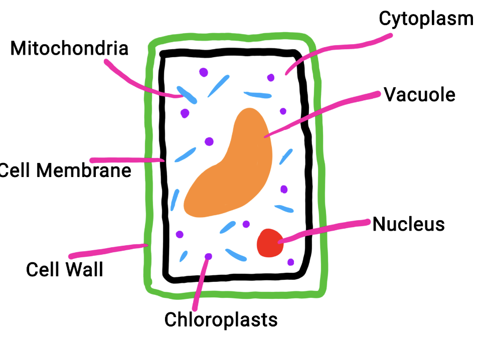
Protects the cell and controlls what gets in and out the cell
3
Which type of cells are the cell wall found in?
Plant Cells and Bacteria Cells
4
what is the chemical that the cell wall is made of? 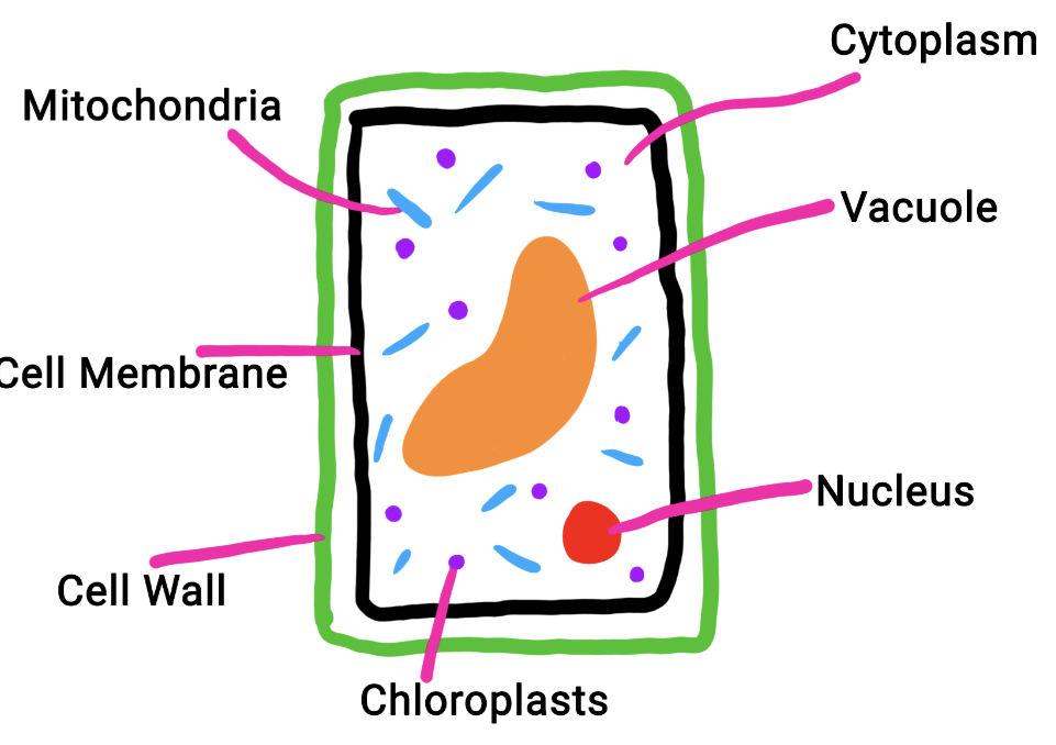
Cellulose
5
Which part of a Plant Cell does photosynthesis take place?
Chloroplast 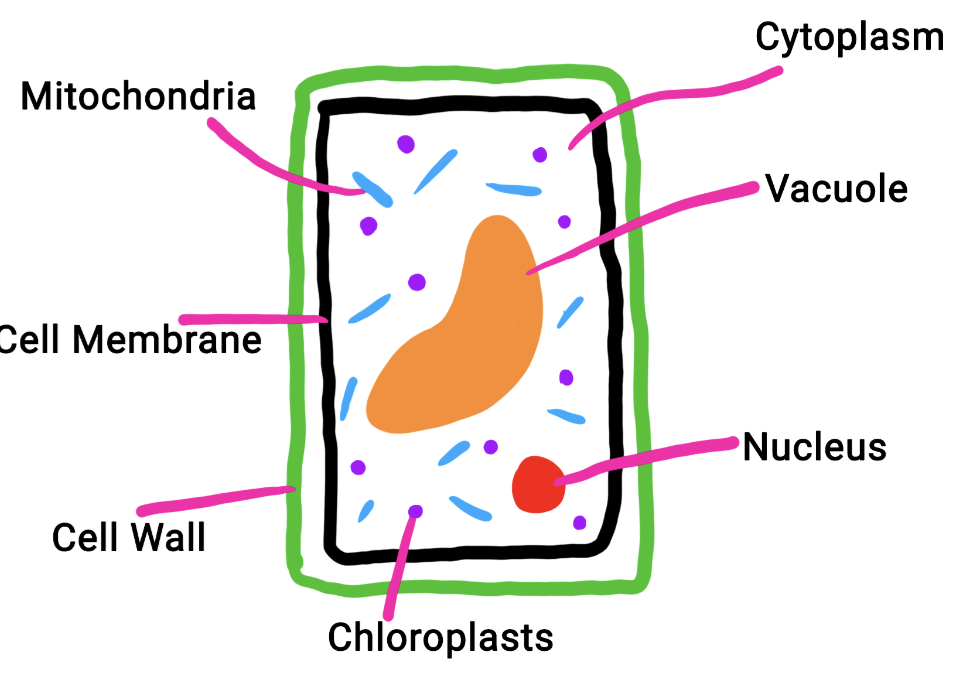
6
In which type of Animal Cells would you find a lot of mitochondria?
Muscle, transfers a lot of energy
7
Name 2 adaptations of the Sperm Cell?
• Tail to swim
•DNA in head
•DNA in head
8
A student prepared a slide of onion epidermis to observe under a microscope.
Describe what is wrong with slide A.
Suggest how this problem can be avoided.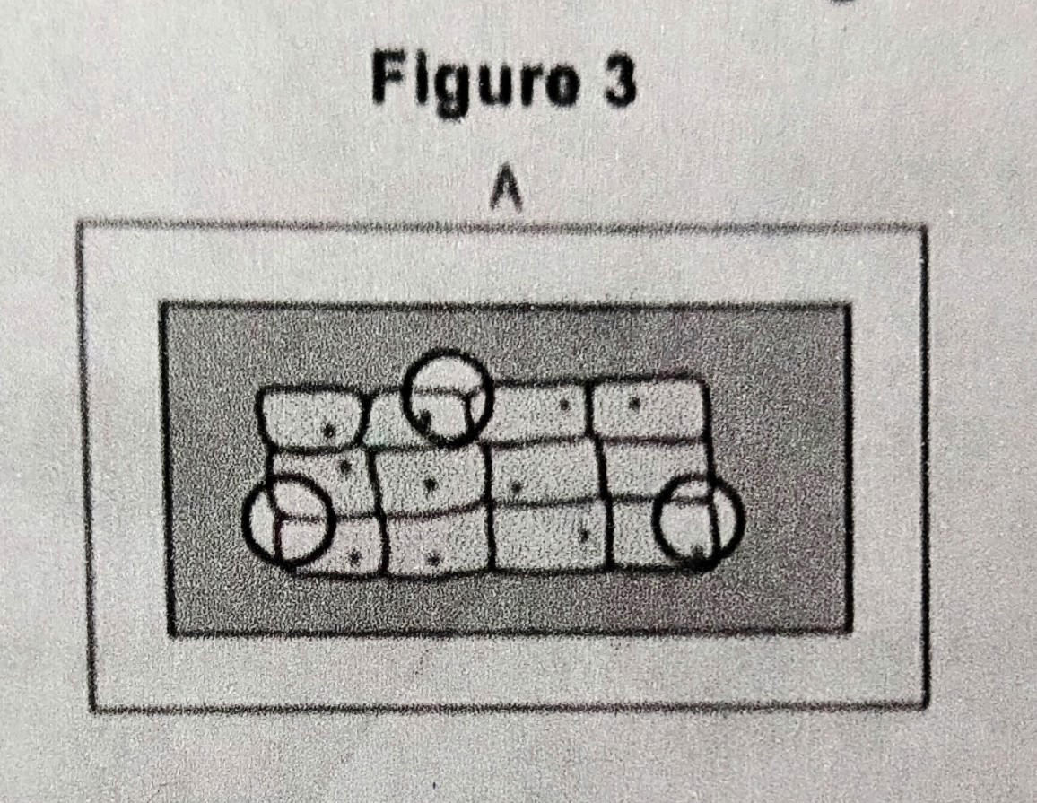
Describe what is wrong with slide A.
Suggest how this problem can be avoided.
Problem: There are air bubbles
Avoid it: Position the Slide carefully with a Mounted Needle to put the cover slip over it.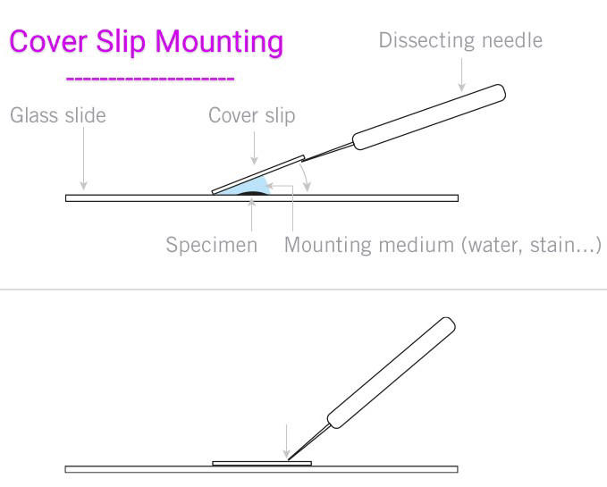
Avoid it: Position the Slide carefully with a Mounted Needle to put the cover slip over it.
9
A student prepared a slide of onion epidermis to observe under a microscope.
Describe what is wrong with slide B.
Suggest how this problem could be corrected.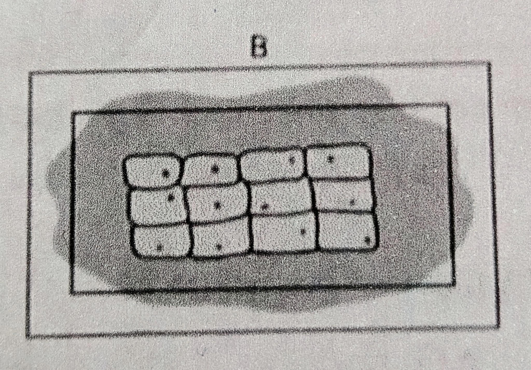
Describe what is wrong with slide B.
Suggest how this problem could be corrected.
Problem: The stain had seeped from the Cover Slip
Fix it: Use filter paper/tissue to soak up the chemical
Fix it: Use filter paper/tissue to soak up the chemical
10
When observing a slide under the microscope you must first use the lower objective lens.
Explain why?
Explain why?
This is to get a bigger field of view
11
What two gases move during Aerobic Respiration?
Oxygen and Carbon Dioxide
12
What does the Nucleus do?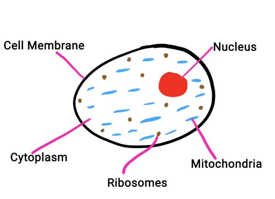
Stores DNA and therefore Controls Cell Function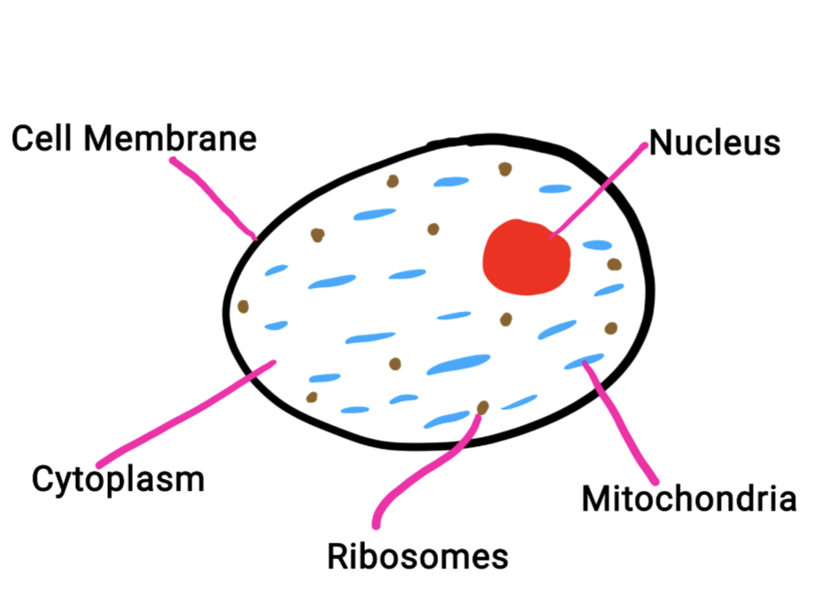
13
What does the Mitochondria do?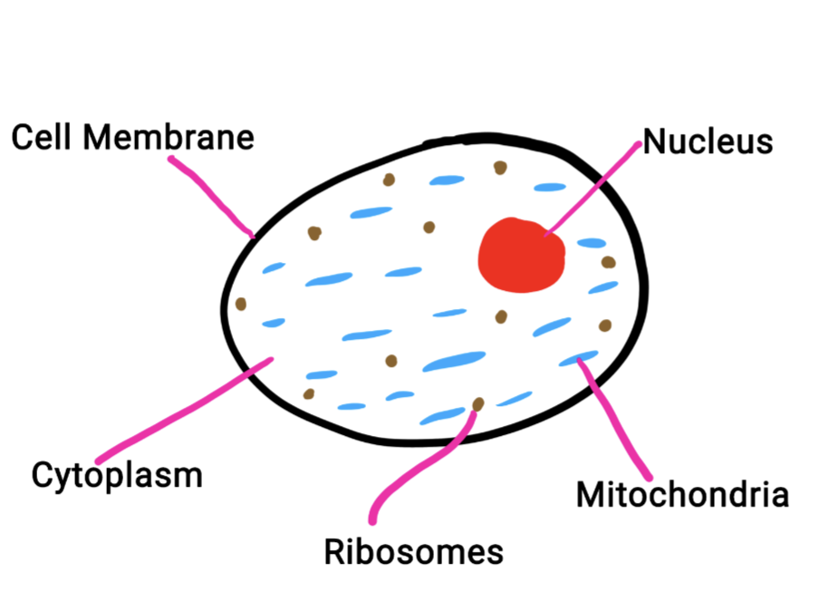
Respiration happens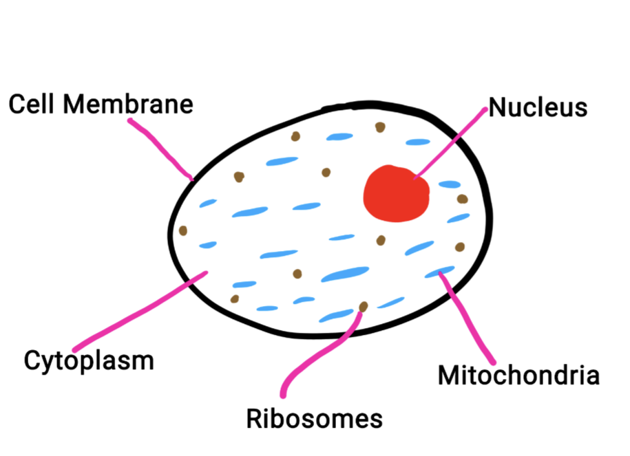
14
What does the Cytoplasm do?
Chemical Reactions are Happening 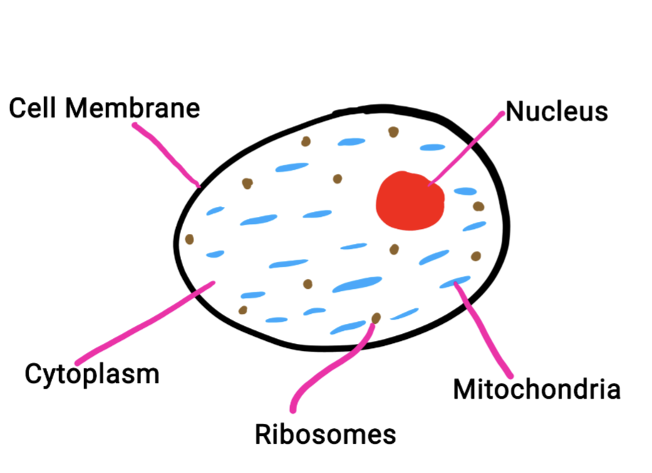
15
What does the Cell Wall do?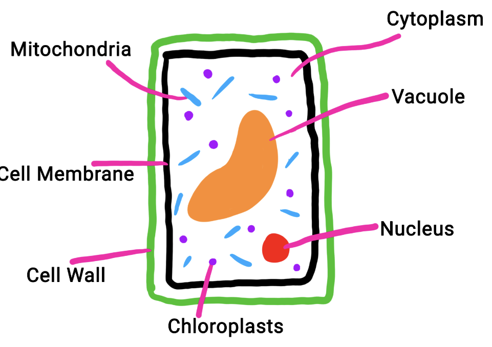
Provides Structure, Protection and Support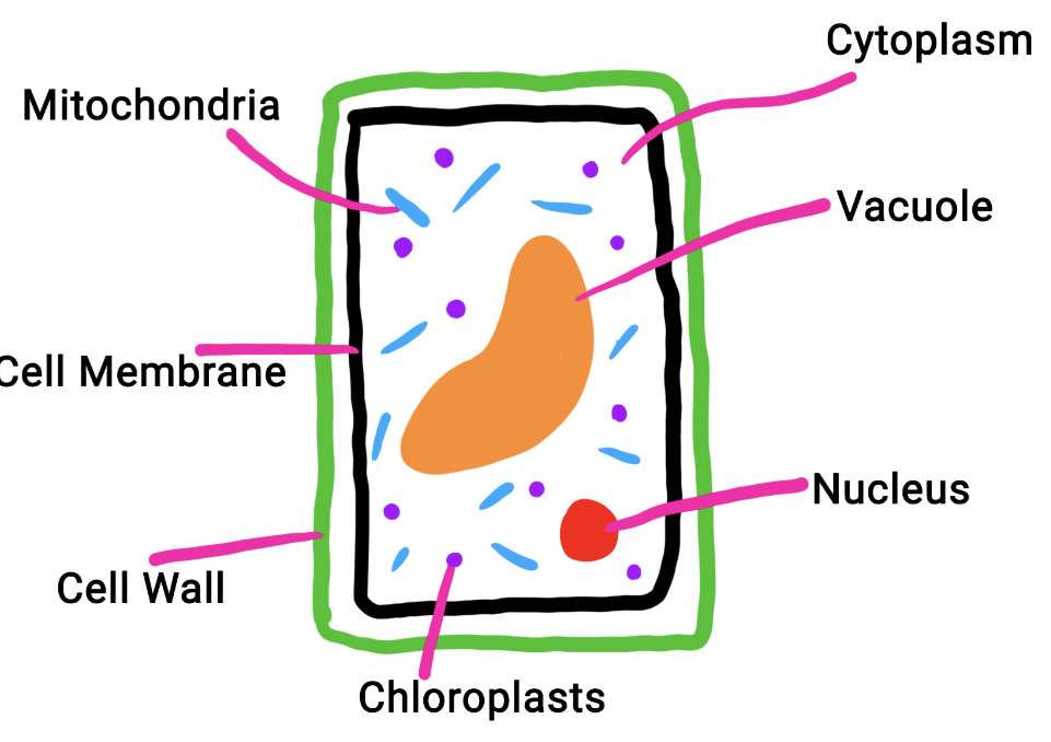
16
What does the Vacuole do?
Contains Cell Sap used for Storage & Keeps Cell Turgid
17
what are Ribosomes ?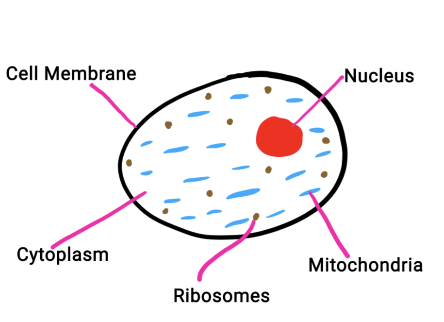
where Protein Synthesis takes place 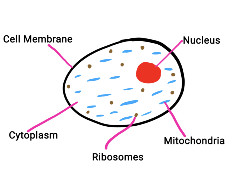
18
what is the equation for magnification of the microscope(for practicals)?
magnification of eyepiece × magnification of objective= magnification of the microscope
19
What is magnification?
How Much a Lens Can Magnify
20
What is resolution?
Measure of Microscope's Ability To Distinguish Between Two Points which are Close Together On an Object
21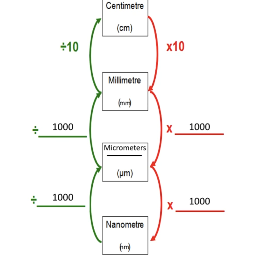
fill in the missing gaps 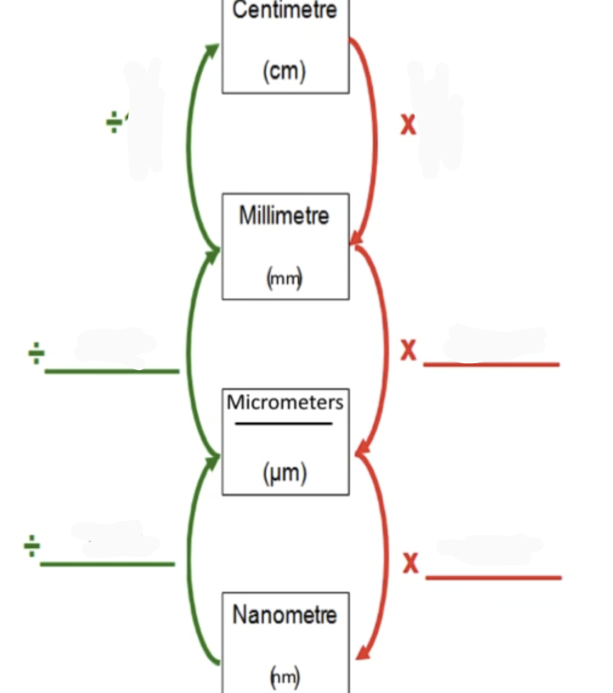
22
What is the equation for magnification?
size of image
--------------
actually size of the object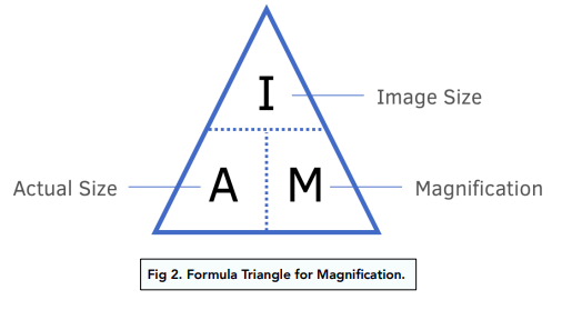
--------------
actually size of the object
23
Properties of Eukaryotic Cells
• Have a Membrane
• Have a Nucleus
• Have a Mitochondria and a Chloroplast
• They include Animals, Plants and Fungi
• Have a Nucleus
• Have a Mitochondria and a Chloroplast
• They include Animals, Plants and Fungi
24
Properties of Prokaryotic Cells
• No Nucleus
• Not Membrane-bound
• Mainly Unicellular Organisms
(1 cell)
• Less complex than Eukaryotic Cells
• Components Float Freely Within Cytoplasm
• The include Bacteria Cell and Archae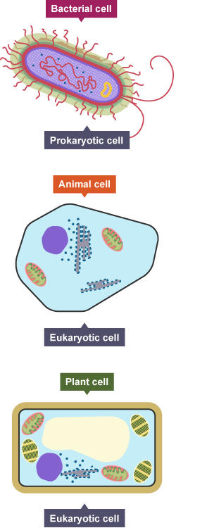
• Not Membrane-bound
• Mainly Unicellular Organisms
(1 cell)
• Less complex than Eukaryotic Cells
• Components Float Freely Within Cytoplasm
• The include Bacteria Cell and Archae
25
What is a Specialised Cell?
Cell has Special Shape and Features that Help It Do Its Job
26
What is special about the Ciliated Cell?
what is the function of the cell?
where is it found?
what is the function of the cell?
where is it found?
CILIATED CELLS
Specialisation: Ciliated Cells have Cillia (tiny hairs).
Function: to Prevent Lung Damage.
Where: Found in Lungs.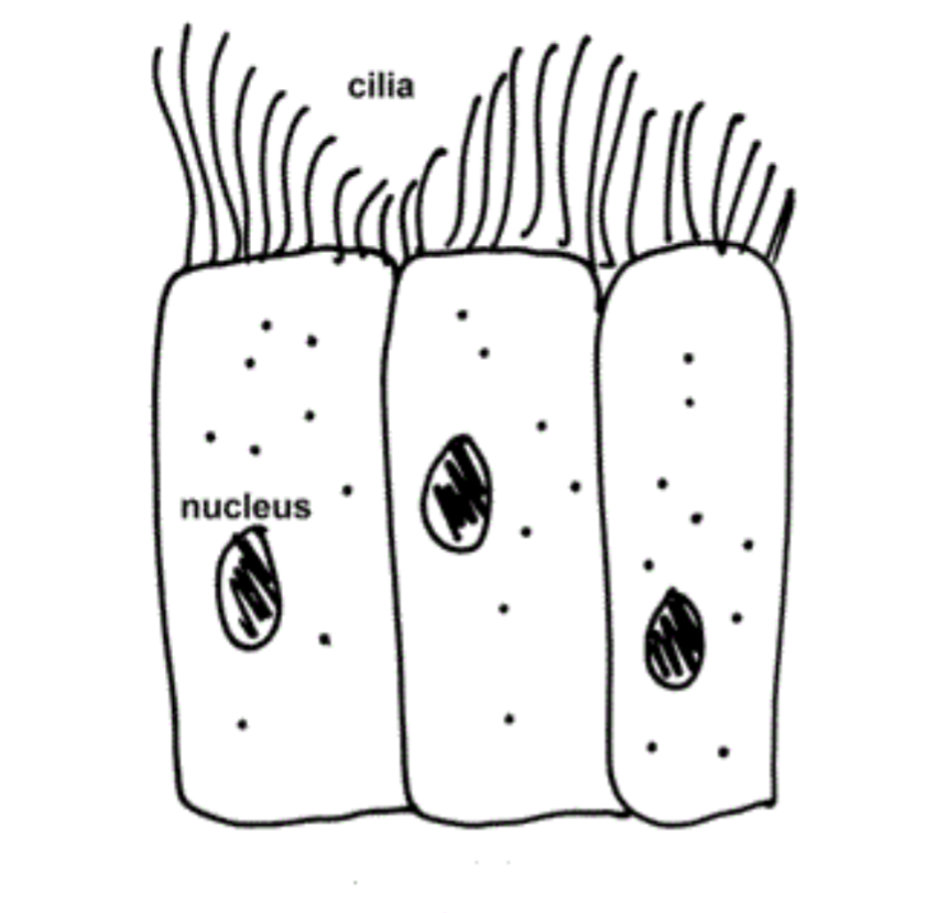
Specialisation: Ciliated Cells have Cillia (tiny hairs).
Function: to Prevent Lung Damage.
Where: Found in Lungs.
27
What is special about the Root Hair Cell?
what is the function of the cell?
where is it found?
what is the function of the cell?
where is it found?
ROOT HAIR CELL
Specialisation - Large Surface Area with Thin Cell Wall and a Large Vacuole For Turdgity. No Chloroplast
Function - Absorbs Mineral Ions and Water By Osmosis
Where - the Roots (plant)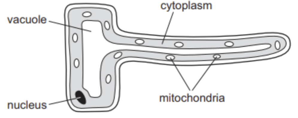
Specialisation - Large Surface Area with Thin Cell Wall and a Large Vacuole For Turdgity. No Chloroplast
Function - Absorbs Mineral Ions and Water By Osmosis
Where - the Roots (plant)
28
What is special about the Red Blood Cell?
what is the function of the cell?
where is it found?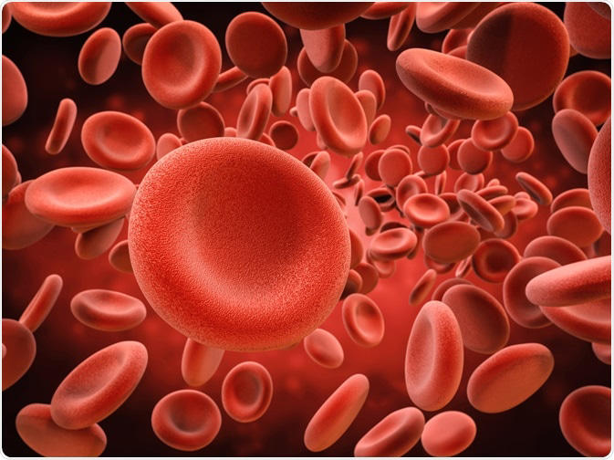
what is the function of the cell?
where is it found?
RED BLOOD CELL
Specialisation - Large Surface Area, No Nucleus, Lots Of Haemoglobin.
Function - Transport Oxygen and Carbon Dioxide Around the Body.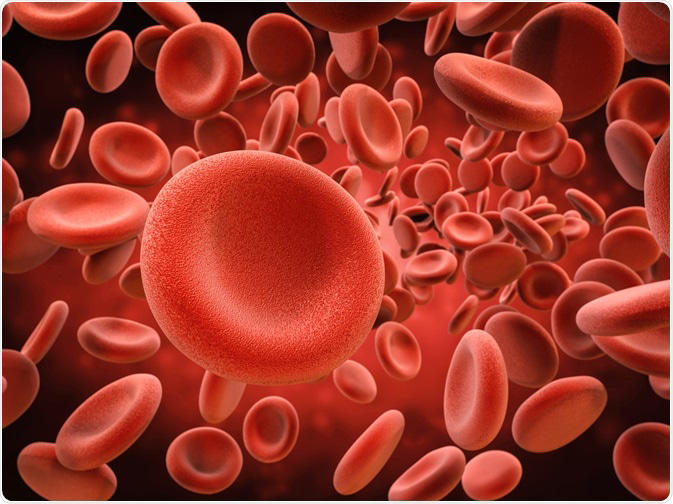
Specialisation - Large Surface Area, No Nucleus, Lots Of Haemoglobin.
Function - Transport Oxygen and Carbon Dioxide Around the Body.
29
What is special about the Sperm Cell?
what is the function of the cell?
where is it found?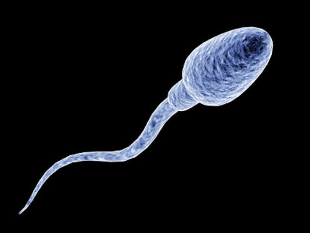
what is the function of the cell?
where is it found?
SPERM CELL
Specialisation - Tail For Swimming, Acrosome & (enzyme) DNA in Head to Enter Egg, Mitochondria In Middle For Energy.
Function - Carry Father's Genetic Information
Where - Testes
Specialisation - Tail For Swimming, Acrosome & (enzyme) DNA in Head to Enter Egg, Mitochondria In Middle For Energy.
Function - Carry Father's Genetic Information
Where - Testes
30
What is special about the Egg Cell?
what is the function of the cell?
where is it found?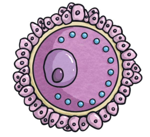
what is the function of the cell?
where is it found?
EGG CELL
Specialisation - Larger Than Any Other Animal Cell, Holds Energy Source For Embryo
Function - Holds Mothers Genetic Information
Where - Ovaries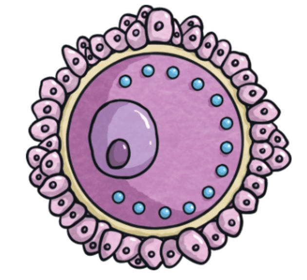
Specialisation - Larger Than Any Other Animal Cell, Holds Energy Source For Embryo
Function - Holds Mothers Genetic Information
Where - Ovaries
31
What is special about the Palisade Cell?
what is the function of the cell?
where is it found?
what is the function of the cell?
where is it found?
PALISADE CELL
Specialisation - Tall and Large Surface Area for More Sunlight. Lots of Chloroplast for Absorption of Sunlight.
Function - Photosynthesis
Where - Upper Side Of Leaf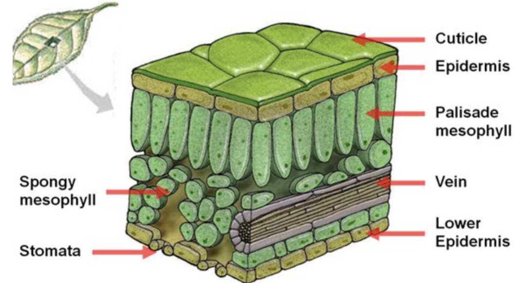
Specialisation - Tall and Large Surface Area for More Sunlight. Lots of Chloroplast for Absorption of Sunlight.
Function - Photosynthesis
Where - Upper Side Of Leaf
32
What is special about the Nerve Cell?
what is the function of the cell?
what is the function of the cell?
NERVE CELL
Specialisation - Long Cells with Branches at Ends so can Connect.
Axon is Middle Bit.
Covered by Myelin Sheath for Insulation.
Function - Transfer Nerve Impulses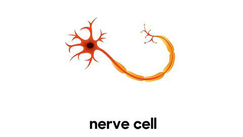
Specialisation - Long Cells with Branches at Ends so can Connect.
Axon is Middle Bit.
Covered by Myelin Sheath for Insulation.
Function - Transfer Nerve Impulses
33
What is special about the White Blood Cell?
what is the function of the cell?
where is it found?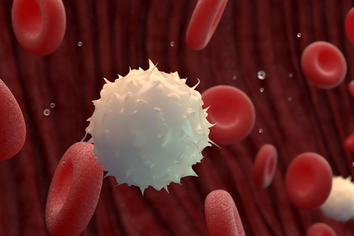
what is the function of the cell?
where is it found?
WHITE BLOOD CELLS
Function - Fight Pathogens By Engulfing(eat) or Releasing Antibodies. Part of Immune System.
Where - (usually Blood Vessel)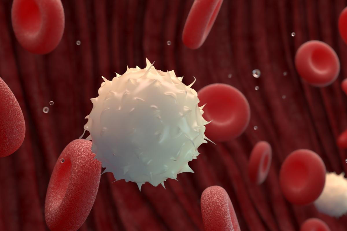
Function - Fight Pathogens By Engulfing(eat) or Releasing Antibodies. Part of Immune System.
Where - (usually Blood Vessel)
34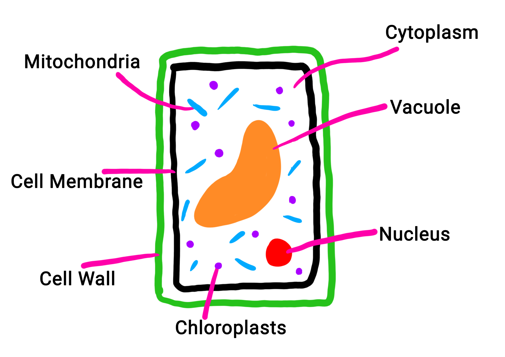
label the Plant Cell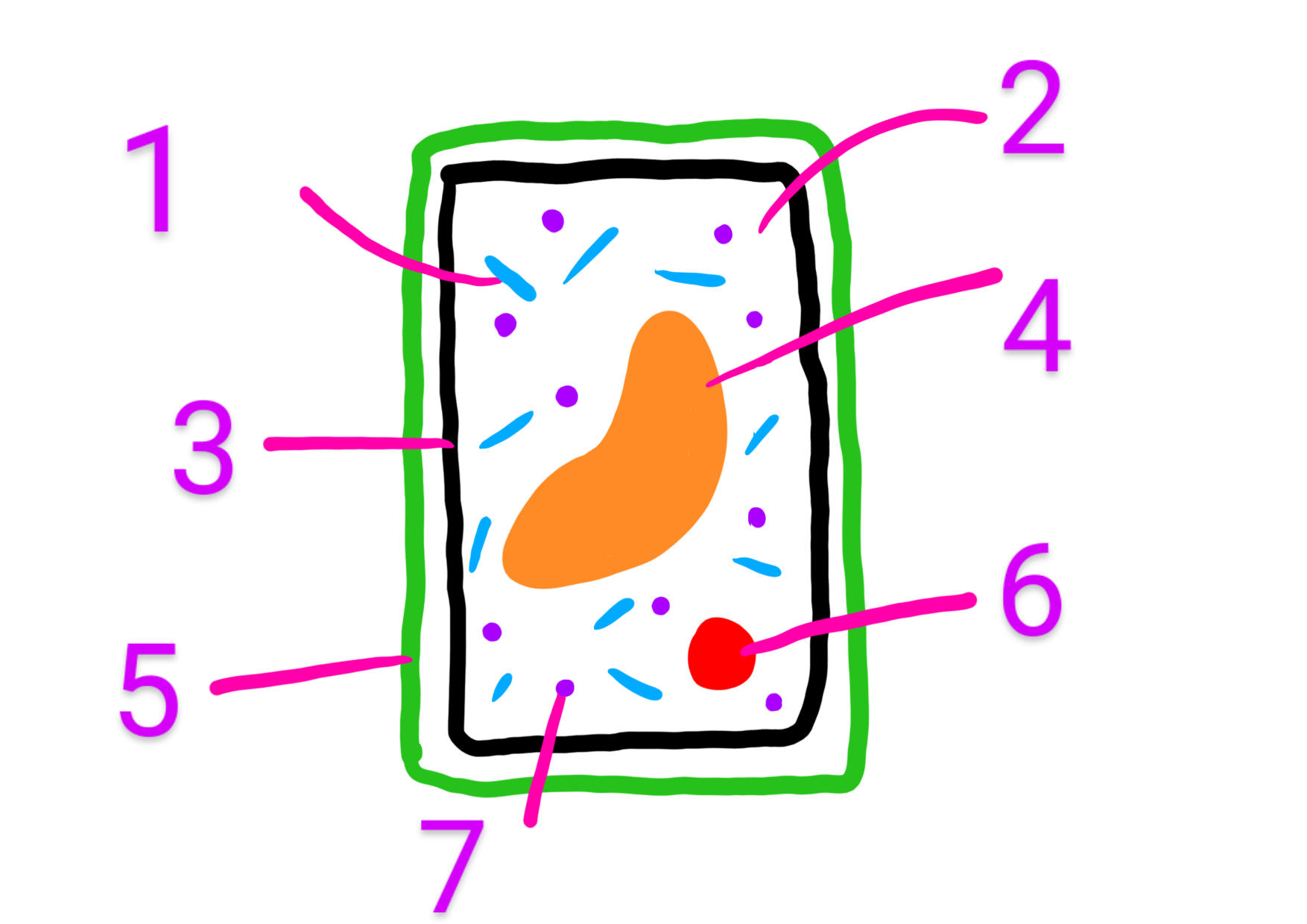
35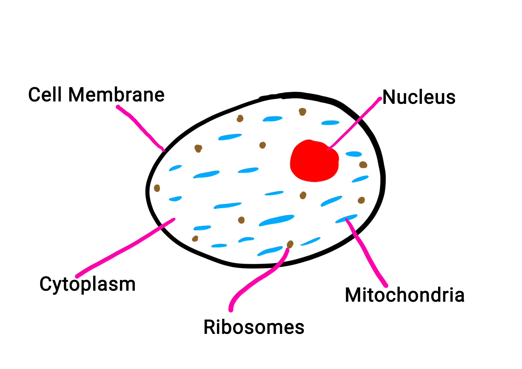
label the Animal Cell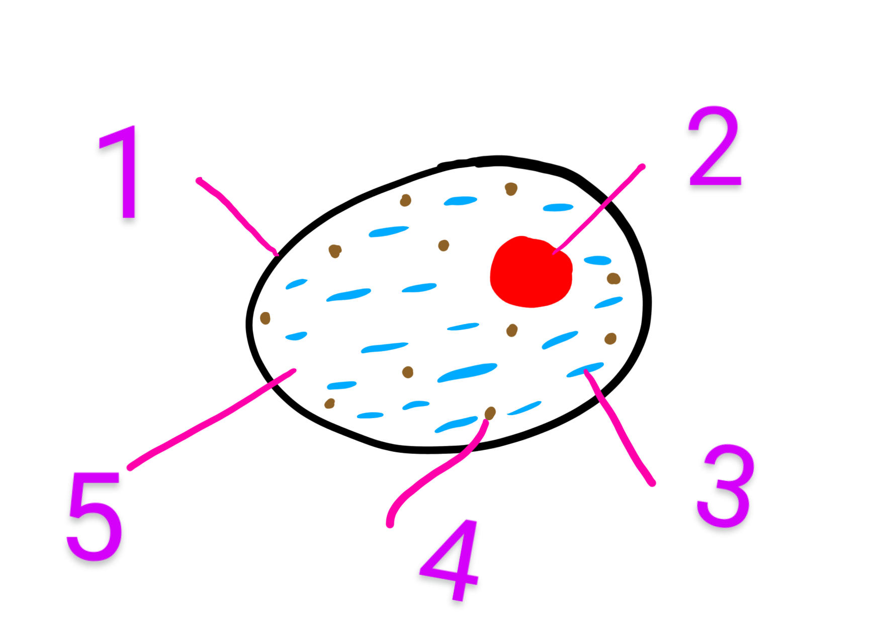
36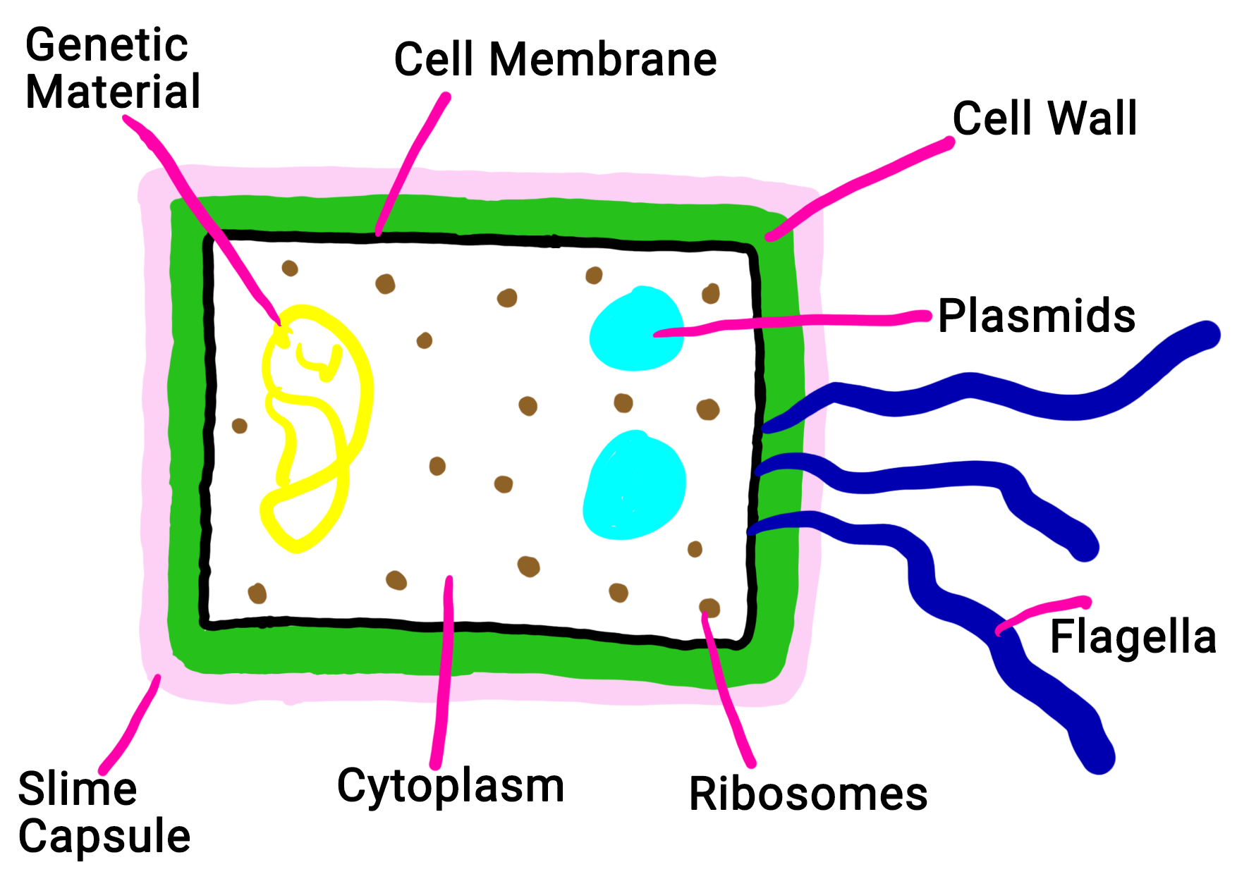
label the Bacterial Cell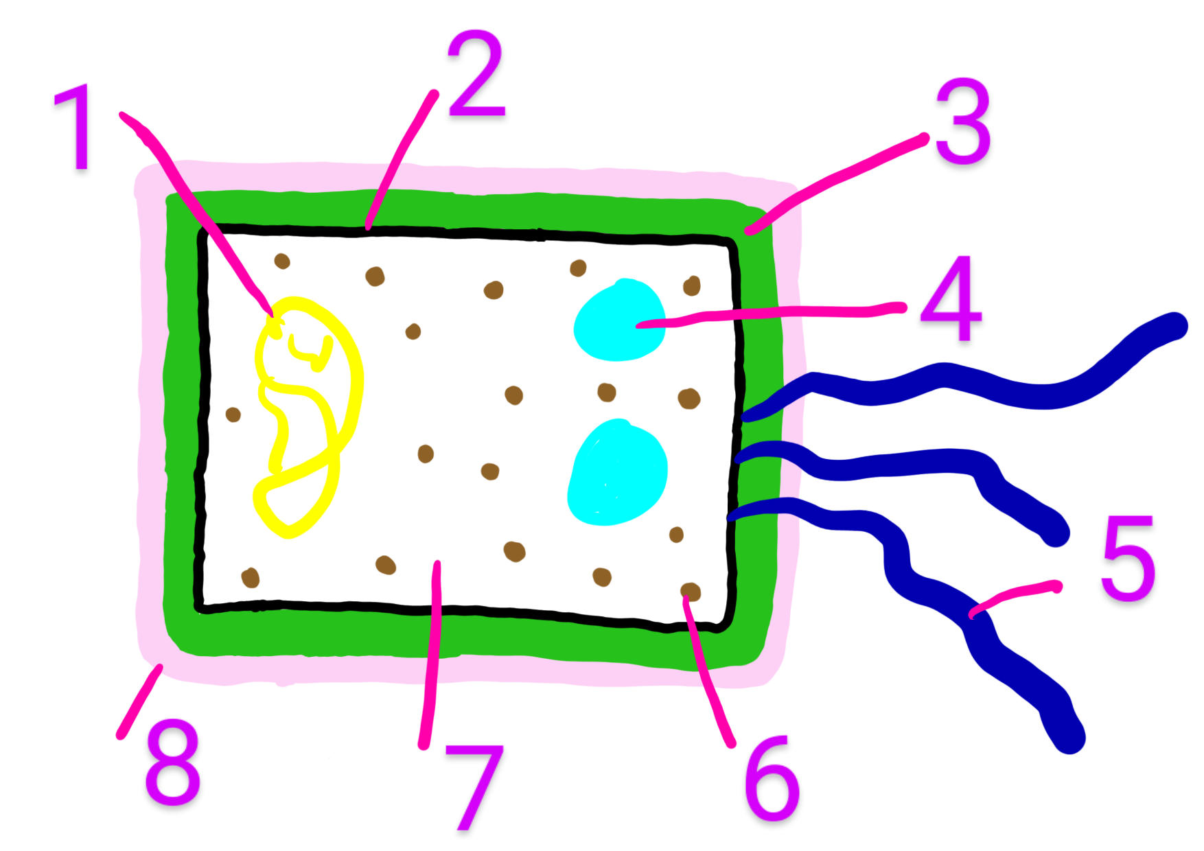
37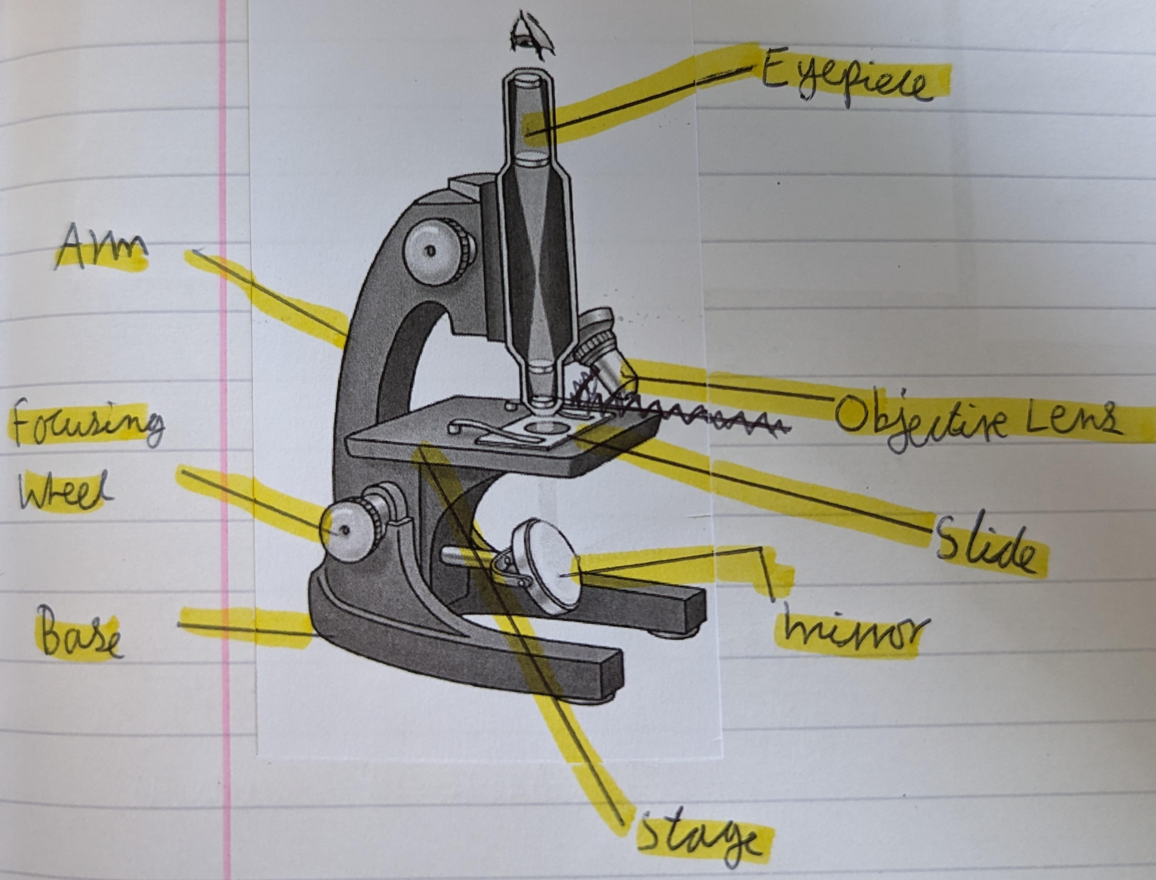
label the Microscope 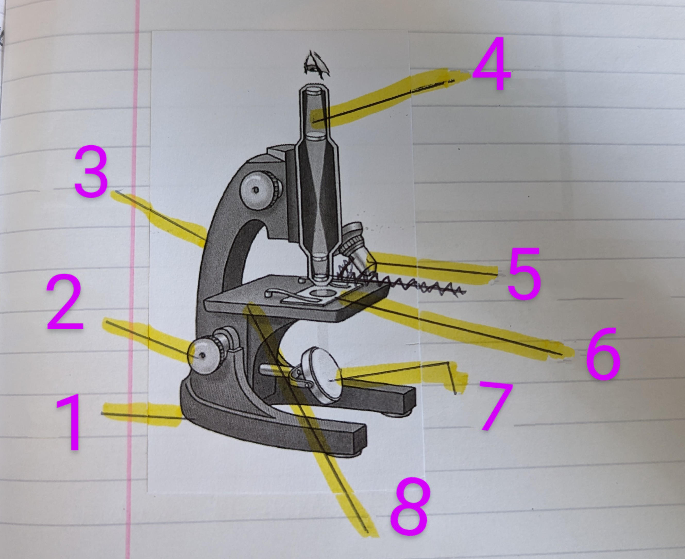
38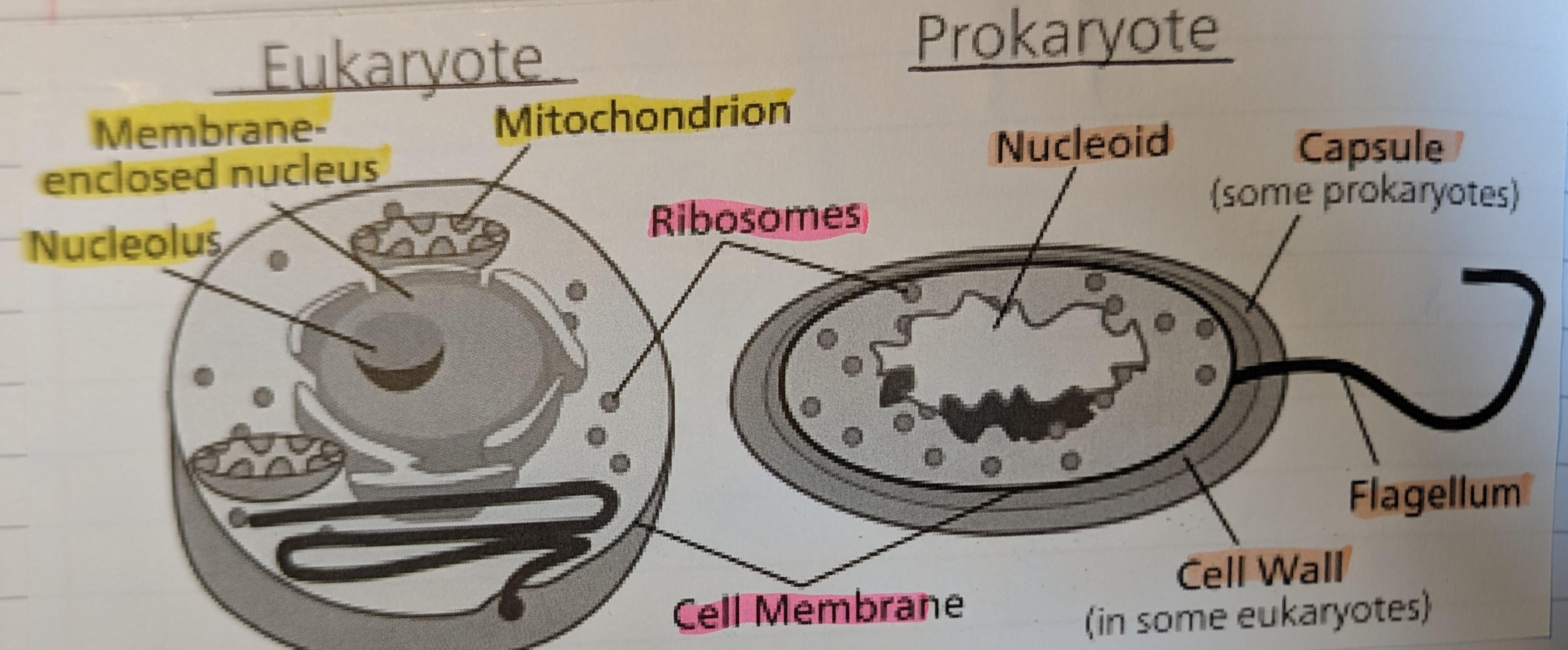
label the Eukaryotic and Prokaryotic Cell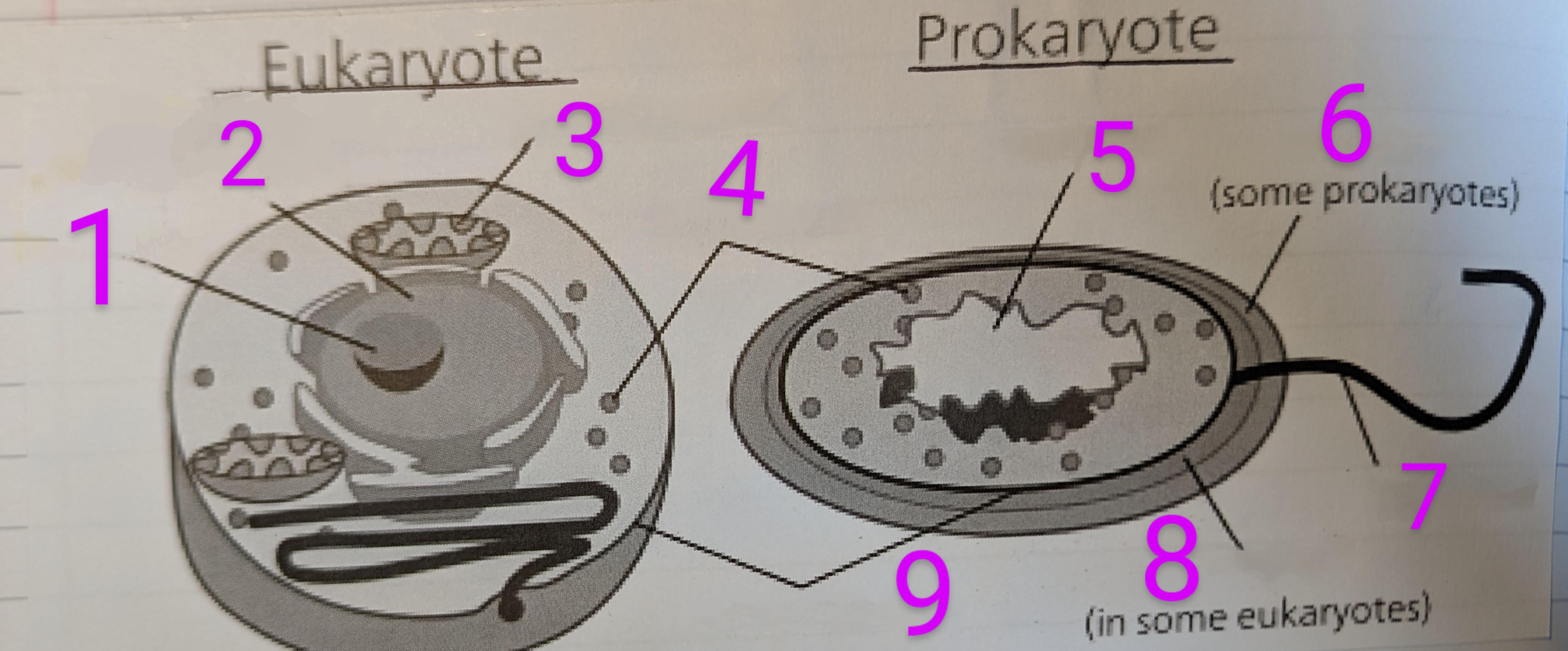
39
Where are Prokaryotic Cells found in?
Bacteria
40
Where are Eukaryotic Cells found in?
•Protista
•Fungi
•Plants
•Animals
•Fungi
•Plants
•Animals
41
What are the Propertie a cell membrane?
Cell membrane are Partially Permeable
42
what is Osmosis?
Movement of Water Particles From a High Concentration to Low Concentration Through Partly Permeable Membrane.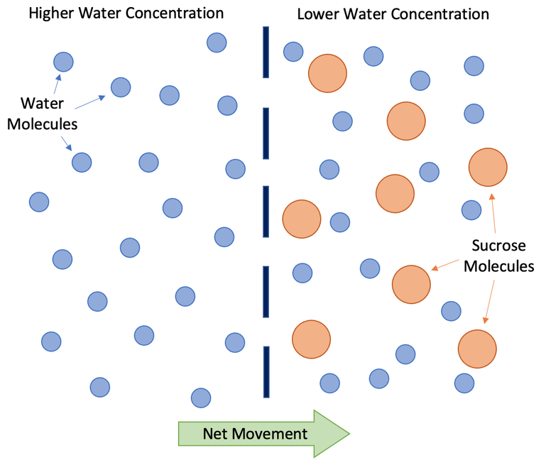
43
how many water molecules are in a Dilute Solution (high or low)
Dilute solutions have a high concentration of water molecules
44
how many water molecules are in a concentrated solution (high or low)
Concentrated solutions have a low concentration of water molecules
45
Define Diffusion
Movement of a Substance from a High Concentration To a Low Concentration.
46
What does the rate of diffusion depend on?
• Distance the Particles Have Travelled
• Difference in Concentration
• Difference in Concentration
47
Define Active Transport
Substances have Transported without Energy From Low Concentration to High Concentration - Against a Concentration Gradient.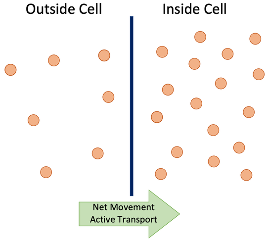
48
where are villi?
They are in the Small Intestine
49
why are villi good for diffusion?
• Lots Of Them so Large Surface Area.
• Good Blood Supply to Maintain the Concentration Gradient.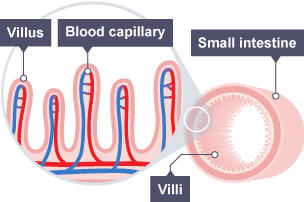
• Good Blood Supply to Maintain the Concentration Gradient.
50
how and why are fish gills adapted for diffusion?
• Large surface area Each Gill Has Lots Of Filaments which have Lamellae at 90° to Catch More Water.
• Lamellae have Good Blood Supply to Maintain Concentration Gradient.
• Constantly Ventilated by Water which Maintains a Concentration Gradient.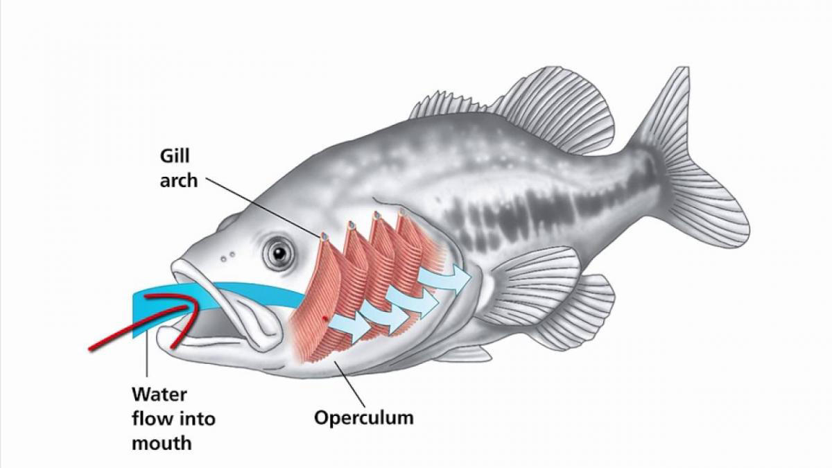
• Lamellae have Good Blood Supply to Maintain Concentration Gradient.
• Constantly Ventilated by Water which Maintains a Concentration Gradient.
51
how are root hair cells adapted for diffusion?
Large surface area because there are lots of them and they are thin and flat.
52
what is the process of gas exchange of an alveoli
Deoxygenated Blood Enters Capillaries.
Oxygen Moves Out of Alveoli and Carbon Dioxide Moves Into the Alveoli.
Oxygenated Blood Leaves by Capillaries.
Oxygen is Transported By Red Blood Cells.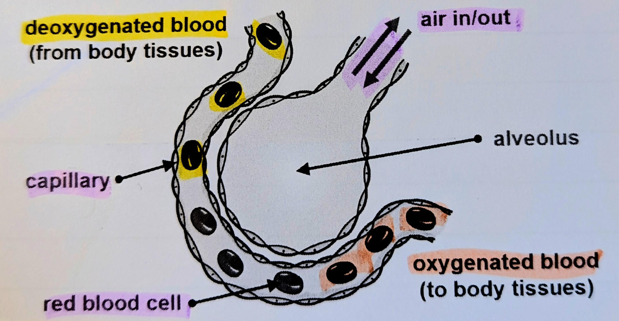
Oxygen Moves Out of Alveoli and Carbon Dioxide Moves Into the Alveoli.
Oxygenated Blood Leaves by Capillaries.
Oxygen is Transported By Red Blood Cells.
53
Where is the Alveoli?
End of the Bronchioles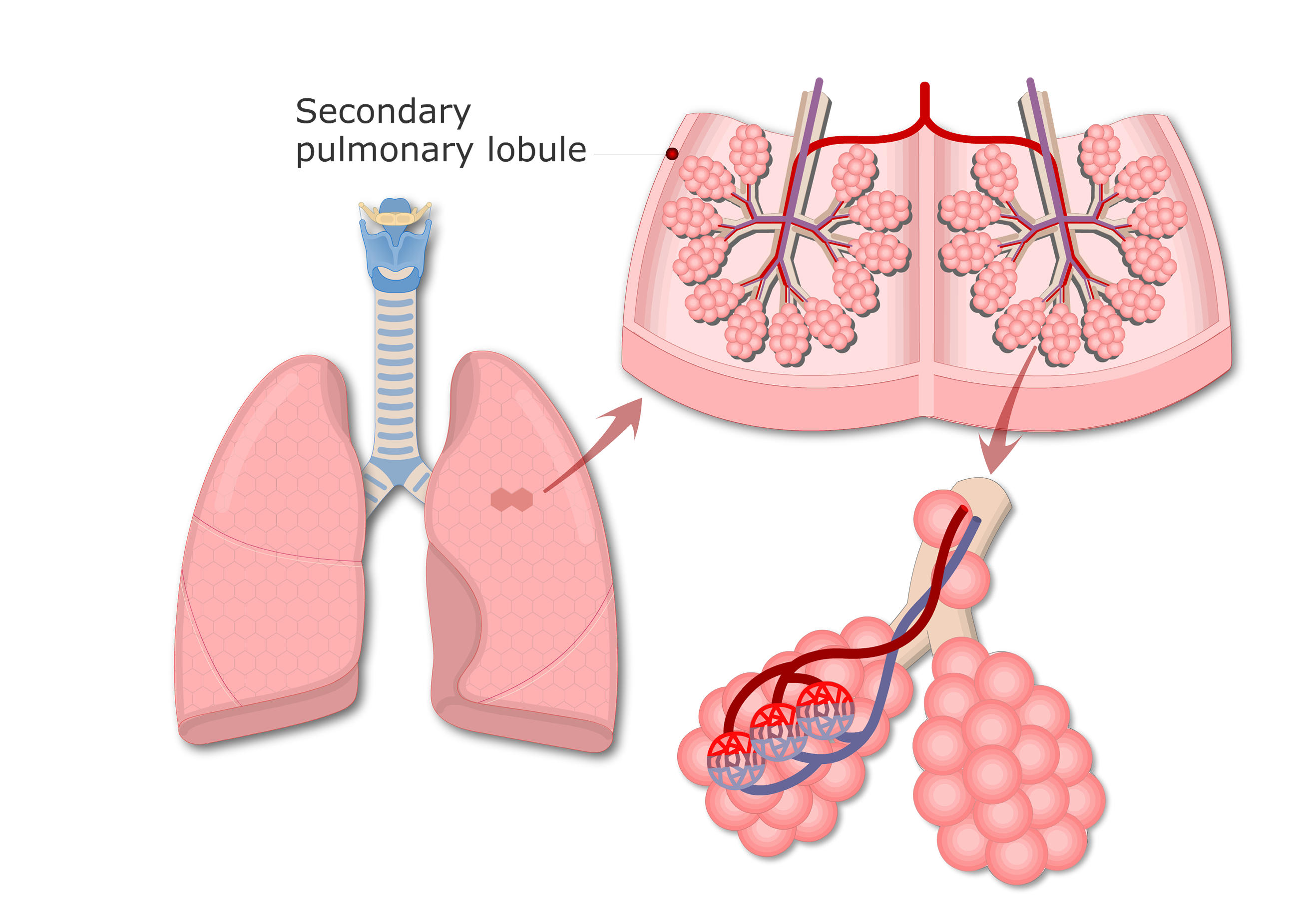
54
What are the adaptations of Alveoli?
• Very Thin Walls
• Covered by a Network Of Capillaries
• Moist
• Large Combined Surface Area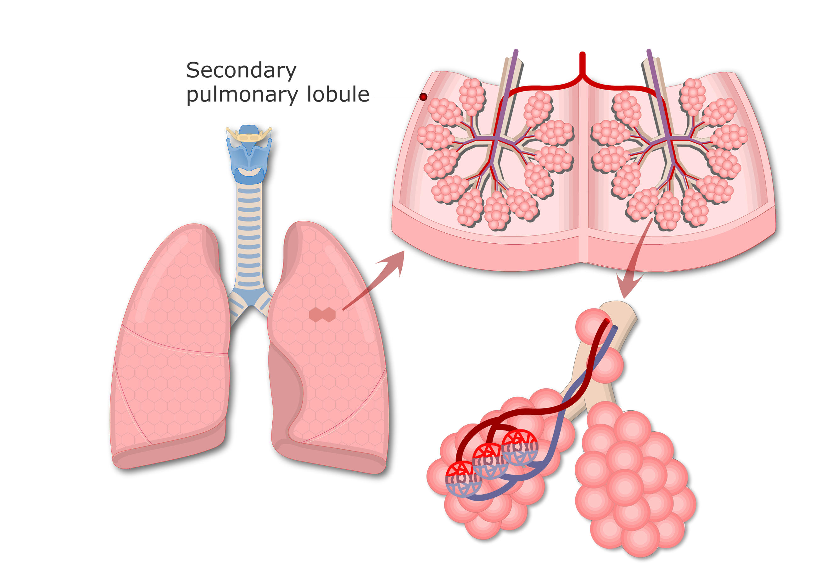
• Covered by a Network Of Capillaries
• Moist
• Large Combined Surface Area
55
Define Concentrated Solution
A solution that contains a large amount of solute.
56
Define concentration
A Measure of the Amount of Solute In A Solution.
57
Define Concentration Gradient
Difference in Concentration Between Two Solutions On Either Side Of a Membrane. 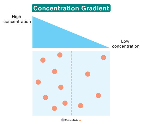
58
Define Partially permeable memebrane
Membrane that Allows Small Molecules to Pass Across it but Not Allow the Passage of large Molecules. 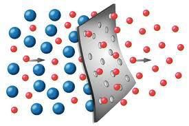
59
REQUIRED PRACTICAL 1 Microscopy
REQUIRED PRACTICAL 1 Microscopy
Method
1. Peel off an Epidermal Layer On Onion Using Forceps.
2. Mount Onto Slide With a Drop Of Water Using Pipette
3. Add 2 drops of Iodine Solution to Stain Cells.
4. Place Cover Slip On By Using Forceps at an Angle to Make Sure No Air Bubbles Trapped.
5. Remove Any Excess Stain With Paper Towel
6. Place on Stage & Select Low Power Objective Look Through Eye Piece & Turn Fine Adjustment Knob Until the Image Comes into Focus. .
7. When Selecting Higher Power Objective Adjust Stage & Turn Knob to Move Stage Away
8. Make a Labelled Drawing of Cells You can See & Write down the magnification
13. Repeat using a prepared slide.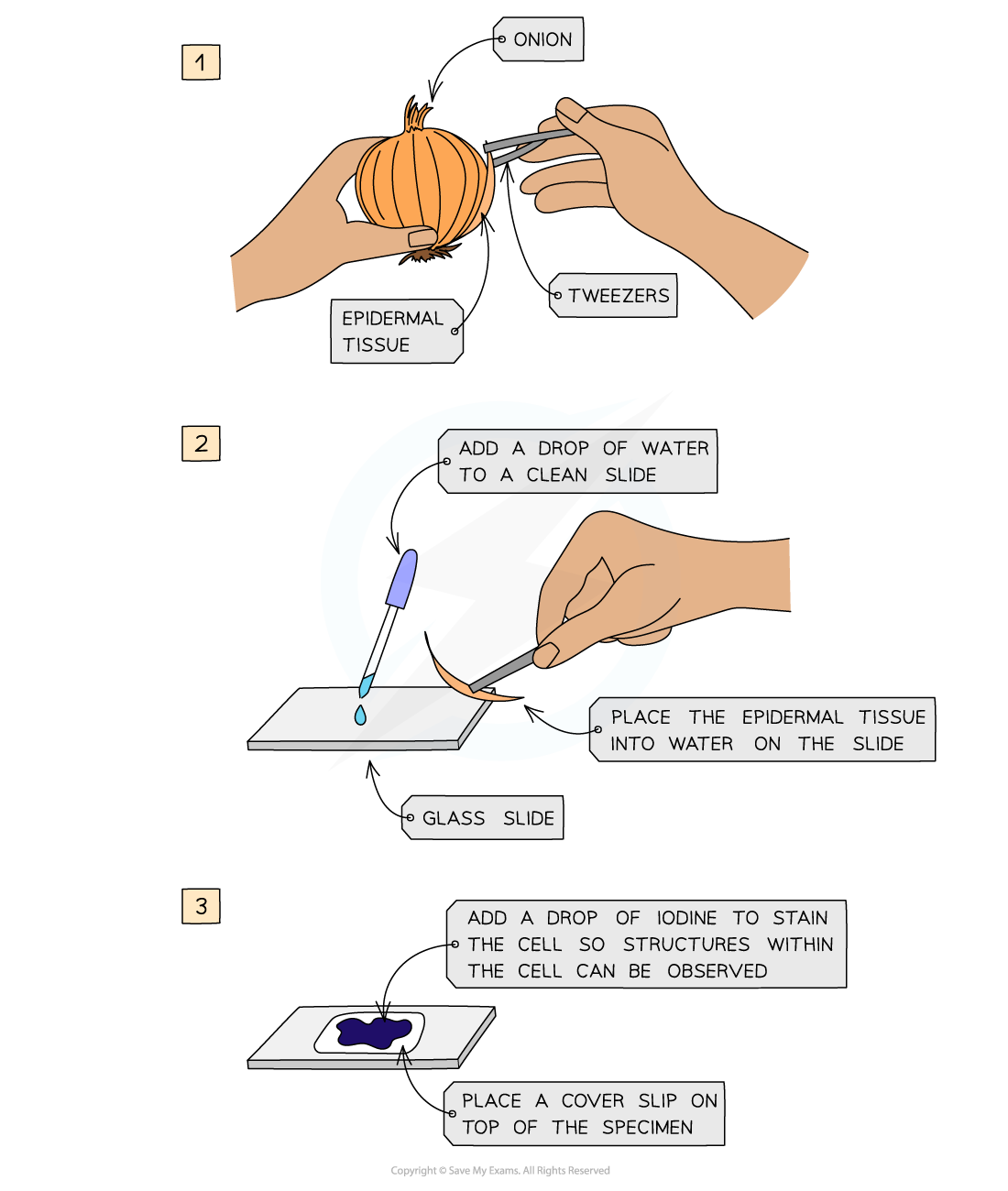
1. Peel off an Epidermal Layer On Onion Using Forceps.
2. Mount Onto Slide With a Drop Of Water Using Pipette
3. Add 2 drops of Iodine Solution to Stain Cells.
4. Place Cover Slip On By Using Forceps at an Angle to Make Sure No Air Bubbles Trapped.
5. Remove Any Excess Stain With Paper Towel
6. Place on Stage & Select Low Power Objective Look Through Eye Piece & Turn Fine Adjustment Knob Until the Image Comes into Focus. .
7. When Selecting Higher Power Objective Adjust Stage & Turn Knob to Move Stage Away
8. Make a Labelled Drawing of Cells You can See & Write down the magnification
13. Repeat using a prepared slide.
60
How to prepare a plant cell slide?
1) Onion cell slide: Use a dropping pipette to put one drop of water onto a microscope slide.
2) Separate one of the thin layers of the onion.
3) Peel off a thin layer of epidermal tissue from the inner surface.
4) Use forceps to put this thin layer on to the drop of water that you have placed on the microscope slide.
5) Make sure that the layer of onion cells are flat on the slide.
6) Put two drops of iodine solution onto the onion tissue.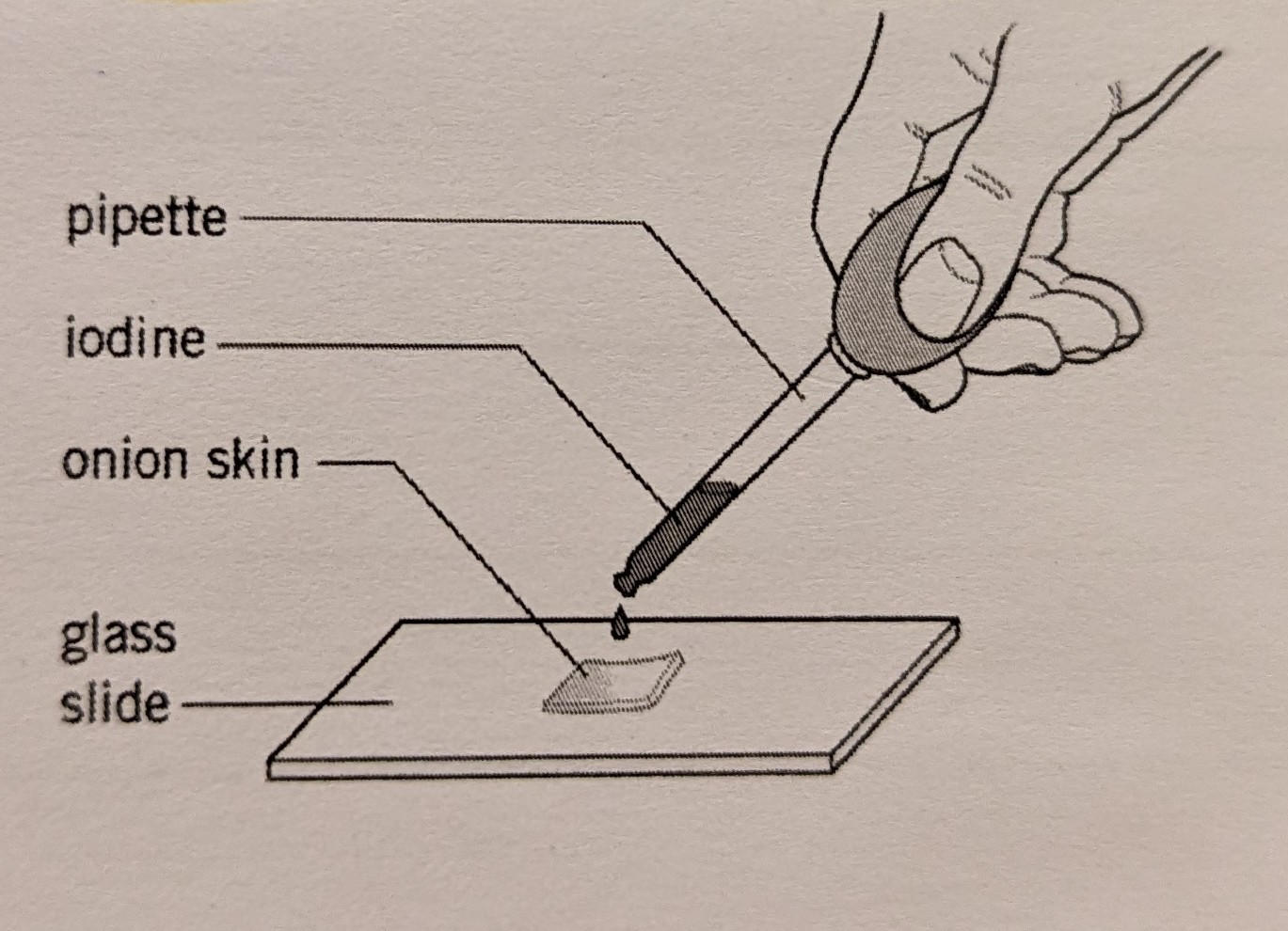
2) Separate one of the thin layers of the onion.
3) Peel off a thin layer of epidermal tissue from the inner surface.
4) Use forceps to put this thin layer on to the drop of water that you have placed on the microscope slide.
5) Make sure that the layer of onion cells are flat on the slide.
6) Put two drops of iodine solution onto the onion tissue.
61
How to prepare an animal cell slide?
1) Cheek cell slide: rub a cotton but inside your cheek
2) Rub the cotton bud on a glass slide to transfer the cells
3) Add a drop of methyl blue on the slide
4) Carefully lower a coverslip onto the slide using a mounting needle
5) There may be some liquid around the edge of the coverslip. So use a tissue to soak this liquid up.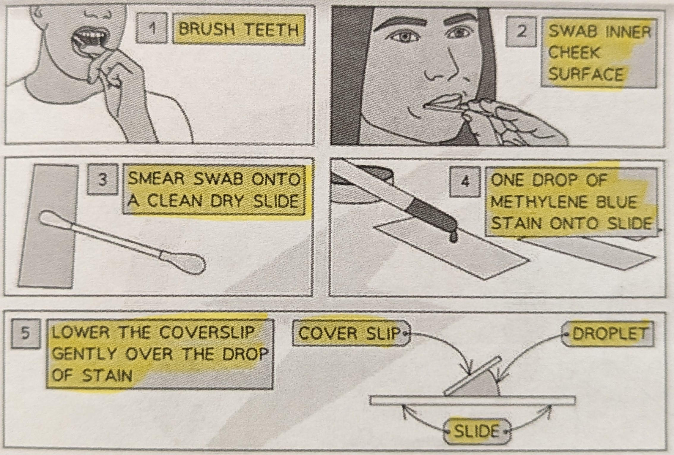
2) Rub the cotton bud on a glass slide to transfer the cells
3) Add a drop of methyl blue on the slide
4) Carefully lower a coverslip onto the slide using a mounting needle
5) There may be some liquid around the edge of the coverslip. So use a tissue to soak this liquid up.
62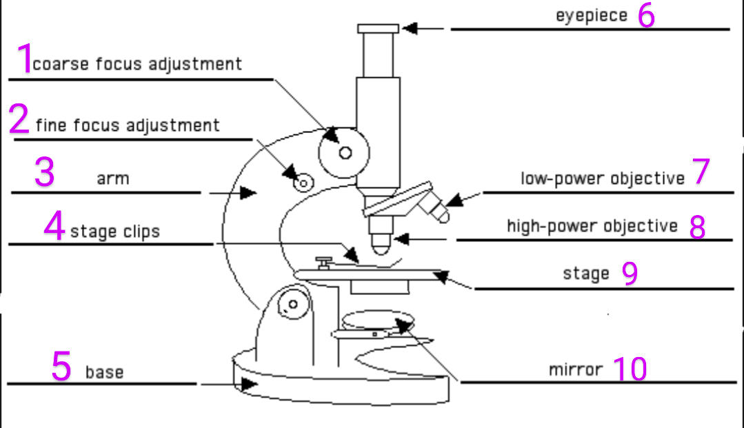
Label the parts of the microscope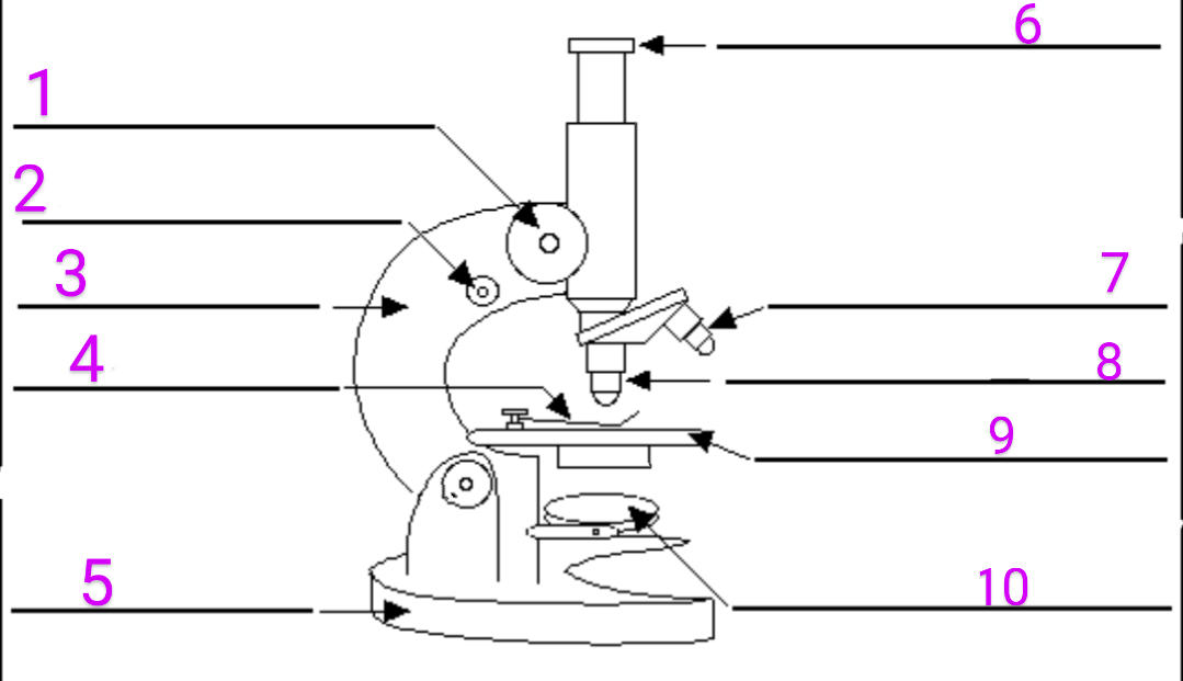
63
Which obejctive lense should you start with?
The lowest objective lense .
So you can see more information/wider view.
So you can see more information/wider view.
64
How do you find the total magnification used?
eyepiece magnification × objective lens magnification
65
Write a method for the osmosis practical for potatoes. Include steps and any calculations needed.
1) Use a Core Borer To Cut 5 Potato Cylinders of the Same Diameter.
2) Trim the Cylinders so all Same Length (About 3 Cm).
3) Accurately Measure and Record the Length and Mass Of Each Potato Cylinder in the Table Provided.
4) Measure 10 cm³ of Distilled Water and put into First Boiling Tube. Label Boiling tube OM
5) Repeat the process for 0.25M, 0.5M, 0.75M, 1M solutions concentrations.
6)Add one potato cylinder to each boiling tube. Make sure Know the Length and Mass of each Potato Cylinder In Each Boiling Tube
7) Leave Potato Cylinders in the boiling Tubes Overnight (or a minimum of 30 minutes) in the Test Tube Rack.
8) Remove the Cylinders from the Boiling Tubes and Carefully Blot Them Dry With Paper Towels.
9) Re-measure Length and Mass Of Each Cylinder (make sure you know which is which). Record your Measurements in the Table. Calculate the Changes in Length & Mass & % Change Of Each Potato Cylinder.
% change=
change in value
--------------- ×100
original value
10) Draw a Line Graph with Smooth Line through Points To Display Results. You will have Some Negative Percentage Changes to Plot so make sure you axes can support this.
2) Trim the Cylinders so all Same Length (About 3 Cm).
3) Accurately Measure and Record the Length and Mass Of Each Potato Cylinder in the Table Provided.
4) Measure 10 cm³ of Distilled Water and put into First Boiling Tube. Label Boiling tube OM
5) Repeat the process for 0.25M, 0.5M, 0.75M, 1M solutions concentrations.
6)Add one potato cylinder to each boiling tube. Make sure Know the Length and Mass of each Potato Cylinder In Each Boiling Tube
7) Leave Potato Cylinders in the boiling Tubes Overnight (or a minimum of 30 minutes) in the Test Tube Rack.
8) Remove the Cylinders from the Boiling Tubes and Carefully Blot Them Dry With Paper Towels.
9) Re-measure Length and Mass Of Each Cylinder (make sure you know which is which). Record your Measurements in the Table. Calculate the Changes in Length & Mass & % Change Of Each Potato Cylinder.
% change=
change in value
--------------- ×100
original value
10) Draw a Line Graph with Smooth Line through Points To Display Results. You will have Some Negative Percentage Changes to Plot so make sure you axes can support this.
66
What would the axis on a graph be for the osmosis practical (watch video)
• X axis is concentration of solution
• Y axis is percentage change in mass/length
(include + and - axis so it )
• Y axis is percentage change in mass/length
(include + and - axis so it )
67
What does the x intercept mean in context to the osmosis practical
(watch video)
(watch video)
Isotonic Point
68
What happens to Plant Cells in a Dilute Solution?
Becomes turgid
69
What happens to a Plant Cell in a concentrated solution
Becomes Flaccid and at Very High Concentrations becomes Plasmolyzed
70
What happens to a Plant Cell in a isotonic solution?
no change
71
What happens to an Red Blood Cel in a dilute solution?
Bursts
72
What happen to an Red Blood Cell in a concentrated solution?
Shrivel/plasmolyzed
73
what happens to a Red Blood Cell in an isotonic solution?
No change.
74
Why do plant cells not burst when placed in a dilute solution?
They have a cell wall which is strong .