Peritoneum and Peritoneal Cavity
Study these flashcardsFree · spaced repetition · iPhone, Android, Mac & web
All cards
1
The parietal and visceral layers are separated from each other by a
small amount of capillary fluid produced by the cells
lining the layers
small amount of capillary fluid produced by the cells
lining the layers
The parietal and visceral layers are separated from each other by a
small amount of capillary fluid produced by the mesothelial cells
lining the layers
small amount of capillary fluid produced by the mesothelial cells
lining the layers
2
The parietal and visceral layers of the peritoneum form the which is a potential space.
The parietal and visceral layers of the peritoneum form the peritoneal
cavity which is a potential space.
cavity which is a potential space.
3
Where is a paracentesis performed?
(1) RLQ or LLQ
(2) 2 cm below umbilicus
(2) 2 cm below umbilicus
4
When quadrant approach is attempted, perform to the rectus
sheath to avoid the
sheath to avoid the
When quadrant approach is attempted, perform lateral to the rectus
sheath to avoid the inferior epigastric vessels
sheath to avoid the inferior epigastric vessels
5
Layers of Penentration RLQ or LLQ
1. Skin
2. Camper's fascia
3. Scarpa's fascia
4. Deep fascia
5. External oblique
6. Internal oblique
7. Tranversus abdominis
8. Fascia transversalis
9. Extraperitoneal fat
10. Pariteal peritoneum
11. Peritoneal cavity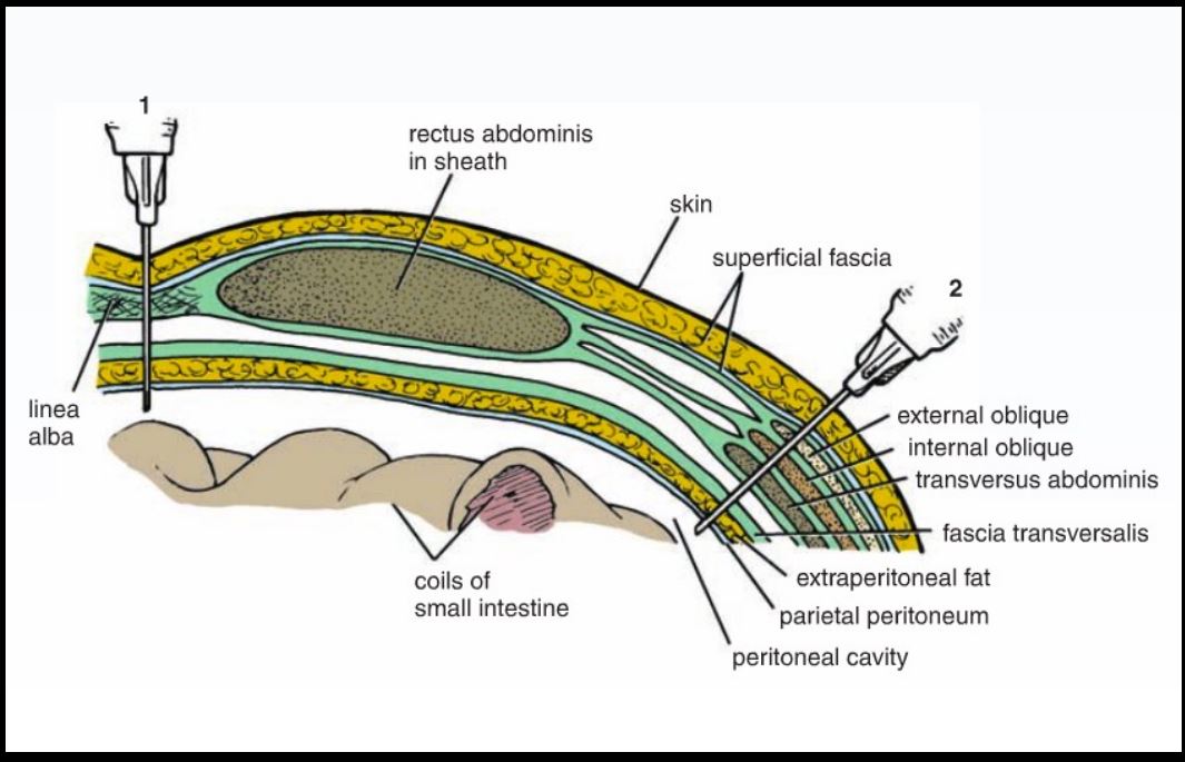
2. Camper's fascia
3. Scarpa's fascia
4. Deep fascia
5. External oblique
6. Internal oblique
7. Tranversus abdominis
8. Fascia transversalis
9. Extraperitoneal fat
10. Pariteal peritoneum
11. Peritoneal cavity
6
Layers of Penentration with Ubilical Approach
1. Skin
2. Camper's fascia
3. Scarpa's fascia
4. Deep Fascia
5. Linea alba
6. Fascia tranversalis
7. Extraperitoneal fat
8. Pariteal peritoneum
9. Peritoneal cavity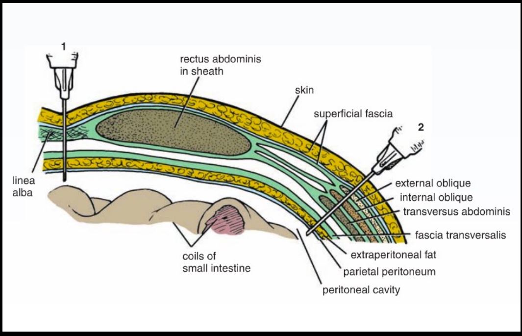
2. Camper's fascia
3. Scarpa's fascia
4. Deep Fascia
5. Linea alba
6. Fascia tranversalis
7. Extraperitoneal fat
8. Pariteal peritoneum
9. Peritoneal cavity
7
The peritoneal cavity is closed in but open in
The peritoneal cavity is closed in males but open in females
8
Greater peritoneal sac = extending from the
to the
to the
Greater peritoneal sac = extending from the diaphragm
to the pelvic floor.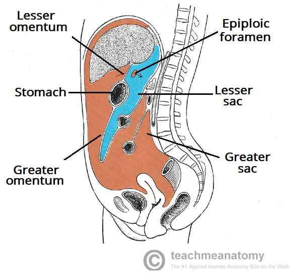
to the pelvic floor.
9
Lesser peritoneal sac = A.K.A. = lies
posterior to the and
posterior to the and
Lesser peritoneal sac = A.K.A. omental bursa = lies
posterior to the stomach and
liver.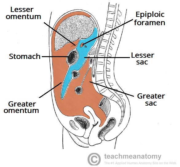
posterior to the stomach and
liver.
10
= double layer of
peritoneum that encloses an
organ and connects it to the
abdominal wall.
peritoneum that encloses an
organ and connects it to the
abdominal wall.
Mesentry = double layer of
peritoneum that encloses an
organ and connects it to the
abdominal wall.
peritoneum that encloses an
organ and connects it to the
abdominal wall.
11
= double
layer of peritoneum that
connects an organ to another
organ or the abdominal wall.
layer of peritoneum that
connects an organ to another
organ or the abdominal wall.
Peritoneal ligament = double
layer of peritoneum that
connects an organ to another
organ or the abdominal wall.
layer of peritoneum that
connects an organ to another
organ or the abdominal wall.
12
Omentum = sheet of
peritoneum that attaches
the to the
or other
peritoneum that attaches
the to the
or other
Omentum = doubled layered sheet of
peritoneum that attaches
the stomach to the
abdominal wall or other
abdominal organs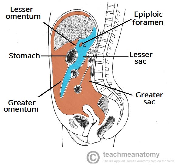
peritoneum that attaches
the stomach to the
abdominal wall or other
abdominal organs
13
**TEST QUESTION
Lesser Omentum = it
connects the lesser
curvature of the
and proximal part of the
to the liver.
Individually these
connections are refered to
as and
ligaments
Lesser Omentum = it
connects the lesser
curvature of the
and proximal part of the
to the liver.
Individually these
connections are refered to
as and
ligaments
**TEST QUESTION
Lesser Omentum = it
connects the lesser
curvature of the stomach
and proximal part of the
duodemun to the liver.
Individually these
connections are refered to
as hepatogastric and
hepatoduodenal ligaments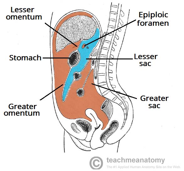
Lesser Omentum = it
connects the lesser
curvature of the stomach
and proximal part of the
duodemun to the liver.
Individually these
connections are refered to
as hepatogastric and
hepatoduodenal ligaments
14
Greater omentum = fat-laden
fold of peritoneum that hangs
down from the greater
curvature of the stomach like
an and connects the
with the ,
and the
colon
fold of peritoneum that hangs
down from the greater
curvature of the stomach like
an and connects the
with the ,
and the
colon
Greater omentum = fat-laden
fold of peritoneum that hangs
down from the greater
curvature of the stomach like
an apron and connects the
stomach with the diaphragm,
spleen and the transverse
colon
fold of peritoneum that hangs
down from the greater
curvature of the stomach like
an apron and connects the
stomach with the diaphragm,
spleen and the transverse
colon
15
Liagments of the Greater Omentum
- = stomach
to transverse colon
- =
stomach to spleen
- =
stomach to diaphragm (superior)
- = stomach
to transverse colon
- =
stomach to spleen
- =
stomach to diaphragm (superior)
Liagments of the Greater Omentum
-Gastrocolic ligament = stomach
to transverse colon
-Gastrosplenic ligament =
stomach to spleen
- Gastrophrenic ligament =
stomach to diaphragm (superior)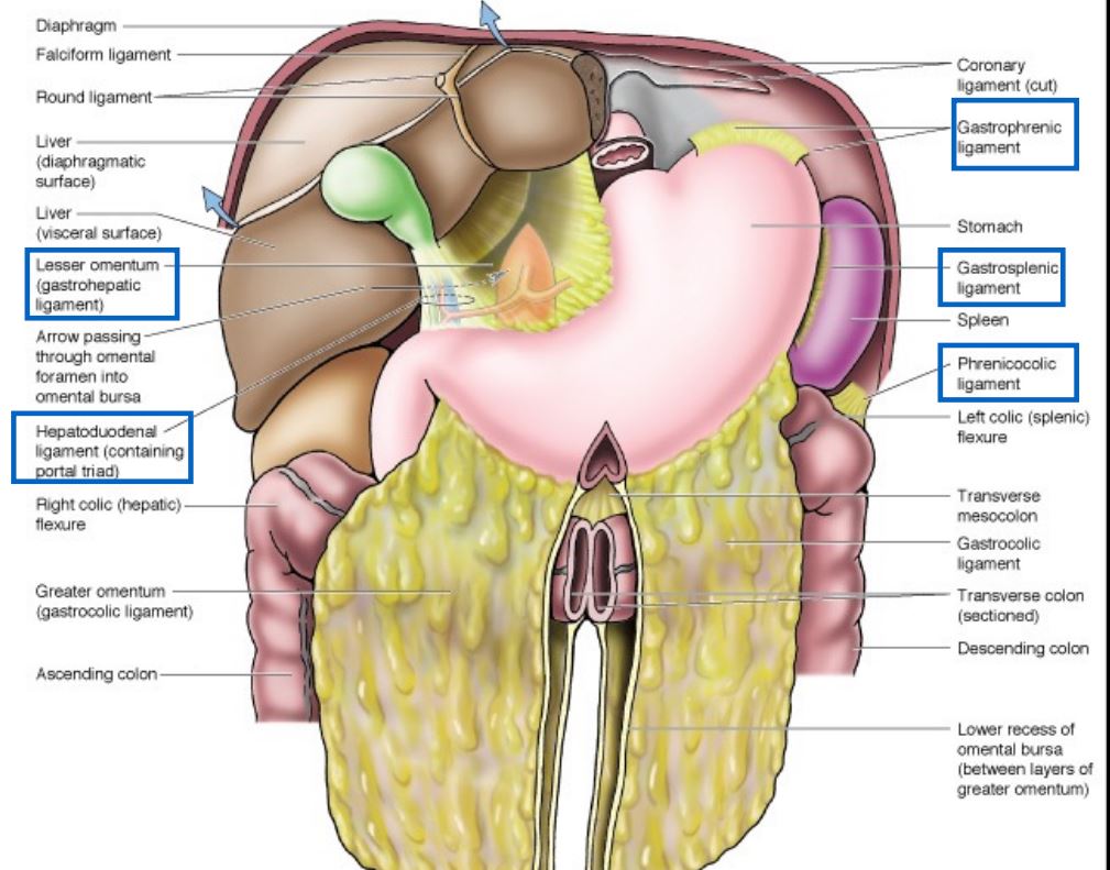
-Gastrocolic ligament = stomach
to transverse colon
-Gastrosplenic ligament =
stomach to spleen
- Gastrophrenic ligament =
stomach to diaphragm (superior)
16
Ligaments of the Lesser Omentum
- = between the stomach and liver
- = between the liver and teh duodenum
- = between the stomach and liver
- = between the liver and teh duodenum
Ligaments of the Lesser Omentum
-Gastrohepatic ligament = between the stomach and liver
-Hepatoduodenal ligament = between the liver and teh duodenum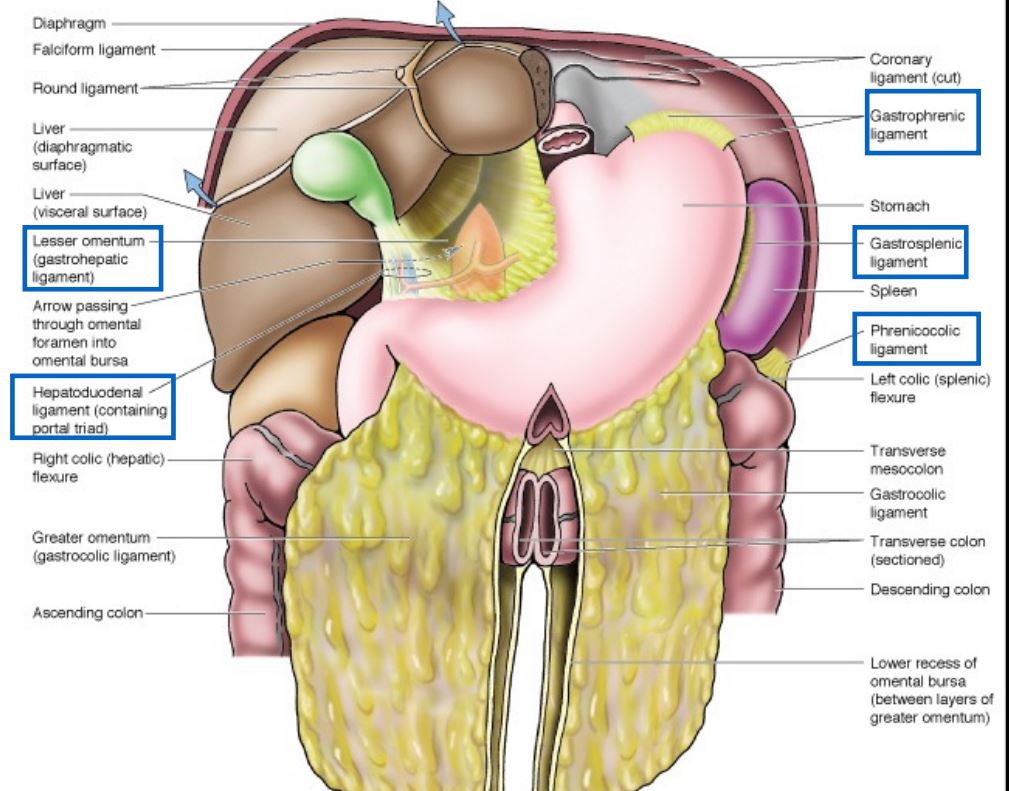
-Gastrohepatic ligament = between the stomach and liver
-Hepatoduodenal ligament = between the liver and teh duodenum
17
= peritoneal
reflections as the peritoneum
cover the abdominal
structures
reflections as the peritoneum
cover the abdominal
structures
Peritoneal folds = peritoneal
reflections as the peritoneum
cover the abdominal
structures
reflections as the peritoneum
cover the abdominal
structures
18
Peritoneal recesses =
peritoneal folds that form
or tubular
cavities that are closed on one
end and open into the
abdominal cavity.
peritoneal folds that form
or tubular
cavities that are closed on one
end and open into the
abdominal cavity.
Peritoneal recesses =
peritoneal folds that form
blind pouches or tubular
cavities that are closed on one
end and open into the
abdominal cavity.
peritoneal folds that form
blind pouches or tubular
cavities that are closed on one
end and open into the
abdominal cavity.
19
The supracolic compartment is found above the and is divided by the into
The supracolic compartment is found above the transverse mesocolon and is divided by the falciform ligament into right/left subphrenic recess
20
The infracolic compartment is between the right/left spaces. The longitudinal channels in the compartment are called the
The infracolic compartment is between the right/left infracolic spaces. The longitudinal channels in the compartment are called the paracolic gutters
21
The is on the lateral portion and the is on the medial portion of asending colon. The left paracolic gutter is on the and the is on the medial of the desending colon. (Look at pic)
The right paracolic gutter is on the lateral portion and the right infracolic space is on the medial portion of asending colon. The left paracolic gutter is on the left lateral side and the left infracolic space is on the medial of the desending colon. (Look at pic) 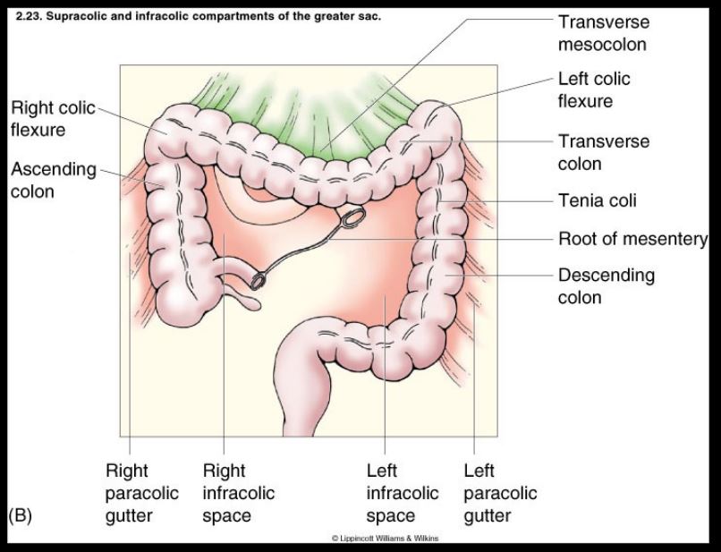
22
Test Question
These gutters slope and , so
when fluid accumulates in the peritoneal cavity, it will
follow these gutters to the when the patient is supine.
These gutters slope and , so
when fluid accumulates in the peritoneal cavity, it will
follow these gutters to the when the patient is supine.
Test Question
These gutters slope superiorly and posteriorly, so
when fluid accumulates in the peritoneal cavity, it will
follow these gutters to the superior part of the
abdomen when the patient is supine.
These gutters slope superiorly and posteriorly, so
when fluid accumulates in the peritoneal cavity, it will
follow these gutters to the superior part of the
abdomen when the patient is supine.
23
The omental bursa is in communication with the through the , or
, which is located posterior to the free edge of the
lesser omentum ( ligament)
, which is located posterior to the free edge of the
lesser omentum ( ligament)
The omental bursa is in communication with the main peritoneal
cavity (greater sac) through the omental foramen, epiploic foramen or
foramen of Winslow, which is located posterior to the free edge of the
lesser omentum (hepatoduodenal ligament)
cavity (greater sac) through the omental foramen, epiploic foramen or
foramen of Winslow, which is located posterior to the free edge of the
lesser omentum (hepatoduodenal ligament)
24
Boundaires of the epiploic foramen? Anterior? Posterior? Superior? Inferior?
Anterior = portal hepaticus (inside the hepatoduodenal ligament- lesser omentum)
Posterior = IVC and right crus of diaphram
Superior= Caudate lobe of liver
Inferior=Superior part of duodenum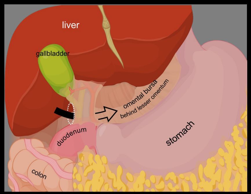
Posterior = IVC and right crus of diaphram
Superior= Caudate lobe of liver
Inferior=Superior part of duodenum
25
Portal hepaticus contents
DAVE Real Life Player
Duct (common bile duct) - Right
Artery (proper hepatic artery) -Left
Vein (portal vein) - Posterior
EPIPLOIC FORAMEN (where its found)
Duct (common bile duct) - Right
Artery (proper hepatic artery) -Left
Vein (portal vein) - Posterior
EPIPLOIC FORAMEN (where its found)
26
What is the nerve supply of the parital layer?
1. Phrenic nerve (subdiaphragmatic branches)
2. Thoracolumbar nerves
3. Subcostal nerve
4. Lumbosacral plexus (L4-S4)
2. Thoracolumbar nerves
3. Subcostal nerve
4. Lumbosacral plexus (L4-S4)
27
What is the nerve supply of the viseral layer?
Insensitive to pain
28
The abdominal cavity had the organs of the , ,
The abdominal cavity had the organs of the GI, UT, great vessels (abdominal aorta, IVC)
29
Peritoneal organs have mesentary and are enclosed in . They are .
Peritoneal organs have mesentary and are enclosed in peritoneum. They are movable.
30
Retroperitoneal organs: are partially covered by
and are or .
and are or .
Retroperitoneal organs: are partially covered by
peritoneum and are immobile or fixed.
peritoneum and are immobile or fixed.
31
What are the primary retroperitoneal organs?
SAD PUCKER
S: Super adrenal gland
A: Aorta
D: Duodenum (second and third part)
P: Pancreas (except tail)
U: Ureters
C: Colon
K: Kindenys
E: Esophagus
R: Rectum
S: Super adrenal gland
A: Aorta
D: Duodenum (second and third part)
P: Pancreas (except tail)
U: Ureters
C: Colon
K: Kindenys
E: Esophagus
R: Rectum
32
Major Secondary
Retroperitoneal
Organs
Retroperitoneal
Organs
-Most of the
duodenum
-Most of the
pancreas (except tail)
-Ascending colon
-Descending colon
-Upper rectum
duodenum
-Most of the
pancreas (except tail)
-Ascending colon
-Descending colon
-Upper rectum
33
Major Peritoneal Organgs
ALL OF THE OTHER STUFF - JUST MEMORIZE FIRST 2
-Stomach
-Liver and
gallbladder
-Spleen
-Beginning of the
duodenum
-Tail of the pancreas
-Jejunum
-Ileum
-Appendix
-Transverse colon
-Sigmoid colon
-Stomach
-Liver and
gallbladder
-Spleen
-Beginning of the
duodenum
-Tail of the pancreas
-Jejunum
-Ileum
-Appendix
-Transverse colon
-Sigmoid colon
34
What are the structures of the foregut?
1.
2.
3.
4.
5.
1.
2.
3.
4.
5.
What are the structures of the foregut?
1. Esophagus
2. Duodenum
3. Liver
4. Biliary passages
5. Gallbladder
1. Esophagus
2. Duodenum
3. Liver
4. Biliary passages
5. Gallbladder
35
What is the parasympathetic nerve of foregut? Sympathetic?
Vagus, Greater splanchic (T5-T9)
36
What is the blood supply of the foregut?
Celiac trunk
37
What are the structures in the midgut?
1.
2.
3.
4.
1.
2.
3.
4.
What are the structures in the midgut?
1. Duodenum (2, 3, 4)
2. Jejunum and ileum
3. Cecum and appendix
4. Transverse colon (prox 2/3)
1. Duodenum (2, 3, 4)
2. Jejunum and ileum
3. Cecum and appendix
4. Transverse colon (prox 2/3)
38
What is the parasympathetic supply of the midgut? Sympathetic?
Vagus, Lesser splanchnic (T10-T11)
39
What is the blood supply of the midgut?
SMA Superior mesenteric artery
40
What are the structures of the hindgut?
1. Transverse colon
(distal 1/3)
2. Descending colon
3. Sigmoid colon
4. Rectum
5. Anal canal (upper part)
(distal 1/3)
2. Descending colon
3. Sigmoid colon
4. Rectum
5. Anal canal (upper part)
41
The is the parasympathetic and sympathetic nerve supply of the hindgut?
Pelvic splanchnic (S2,3,4), Lumbar splanchnic (L1-L2)
42
What is teh blood supply of the hindgut?
Inferior mesenteric artery (IMA)