All cards
1
Auscultation angle of louis =
Auscultation angle of louis = intercostal space 2
2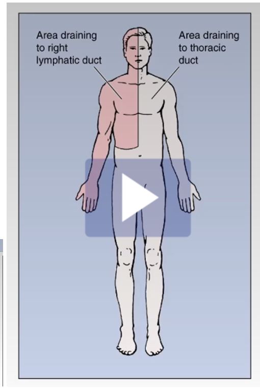
Image occlusion: name the hidden labels
3
What is the cisterna chyli?
cisterna chyli is a large midline lymphatic collecting structure
4
What drains to the right lympathic duct?
The right portion of the upper body, chest, face, and neck
5
What drains to the thoracic duct?
Everything EXCEPT the right portion of upper body, chest, face, and neck
6
Where is the sternal angle of Louis?
Aligns with T4/T5 vertebrae (lower border of T4)
7
What forms the angle of louis?
The angle between the manubrium and body of the sternum
8
What does the sternal angle of louis mark the level of?
The level of the 2nd costal space
9
Costal cartilage of 2nd rib aligns with what?
manubriosternal joint
10
What does the Xiphoid process align with?
T10 or T11 vertebrae
11
What is the costal margin?
Costal cartilage 7-10
12
Where is the infrastenal angle found? Why is it important?
-Located between xiphoid process and left costal margin
-Important for decompressing pericardium when there is too much fluid (cardiac tampenode)
-Important for decompressing pericardium when there is too much fluid (cardiac tampenode)
13
Where is the pericardiac sac?
Found within the left infrasternal angle
14
Auscultation angle of louis =
Auscultation angle of louis = intercostal space 2
15
What are the 9 things that the sternal angle/angle of Louis corresponds to?
1. Rib 2
2. Aortic arch
3. Tracheal bifurcation
4. Pulmonary trunk
5. Ligamentum ateriosum
6. Azygos vein into SVC
7. Nerves (cardiac plexus)
8. Thoracic duct
9. T4/T5 || notes:RAT PLANTT
2. Aortic arch
3. Tracheal bifurcation
4. Pulmonary trunk
5. Ligamentum ateriosum
6. Azygos vein into SVC
7. Nerves (cardiac plexus)
8. Thoracic duct
9. T4/T5 || notes:RAT PLANTT
16
What are the boundaries of the thoracic Inlet?
1. Anteriorly - superior border of manubrium
2. Posteriorly - T1 vertebral body
3. Laterally - 1st rib and costal cartilage
2. Posteriorly - T1 vertebral body
3. Laterally - 1st rib and costal cartilage
17
What are the contents of the thoracic inlet?
1. Trachea
2. Apex of lungs (CUPLA is the pariteal pleura OVER this)
3. Esophogus
4. Vagus nerve (CN X)
5. Prenic nerve (C3,C4,C5) || notes:Troy Always Eats Vegan Pizza
2. Apex of lungs (CUPLA is the pariteal pleura OVER this)
3. Esophogus
4. Vagus nerve (CN X)
5. Prenic nerve (C3,C4,C5) || notes:Troy Always Eats Vegan Pizza
18
WHat are the boundaries for the inferior thoracic aperture (outlet)?
1. Xyphoid process (Anteriorly)
2.Costal cartilage of ribs 7-10 (Anterolaterally)
2. Rib 12 (Posterior laterally)
3. T12 (Posterior)
2.Costal cartilage of ribs 7-10 (Anterolaterally)
2. Rib 12 (Posterior laterally)
3. T12 (Posterior)
19
What are the contents of the thoracic outlet?
1. Aortic opening (T12)
2. Espohageal opening (T10)
3. Caval opening (T8)
2. Espohageal opening (T10)
3. Caval opening (T8)
20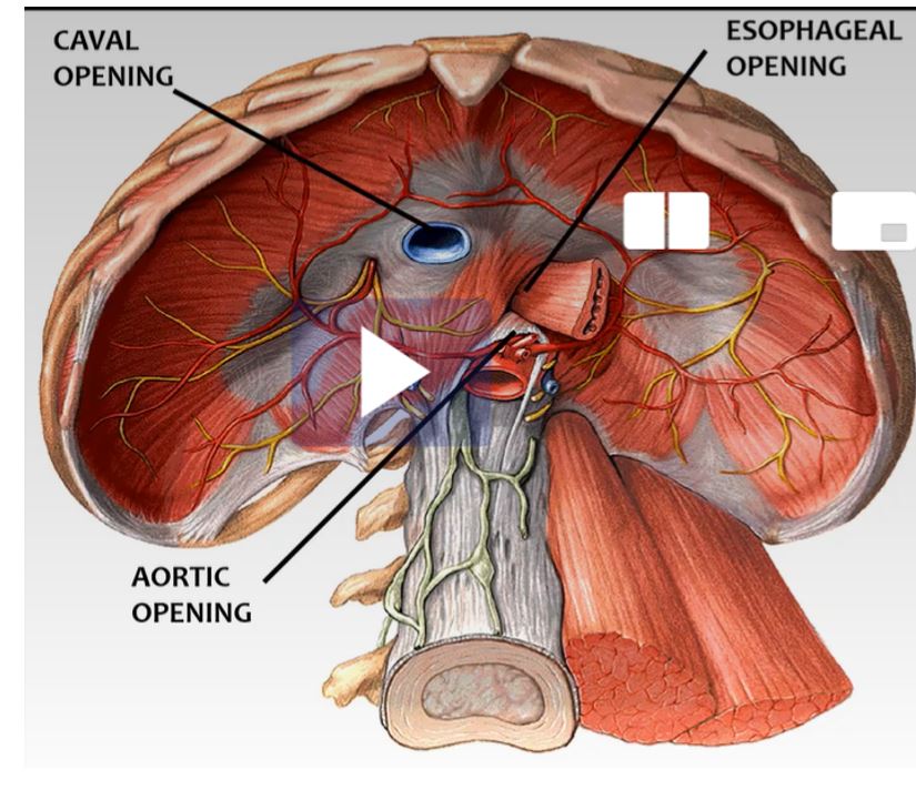
Image occlusion: name the hidden labels
21
What are the contents of the caval opening?
-At T8
-IVC, Phrenic nerves (C3,C4,C5), lympathic vessels
-IVC, Phrenic nerves (C3,C4,C5), lympathic vessels
22
What are the contents of the esophageal opening?
-T10
-Esophoagus, right and left vagus nerve, esophageal branch of left gastric artery + vein
-Esophoagus, right and left vagus nerve, esophageal branch of left gastric artery + vein
23
What are the contents of the aortic opening?
-T12
-Thoracic aorta, thoracic duct, greater sphlanchnic nerves (T5-T9), azygos vein
-Thoracic aorta, thoracic duct, greater sphlanchnic nerves (T5-T9), azygos vein
24
How do the fibers of the external intercostal muscles run?
Run downward and forward; extenal intercostal membrane at chondral joint
25
What is the action of the external intercostal muscles?
Elevates the rib cage, inspiration
26
How do the fibers of the interal muscles run?
Fibers run down and backward; internal intercostal membrane near vertebrae
27
What is the action of the interal muscles?
Expiration
28
What are the three parts of the innermost muscles?
1. Inner most proper= between ribs
2. Subcostalis: angle
3. Sternocostalis: near sternum
2. Subcostalis: angle
3. Sternocostalis: near sternum
29
What is the function of the innermost muscle?
Expiration
30
What are the contents of the intercostal space>
1. Ventral primary ramus of spinal nerve
2. Internal intercostal
3. Subcostal
2. Internal intercostal
3. Subcostal
31
How are instruments passed through the ICS?
-Throygh lower part of space to avoid neurovascular bundle
32
How is an intercostal nerve block adminstered?
Done in upper portion of intercostal space
33
Where is the costal groove located?
Inferior aspect of the rib
34
What does the costal groove protect?
shelters intercostal neurovascular bundle
35
What two muscles is VAN located between?
Innermost intercostal muscle (deep) and internal intercostal muscles (superficial)
36
What are the contents of the costal groove?
VAN = vein, nerve, artery
37
All ribs have a costal groove except?
Rib 1
38
What is a thoracentessis?
Surgical puncture of chest wall between ribs 9 and 10 via MID-AXILLARY APPROACH
39
What are the 12 layers for midaxillary penetration?
1. Skin
2. Superficial fascia
3. Deep fascia
4. Serrarus anterior
5. External intercostal muscle
6. Internal intercostal muscle
7. Innermost intercostal muscle
8. Endothoracic fascia
9. Parietal pleura
10. Pleural cavity
11. Viceral pleura
12. Lungs
2. Superficial fascia
3. Deep fascia
4. Serrarus anterior
5. External intercostal muscle
6. Internal intercostal muscle
7. Innermost intercostal muscle
8. Endothoracic fascia
9. Parietal pleura
10. Pleural cavity
11. Viceral pleura
12. Lungs
40
What is a pericardiocentesis?
-Done to decompress a cardiac tamponade
-Done at left infrasternal angle through cardial notch of left lung
-ISC 4 (can also be done at 5 or 6)
-Done at left infrasternal angle through cardial notch of left lung
-ISC 4 (can also be done at 5 or 6)
41
What is cardia tamponade?
Fluid compresses heart which screws up cardiac venous filling in diastole and decreases cardiac output
42
Where does the anterior aspect of the thorax drain?
Internal thoracic vein into brachiocephalic
43
Where does the posterior apect of the thorax drain?
Into the azygos system
44
Where does the posterior apect of the thorax drain?
Into the azygos system
45
The right side of the azygous system drains?
1st - highest intercostal vein -> brachiochephalic
2nd + 3rd = higher intercostal -> azygos
4 th - 11th -> azygos
12th (subcostal veins) -> azygos
2nd + 3rd = higher intercostal -> azygos
4 th - 11th -> azygos
12th (subcostal veins) -> azygos
46
The left side of the azygous system drains where?
1st - highest intercostal vein -> brachiochephalic
2nd + 3rd +4th = accessory hemiazygous (can drain into brachiocephalic)
5th -11th +12th = hemiazygos vein
2nd + 3rd +4th = accessory hemiazygous (can drain into brachiocephalic)
5th -11th +12th = hemiazygos vein
47
Thoracic cage =
Thoracic cage = osseocartilaginous
48
Thoracic cage attachment
Left hand side= of rib 5
Right hand side= , right side higher bc of liver
Left hand side= of rib 5
Right hand side= , right side higher bc of liver
Thoracic cage attachment
Left hand side= lower border of rib 5
Right hand side= upper border of rib 5, right side higher bc of liver
Left hand side= lower border of rib 5
Right hand side= upper border of rib 5, right side higher bc of liver
49
Thoracic cage =
Thoracic cage = osseocartilaginous
50
Organs in the thoracic cavity
thymus, heart, lungs, distal part of the trachea, and most of the
esophagus
esophagus
51
Bones of sternum = , ,
Bones of sternum = Manubrium sterni, body, xiphoid process
52
Ribs join by common costocartilage
Ribs 7,8, 9, 10 join by common costocartilage
53
= false ribs
= floating ribs
= floating ribs
8,9,10 = false ribs
11, 12 = floating ribs
11, 12 = floating ribs
54
Because the sensory and motor innervation of the diaphragm is primarily from
the pain arising from the diaphragm is referred to the
dermatomes in the cervical region (referred pain).
the pain arising from the diaphragm is referred to the
dermatomes in the cervical region (referred pain).
Because the sensory and motor innervation of the diaphragm is primarily from
the phrenic nerve (C3, 4, 5), pain arising from the diaphragm is referred to the
dermatomes in the cervical region (referred pain).
the phrenic nerve (C3, 4, 5), pain arising from the diaphragm is referred to the
dermatomes in the cervical region (referred pain).
55
congenital diaphragmatic hernia (CDH) =
side of the thoracic cavity
side of the thoracic cavity
congenital diaphragmatic hernia (CDH) = left posterolateral
side of the thoracic cavity
side of the thoracic cavity
56
esophageal hiatal hernia is a herniation of the stomach into the
esophageal hiatal hernia is a herniation of the stomach into the pleural cavity
57
No anterior intercostal
arteries in
arteries in
No anterior intercostal
arteries in ICS 10-11
arteries in ICS 10-11
58
Anterior intercostal arteries to
ICS 7,8,9 come from the
ICS 7,8,9 come from the
Anterior intercostal arteries to
ICS 7,8,9 come from the musculophrenic
artery (internal throracic)
ICS 7,8,9 come from the musculophrenic
artery (internal throracic)
59
Anterior intercostal arteries to
ICS 1-6 come from the
ICS 1-6 come from the
Anterior intercostal arteries to
ICS 1-6 come from the internal thoracic
artery.
ICS 1-6 come from the internal thoracic
artery.
60
What two veins unite to form superior vena cava? Right and left
What two veins unite to form superior vena cava? Right and left brachiocephalic veins
61
= connective
tissue layer that functions to prevent
the parietal pleura from rubbing against
the innermost intercostal muscles. This
fascia is thickened over the apex of
the lungs:
tissue layer that functions to prevent
the parietal pleura from rubbing against
the innermost intercostal muscles. This
fascia is thickened over the apex of
the lungs:
Endothoracic fascia= connective
tissue layer that functions to prevent
the parietal pleura from rubbing against
the innermost intercostal muscles. This
fascia is thickened over the apex of
the lungs: SIBSON’sfascia
tissue layer that functions to prevent
the parietal pleura from rubbing against
the innermost intercostal muscles. This
fascia is thickened over the apex of
the lungs: SIBSON’sfascia
62
Tramatic injury is rare in children because their ribs are
Tramatic injury is rare in children because their ribs are elastic
63
due to multiple rib fractures leading to
paradoxical chest movement (i.e. inward with
inspiration and outward with expiration); it impairs
ventilation
paradoxical chest movement (i.e. inward with
inspiration and outward with expiration); it impairs
ventilation
Flail chest: due to multiple rib fractures leading to
paradoxical chest movement (i.e. inward with
inspiration and outward with expiration); it impairs
ventilation
paradoxical chest movement (i.e. inward with
inspiration and outward with expiration); it impairs
ventilation
64
Rib dislocation: displacement of the costal
cartilage from the ; contact sports
cartilage from the ; contact sports
Rib dislocation: displacement of the costal
cartilage from the sternum (2-7); contact sports
cartilage from the sternum (2-7); contact sports
65
Rib separation: dislocation of the between the rib and its costal cartilage
Rib separation: dislocation of the costochondral
joint between the rib and its costal cartilage
joint between the rib and its costal cartilage
66
: “funnel chest”: body of the
sternum projects inferiorly and posteriorly
pressing on the hear
sternum projects inferiorly and posteriorly
pressing on the hear
Pectus excavatum: “funnel chest”: body of the
sternum projects inferiorly and posteriorly
pressing on the hear
sternum projects inferiorly and posteriorly
pressing on the hear
67
“pigeon chest”: sternum
projects anteriorly.
projects anteriorly.
Pectus carinatum: “pigeon chest”: sternum
projects anteriorly.
projects anteriorly.
68
= surgical puncture of the chest
wall.
wall.
Thoracentesis = surgical puncture of the chest
wall.
wall.
69
1) needle
2) tube ( at the
anterior axillary line)
2) tube ( at the
anterior axillary line)
Thoracostomy: 1) needle
2) tube (ICS 4 or ICS 5 at the
anterior axillary line)
2) tube (ICS 4 or ICS 5 at the
anterior axillary line)
70
= surgical incision of the chest wall.
Over , extending from the lateral
margin of the sternum to the anterior axillary line.
Over , extending from the lateral
margin of the sternum to the anterior axillary line.
Thoracotomy = surgical incision of the chest wall.
Over ICS 4 or ICS 5, extending from the lateral
margin of the sternum to the anterior axillary line.
Over ICS 4 or ICS 5, extending from the lateral
margin of the sternum to the anterior axillary line.
71
Pleural cavity Vocab
- = lines the thoracic wall and covers surface of diaphragm
- = covers outer surface of lungs
- = open cavity with structures going in and out
- = lines the thoracic wall and covers surface of diaphragm
- = covers outer surface of lungs
- = open cavity with structures going in and out
Pleural cavity Vocab
-Parietal = lines the thoracic wall and covers surface of diaphragm
-Visceral = covers outer surface of lungs
-Hilum = open cavity with structures going in and out
-Parietal = lines the thoracic wall and covers surface of diaphragm
-Visceral = covers outer surface of lungs
-Hilum = open cavity with structures going in and out
72
What is the cupla?
An area of parietal pleura reflected over the apex of the lung
73
What is the costodiaphragmatic recess?
-Junction of costal and diaphragmatic parietal pleura (mid-axillary line)
-Lowest area of the pleural cavity where lungs expand
-Lowest area of the pleural cavity where lungs expand
74
Where does the costal parietal pleura extend?
-Anteriorly at ICS 8
-Mid-axillary ICS 9
-Posteriorly ICS 10
-Mid-axillary ICS 9
-Posteriorly ICS 10