Hist Lab 5 - Muscle
Study these flashcardsFree · spaced repetition · iPhone, Android, Mac & web
All cards
1
What are the three types of muscle? And describe the general characteristics of their fibers.
All three types of muscle (skeletal, cardiac, and smooth) are composed of cells (also
called fibers) specialized to shorten, or contract. All muscle fibers have an elongated
shape, oval euchromatic nuclei, and acidophilic cytoplasm filled with contractile
filaments (actin and myosin). Skeletal and cardiac muscle fibers have visible
cytoplasmic striations due to the complex arrangement of the actin and myosin
filaments into repeating units called sarcomeres and myofibrils.
called fibers) specialized to shorten, or contract. All muscle fibers have an elongated
shape, oval euchromatic nuclei, and acidophilic cytoplasm filled with contractile
filaments (actin and myosin). Skeletal and cardiac muscle fibers have visible
cytoplasmic striations due to the complex arrangement of the actin and myosin
filaments into repeating units called sarcomeres and myofibrils.
2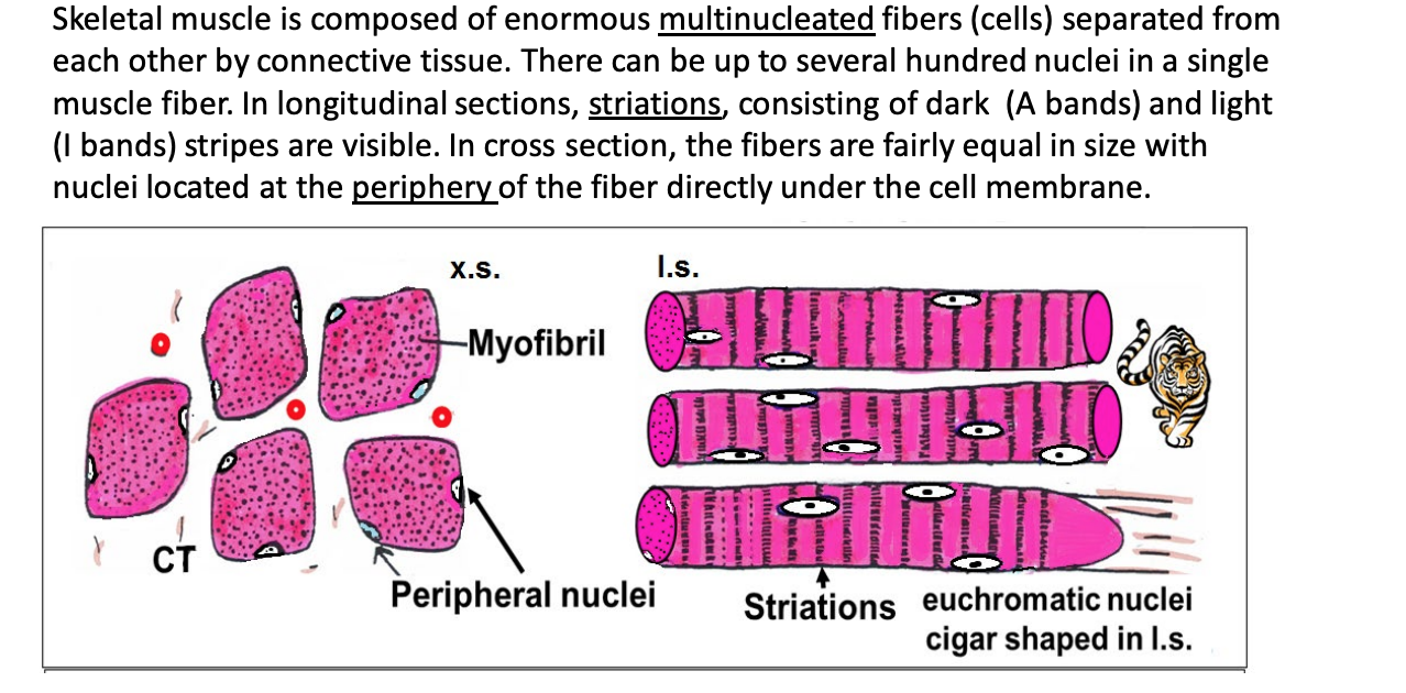
Describe skeletal muscle
3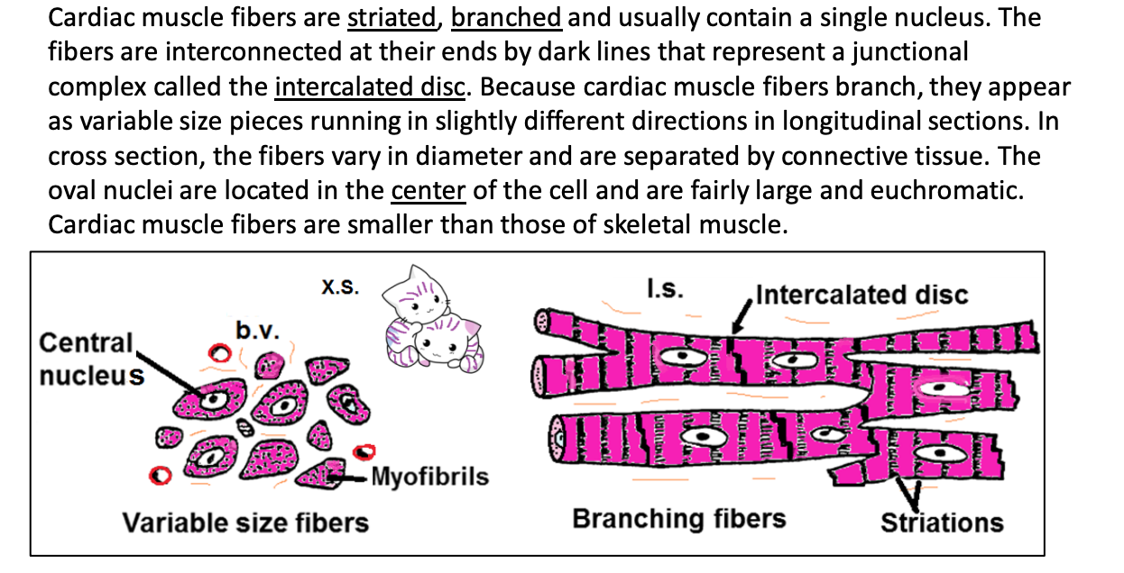
Describe cardiac muscle
4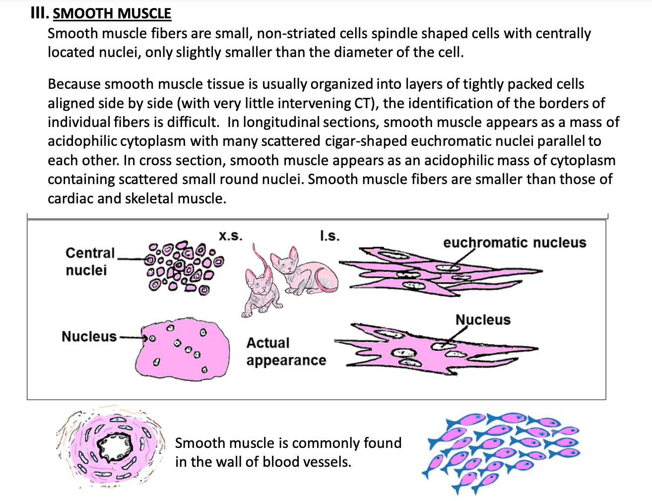
Describe smooth muscle
5
What type of muscle is this?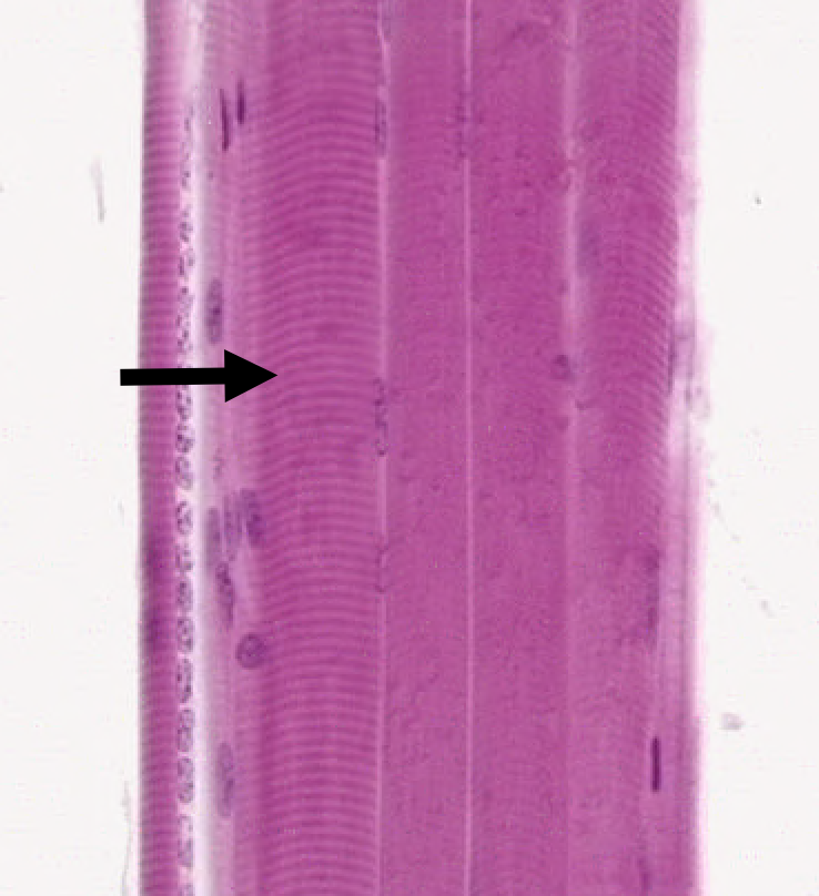
Skeletal muscle (longitudinal section). You can see the cross striations consisting of dark and light lines (known as A and I bands).
*need to know longitudinal section vs. cross section (said in lecture).
-lots of nuclei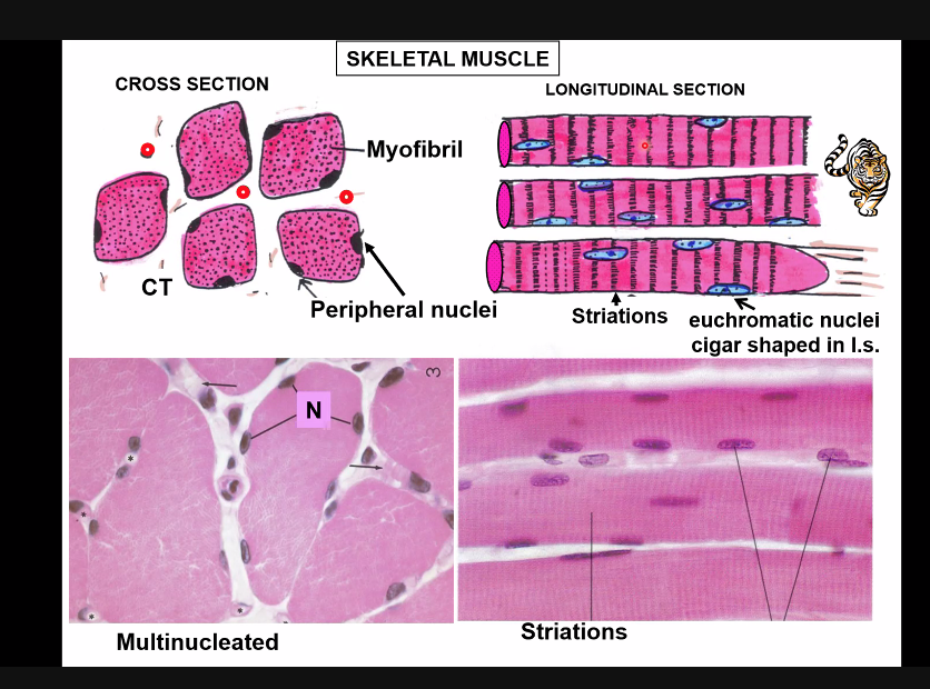
*need to know longitudinal section vs. cross section (said in lecture).
-lots of nuclei
6
What type of muscle is this?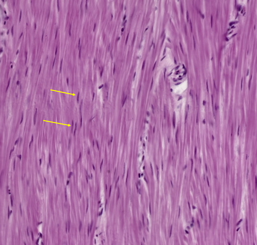
Smooth muscle (longitudinal section). The arrows are pointing to smooth muscle nuclei.
7
What type of muscle is this?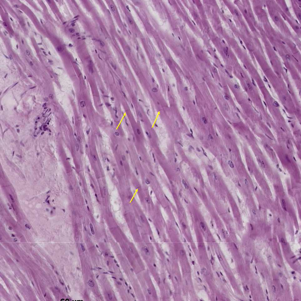
Cardiac muscle (longitudinal section). The arrows are pointing to intercalated discs. The branching cardiac muscle have striations but they are not visible in the slide. 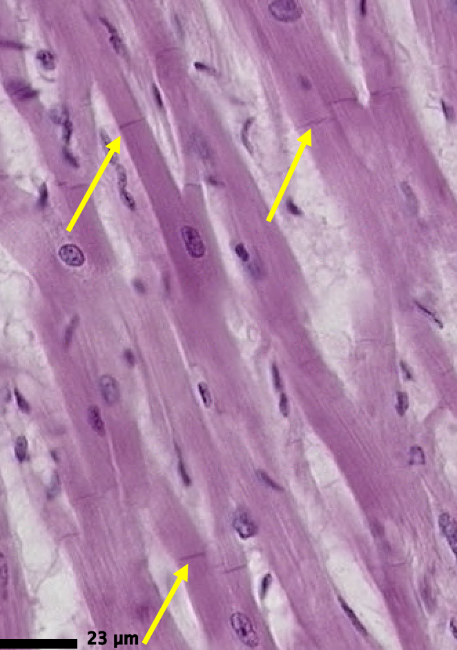
8
What type of muscle is this? And what is being highlighted?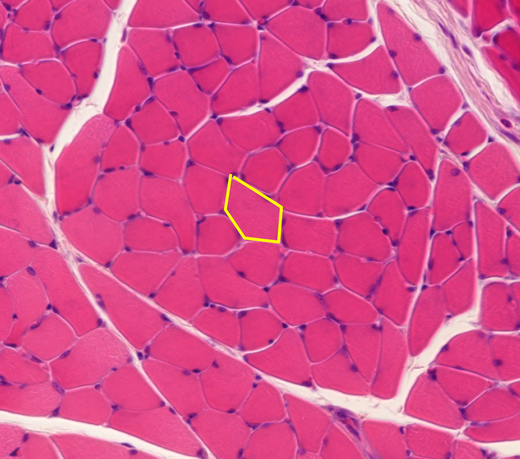
9
What type of muscle is this?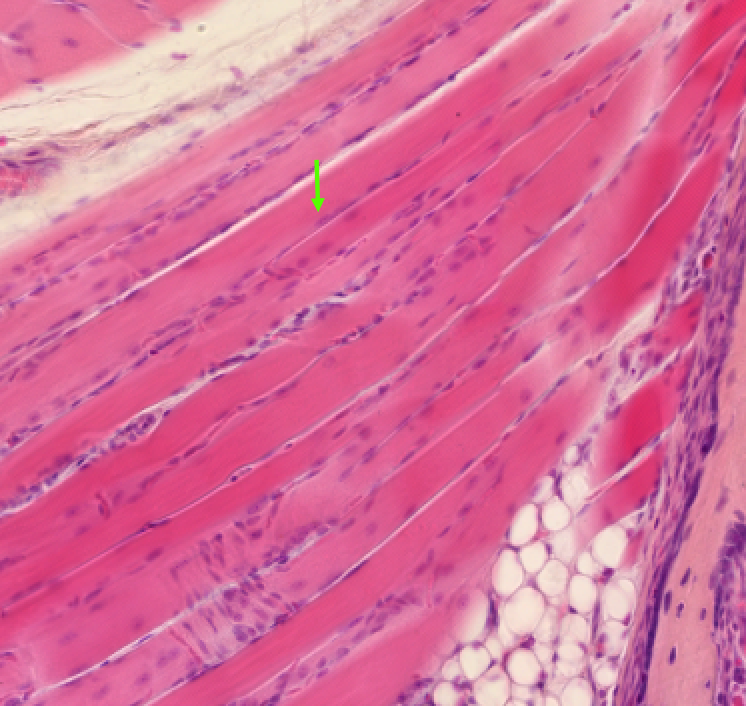
Skeletal muscle (longitudinal)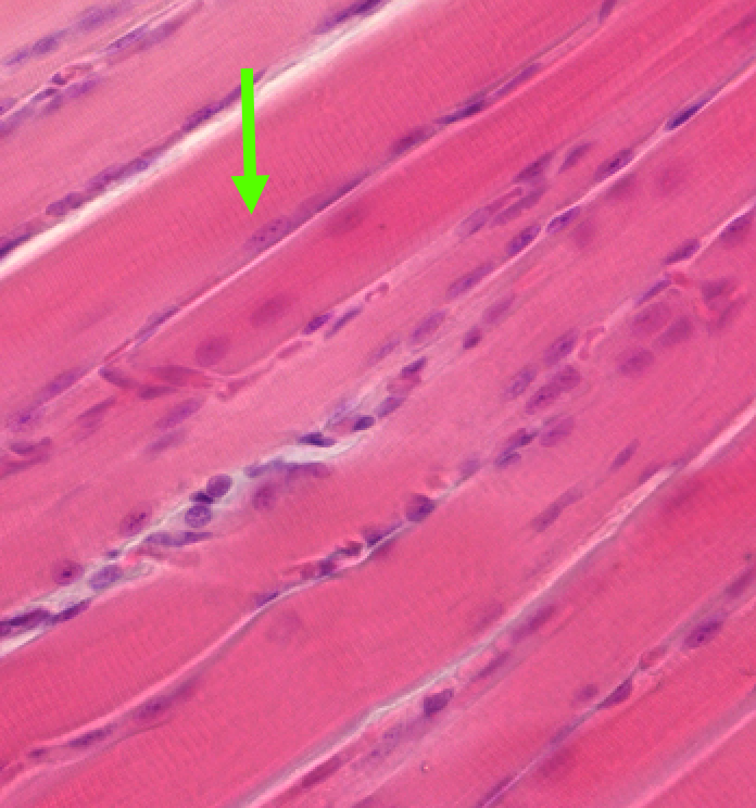
10
Classify the tissue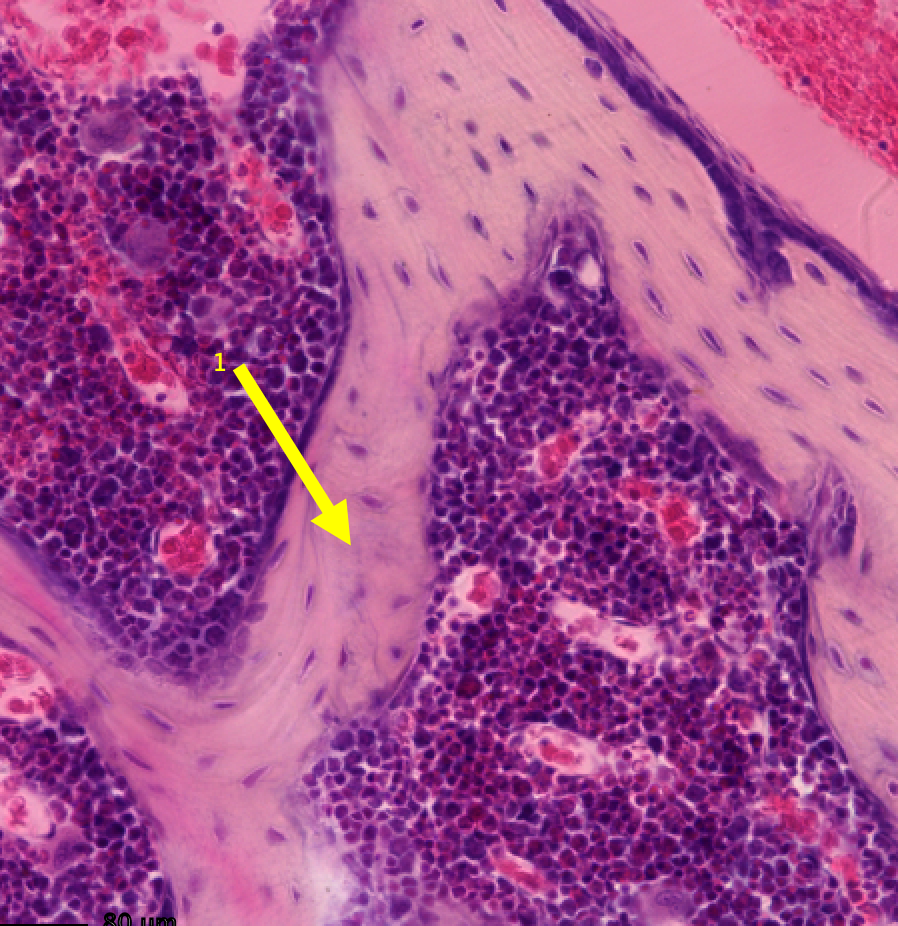
spongy bone
11
Identify the cells 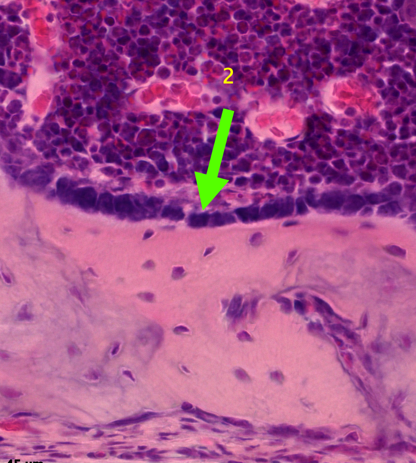
Osteoblasts
12
Classify the tissue 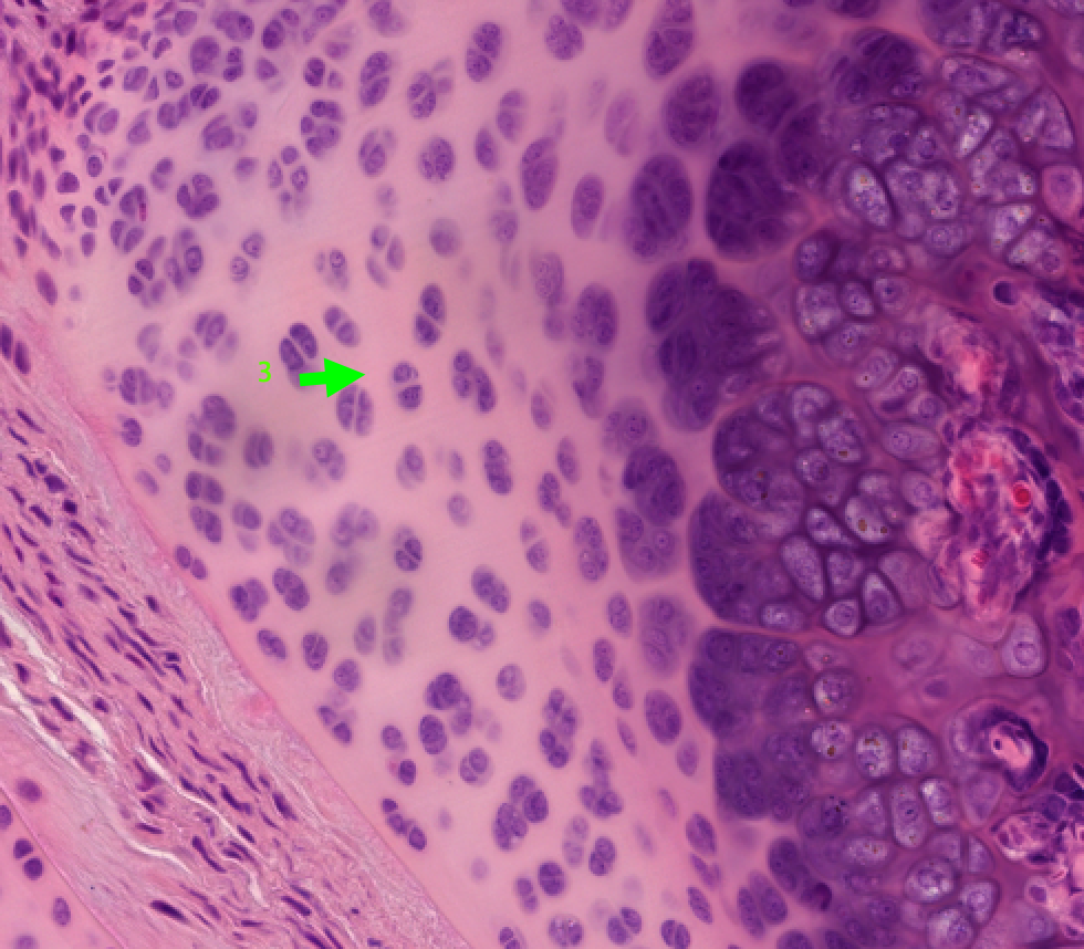
Hyaline cartilage
13
What type of muscle is this and what are the arrows pointing to? 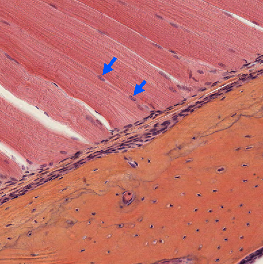
14
Identify structure and classify the tissue 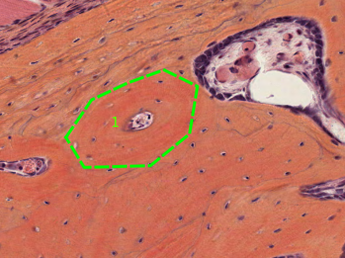
Osteon (Haversian system) in compact bone
15
Identify the cellular lining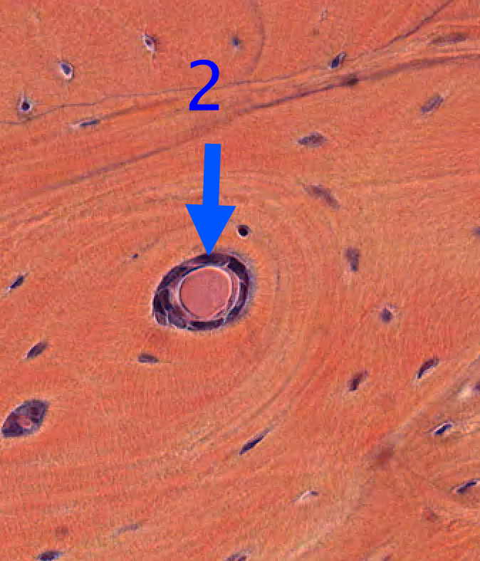
endosteum
16
Classify the tissue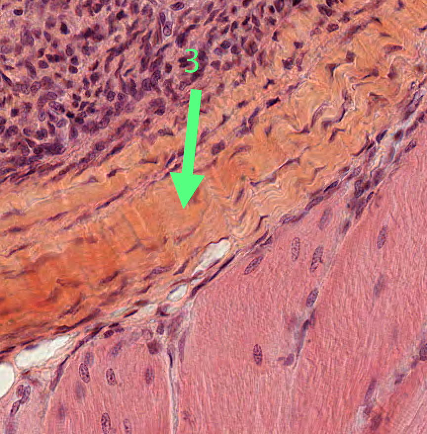
dense regular connective tissue
17
Identify the cell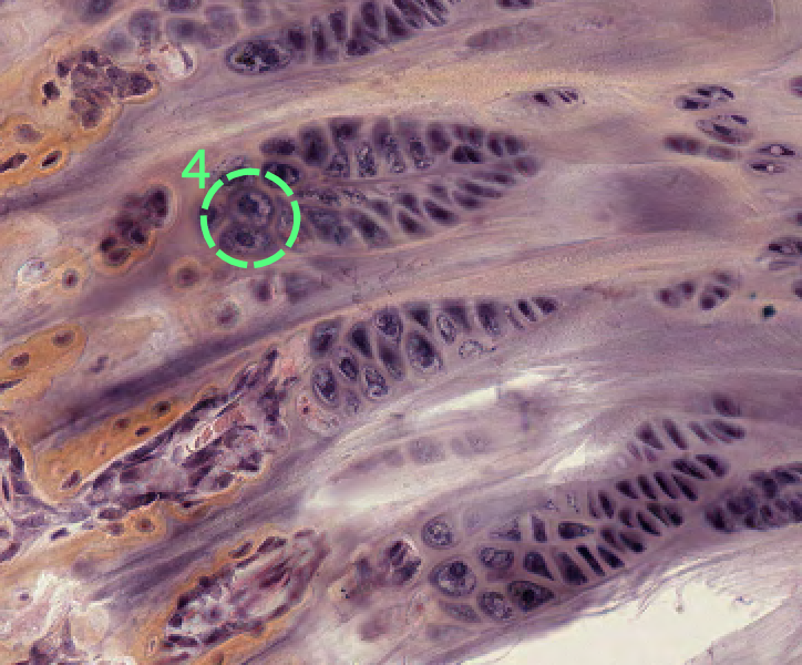
chondrocyte
18
Identify the tissue 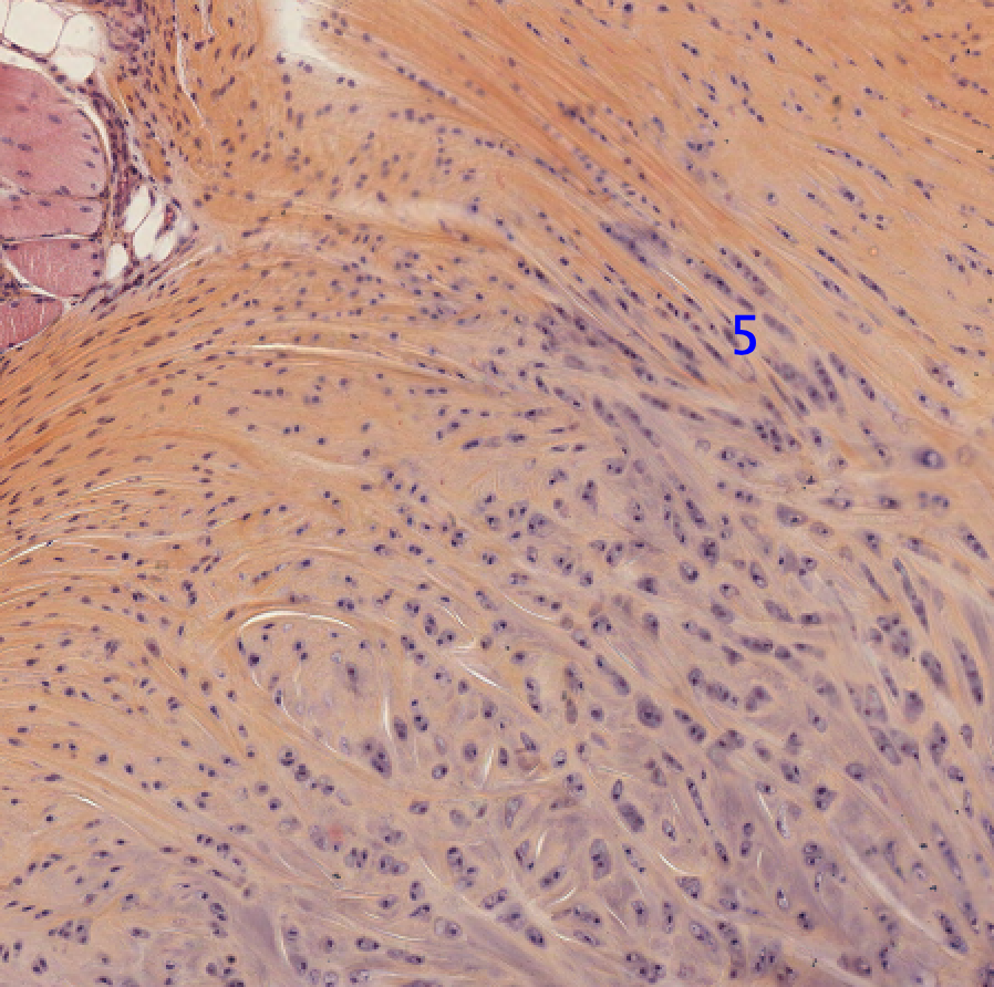
fibrocartilage
19
Classify the tissue 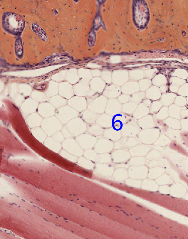
adipose tissue
20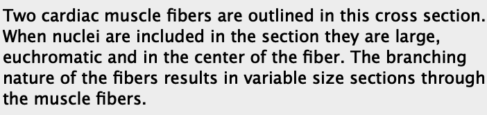
What type of muscle is this and what is being emphasized? 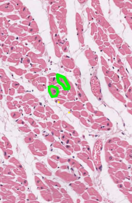
21
What type of muscle is this?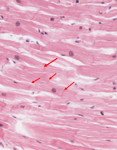
Cardiac Muscle (longitudinal), has striations, and intercalated discs (red arrows). The branching nature of the fibers is evident 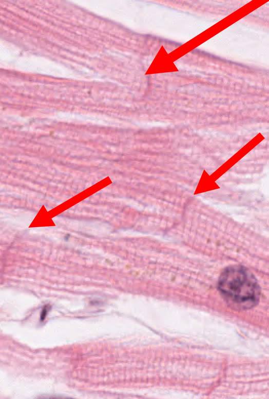
22
General view of a skeletal muscle 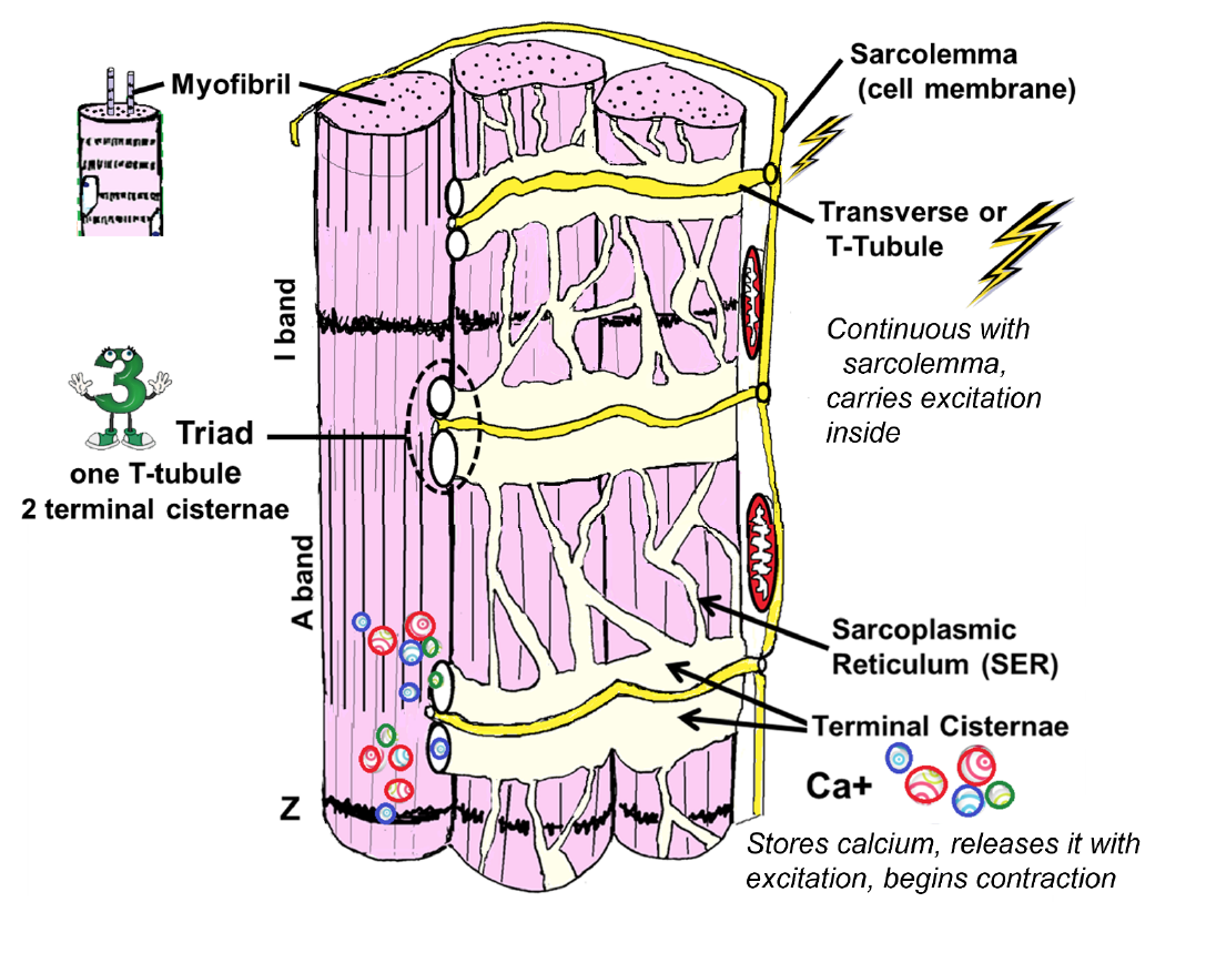
-know bands 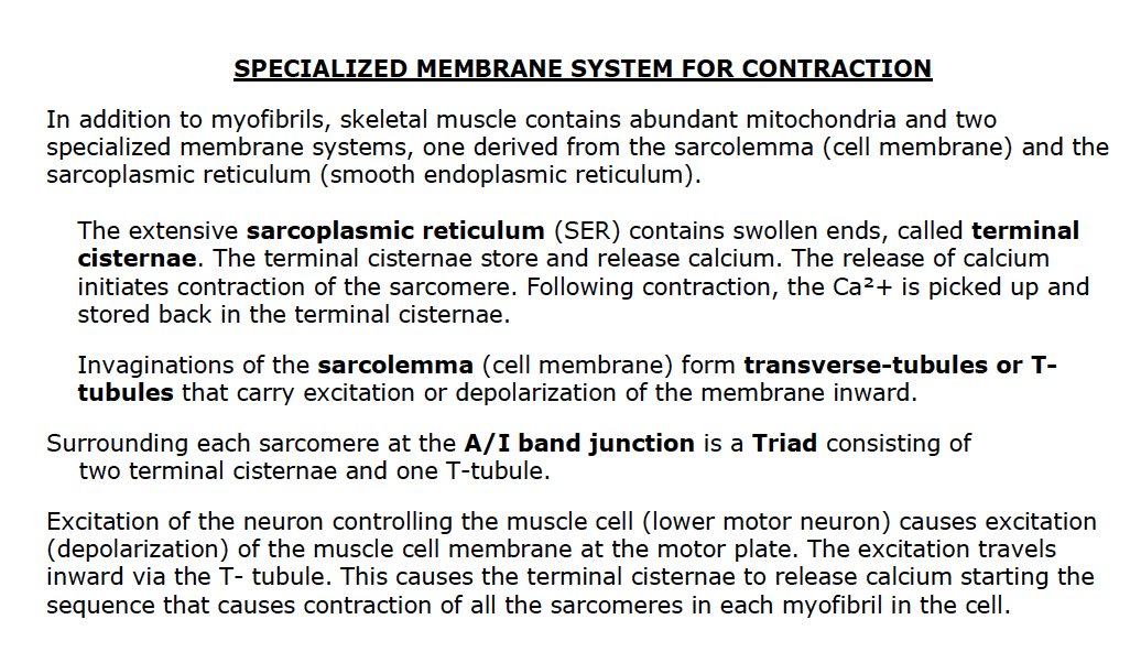
23
Classify tissue 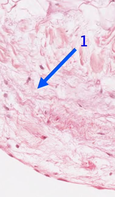
loose connective tissue
24
Classify tissue 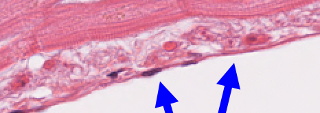
simple squamous epithelium
25
What is the arrow pointing to?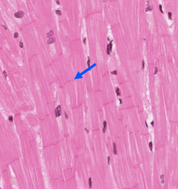
Intercalated disc 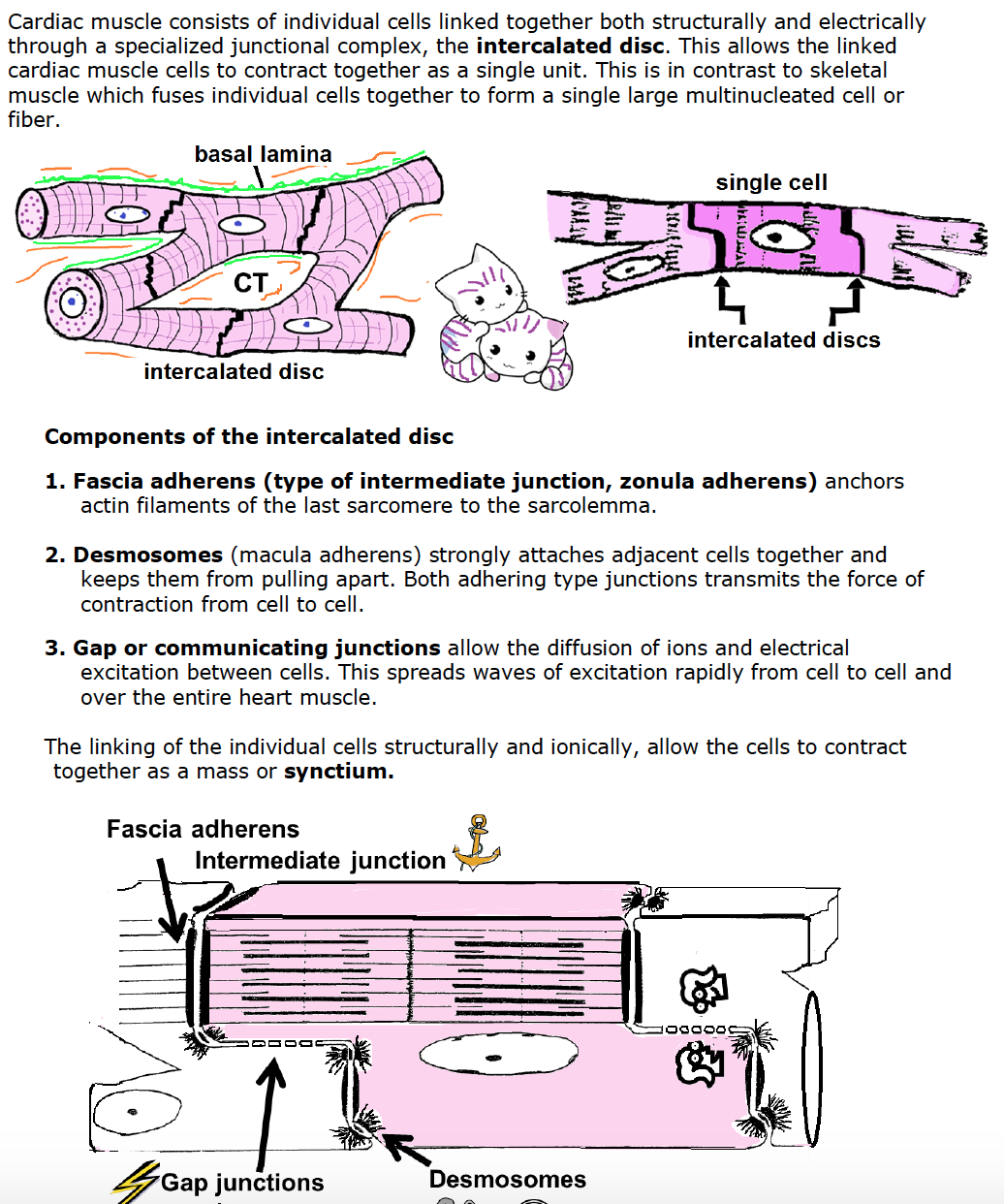
26
What is being highlighted?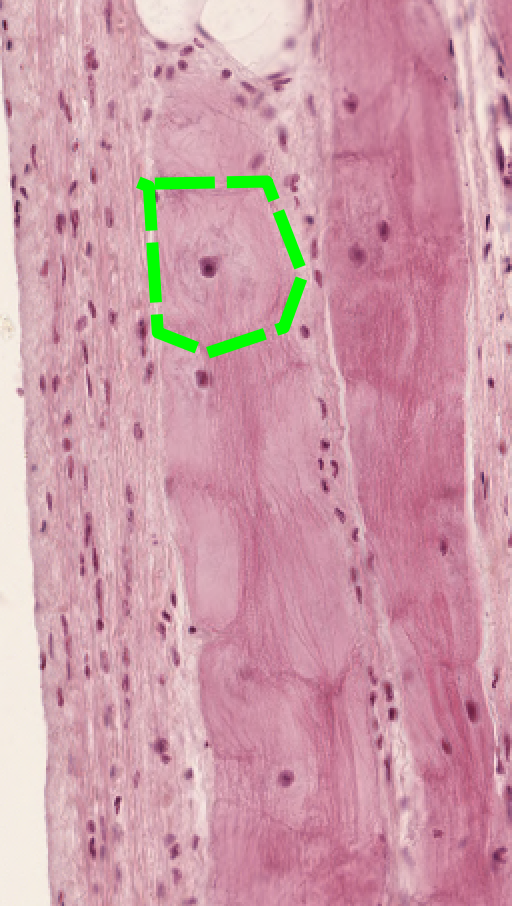
A single Purkinje fiber 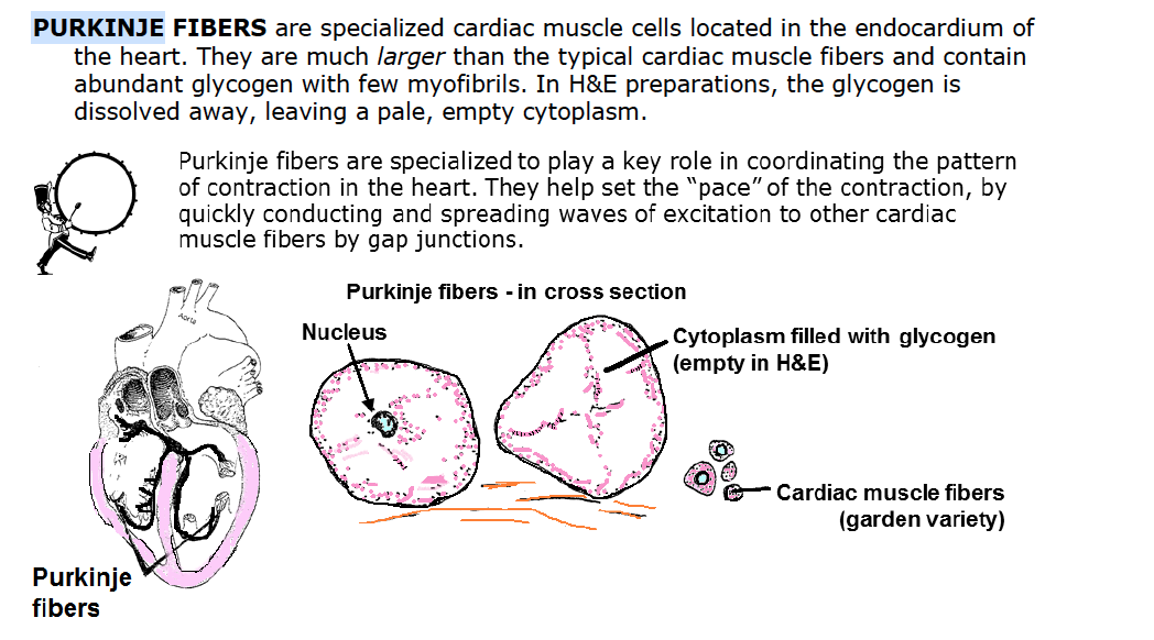
27
Describe the tissue 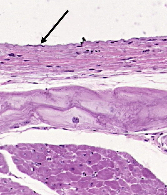
simple squamous endo of the endocardium of the heart
28
WHat is this?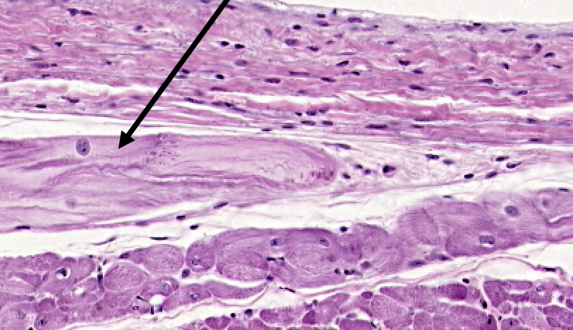
Purkinje fiber in endocardium
29
WHat is this?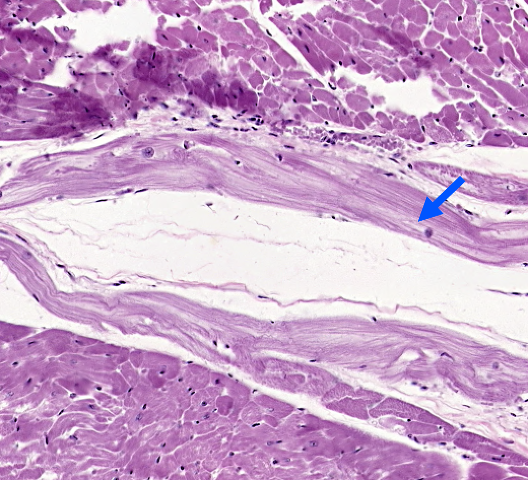
Purkinje fiber
30
what muscle is this and what is the arrow pointing to?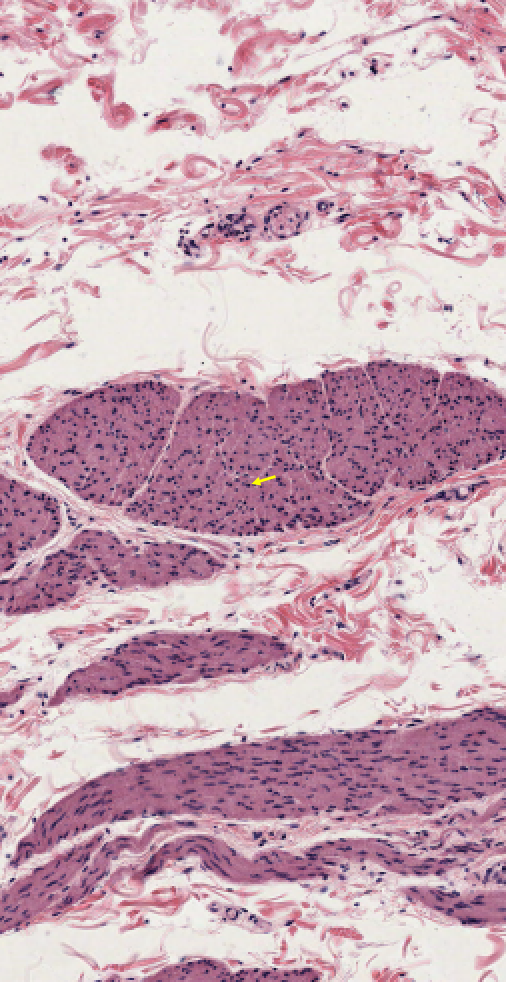
31
What muscle is this?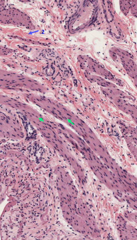
smooth, pointing to cigar shaped nuclei, 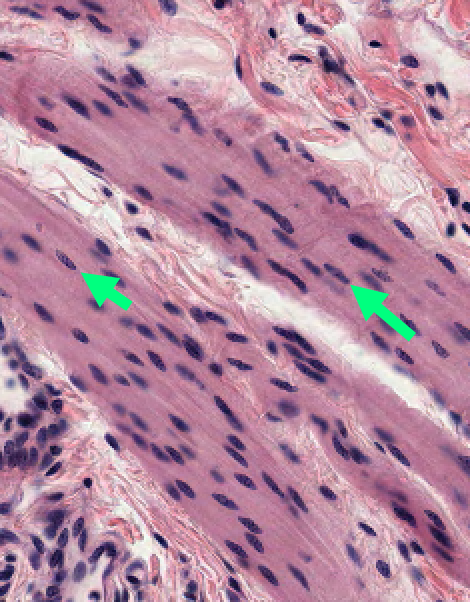
32
Describe what is happening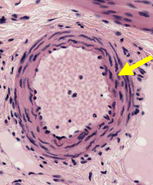
smooth muscle running concentrically in the wall of a muscular artery
33
Classify the tissue 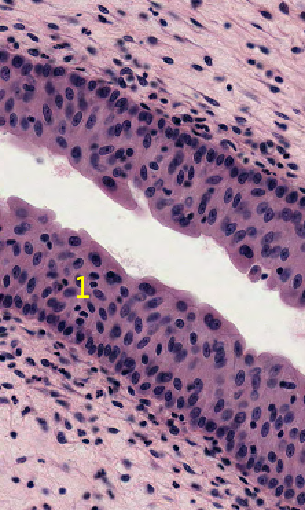
Transitional
34
Classify the fiber 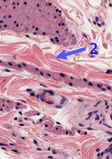
Collagen (type I) fiber
35
Describe what is being shown 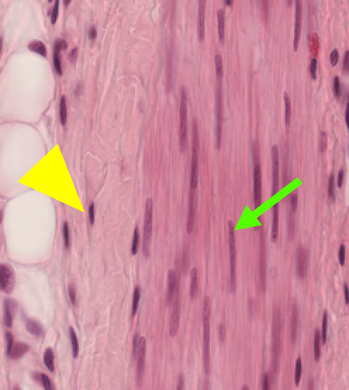
nuclei of smooth muscle (green) vs fibroblast (yellow triangle)
36
Classify the tissue 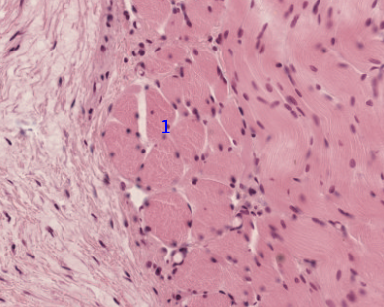
– skeletal muscle (in cross section, x.s.)
37
Classify the tissue 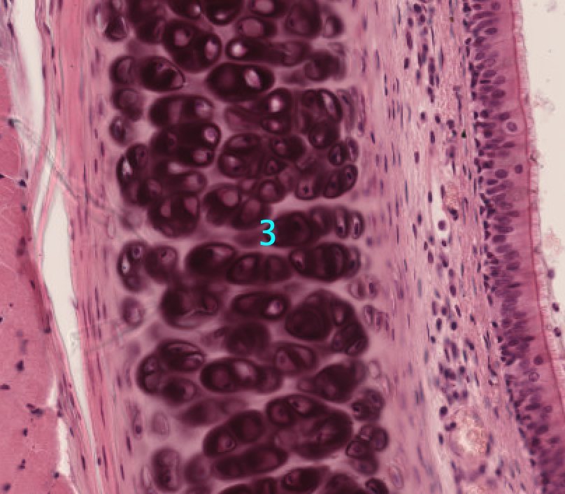
hyaline cartilage (with lots of cells)
38
What type of muscle is this 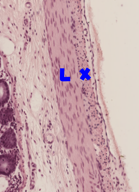
smooth longitudinal is L and cross is X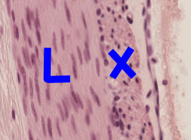
39
Identify the cell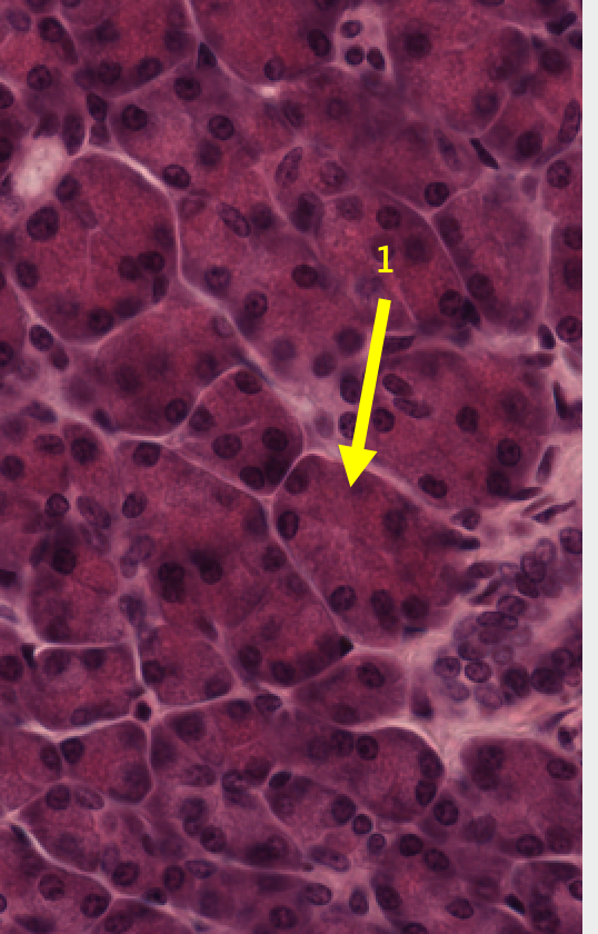
serous cell
40
Classify the tissue 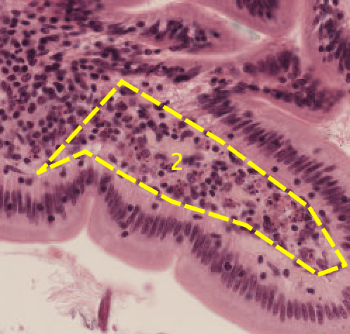
Loose connective tissue with a lot of cells
41
Classify the tissue 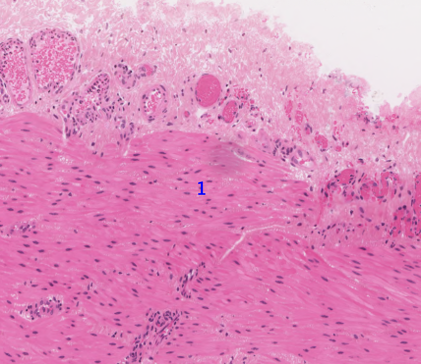
smooth muscle 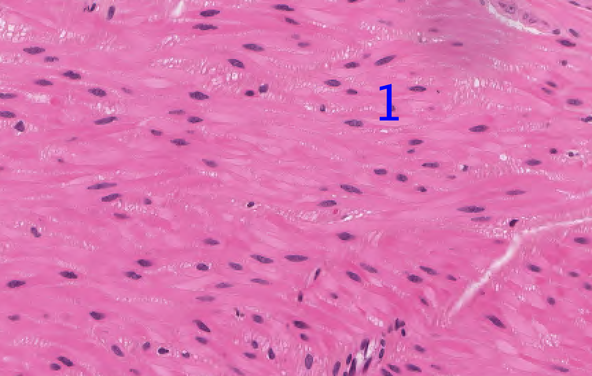
42
Identify the cells 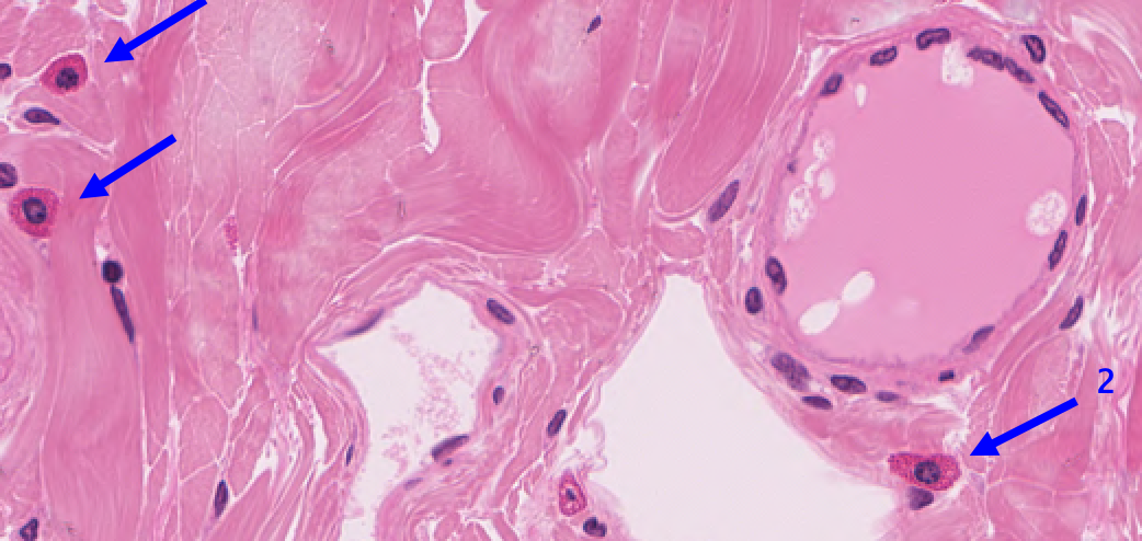
Mast cells
43
Classify the tissue and orientation of section 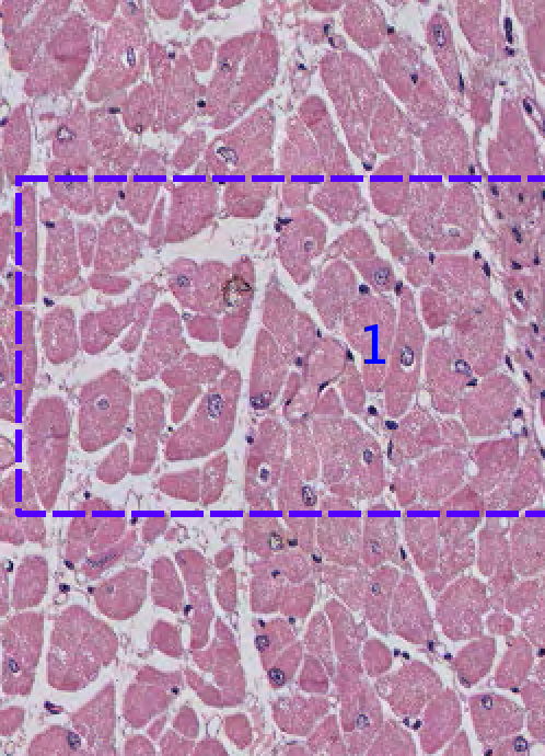
cardiac and cross
44
Identify the structure 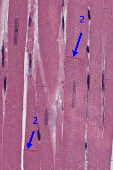
Intercalated discs
45
Classify the tissue 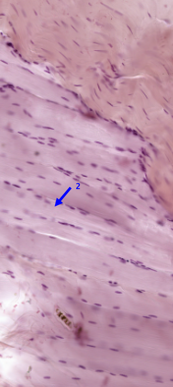
skeletal muscle
46
Classify the tissue 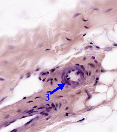
smooth muscle (around a vessel)
47
Classify the tissue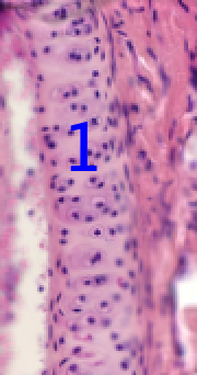
Hyaline cartilage
48
Classify the tissue –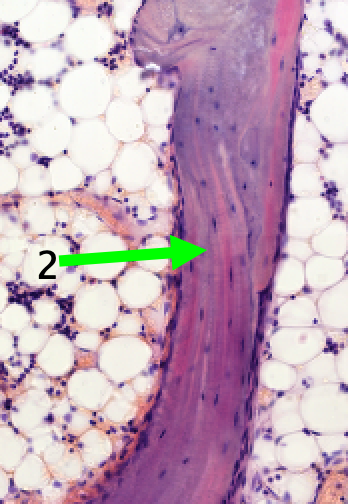
spongy
49
Classify the tissue 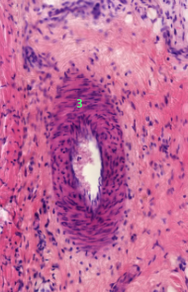
– smooth muscle (in a vessel)
50
Classify the tissue 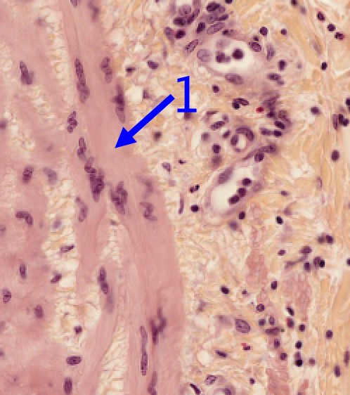
smooth muscle
51
Classify the tissue 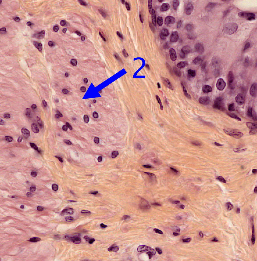
smooth muscle
52
Classify the tissue 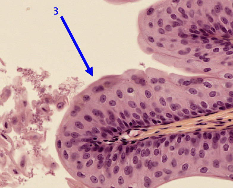
transitional epithelium
53
Classify the tissue 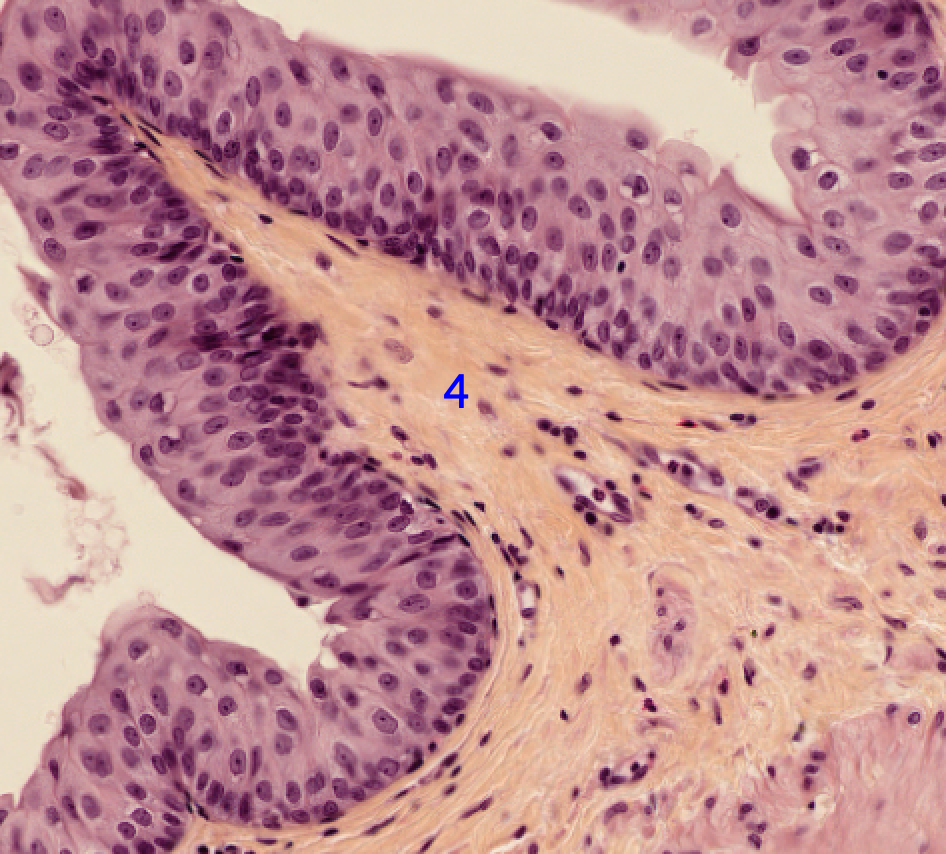
dense irregular connective tissue