Hist Lab 4 - Bone
Study these flashcardsFree · spaced repetition · iPhone, Android, Mac & web
All cards
1
General Characteristics of Bone
2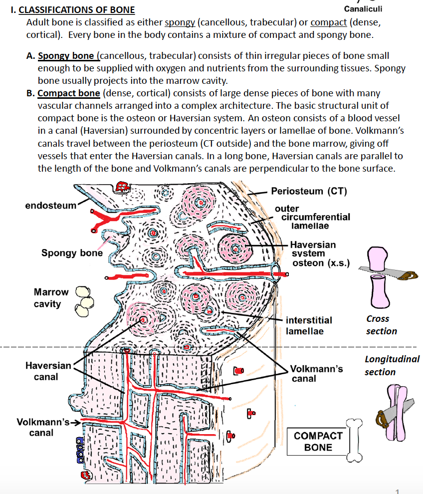
Classification of Bone
3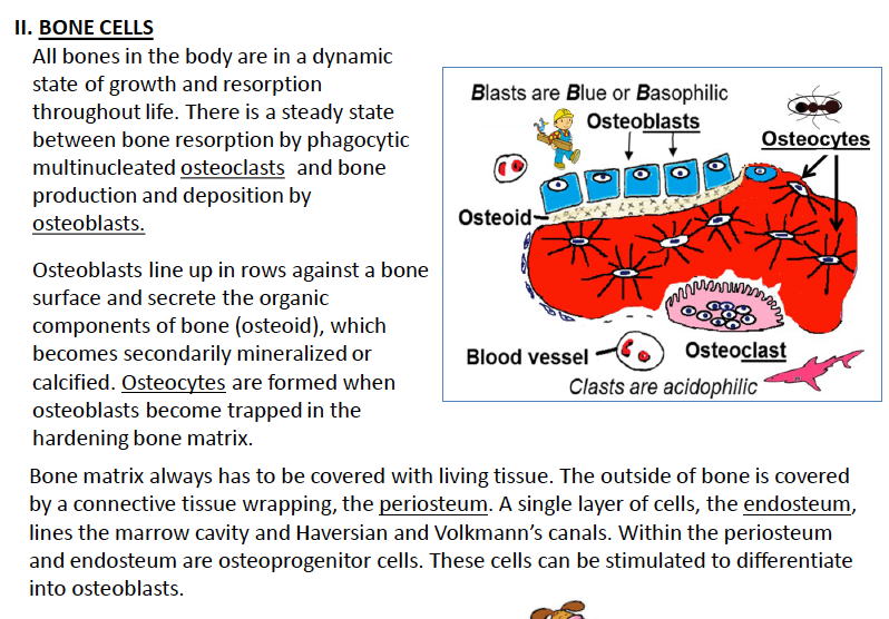
General Characteristics of bone cells
4
WHat are osteoprogenitor cells
1. Osteoprogenitor cells – osteogenic stem cell
a. mesenchymal stem cell hiding in periosteum and endosteum
b. can undergo mitosis (that’s why it is called a stem cell)
c. differentiate into osteoblasts
a. mesenchymal stem cell hiding in periosteum and endosteum
b. can undergo mitosis (that’s why it is called a stem cell)
c. differentiate into osteoblasts
5
what are osteoblasts
2. Osteoblasts – primary producer of bone matrix
a. basophilic cells with round euchromatic nucleus (typical protein secreting cell, RER)
b. lined up in one-cell thick rows on surfaces of bone (appositional growth)
c. secrete organic unmineralized components of bone matrix called osteoid (collagen & ground substance) and matrix vesicles that initiate bone mineralization.
d. terminally differentiated, can NOT undergo mitosis
e. when trapped in mineralizing matrix, becomes an osteocyte
a. basophilic cells with round euchromatic nucleus (typical protein secreting cell, RER)
b. lined up in one-cell thick rows on surfaces of bone (appositional growth)
c. secrete organic unmineralized components of bone matrix called osteoid (collagen & ground substance) and matrix vesicles that initiate bone mineralization.
d. terminally differentiated, can NOT undergo mitosis
e. when trapped in mineralizing matrix, becomes an osteocyte
6
what are osteocytes
Osteocytes – trapped in lacunae (appears like a clear space)
a. trapped in lacunae, with long, thin cell processes extending into tiny channels called canaliculi
b. communicate with each other via gap junctions at the tips of their processes
c. maintain bone matrix (some synthetic and resorptive ability)
d. osteocytes can NOT divide and are terminally differentiated.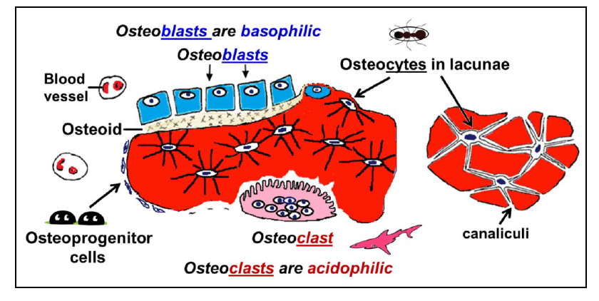
a. trapped in lacunae, with long, thin cell processes extending into tiny channels called canaliculi
b. communicate with each other via gap junctions at the tips of their processes
c. maintain bone matrix (some synthetic and resorptive ability)
d. osteocytes can NOT divide and are terminally differentiated.
7
what are osteoclasts
Osteoclasts – bone resorbing cell
a. phagocytic, derived from monocytes in blood
b. large multinucleated cell with acidophilic cytoplasm (abundant mitochondria) and a ruffled or high folded cell membrane, in a shallow depression on the bone surface (Howship’s lacunae).
c. contain many lysosomes, secretes acid, collagenase, and other lytic enzymes, that break down bone matrix (resorption), releasing calcium & phosphate into blood. Cell membrane is highly folded into a ruffled border.
a. phagocytic, derived from monocytes in blood
b. large multinucleated cell with acidophilic cytoplasm (abundant mitochondria) and a ruffled or high folded cell membrane, in a shallow depression on the bone surface (Howship’s lacunae).
c. contain many lysosomes, secretes acid, collagenase, and other lytic enzymes, that break down bone matrix (resorption), releasing calcium & phosphate into blood. Cell membrane is highly folded into a ruffled border.
8
Identify the structure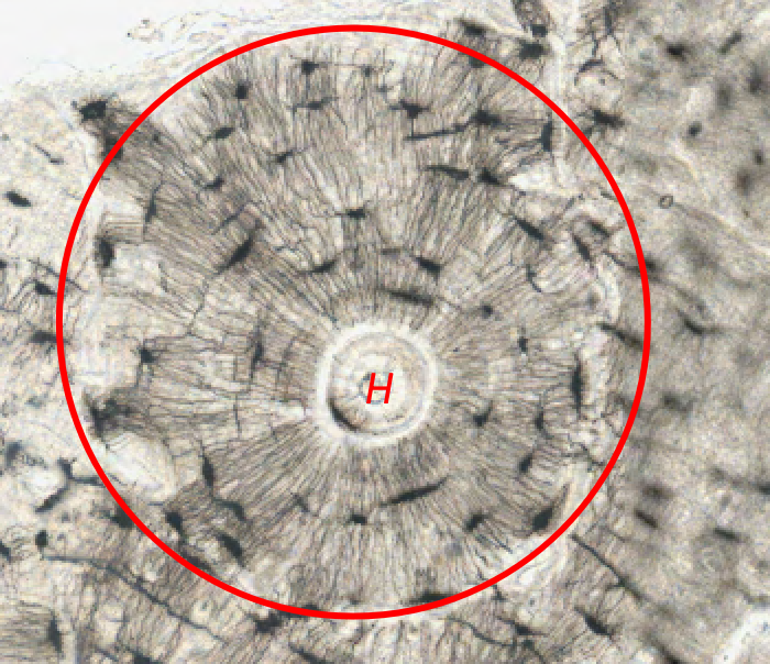
Haversian system inside a osteon
-google - Haversian canals are a series of tubes around narrow channels formed by lamellae. The Haversian canals surround blood vessels and nerve fibers throughout the bone and communicate with osteocytes. The canals and the surrounding lamellae are called a Haversian system (or an osteon).
-google - Haversian canals are a series of tubes around narrow channels formed by lamellae. The Haversian canals surround blood vessels and nerve fibers throughout the bone and communicate with osteocytes. The canals and the surrounding lamellae are called a Haversian system (or an osteon).
9
Identify the structures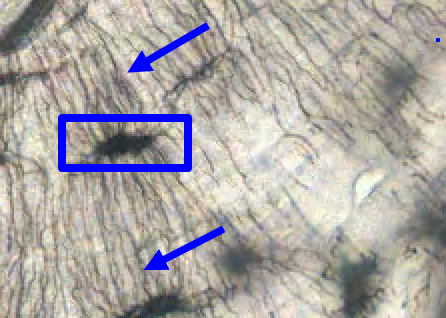
arrows - tiny canaliculi
rectangle - lacunae - dark stain
rectangle - lacunae - dark stain
10
Identify the structure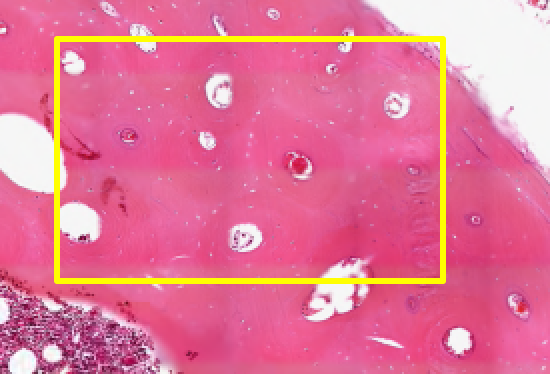
Haversian canal in compact bone (white structure) 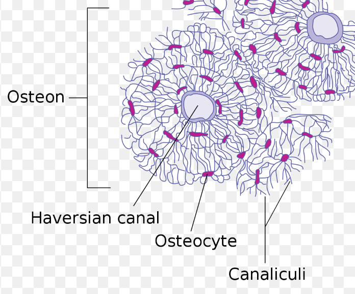
11
Identify the structure 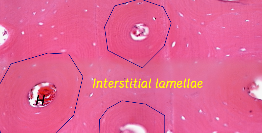
(like the image says) interstitial lamallae that is inbetween osteons
12
Identify the structure 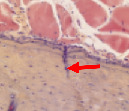
Volkmann's canal entering the bone from the periosteum
13
Identify the structure 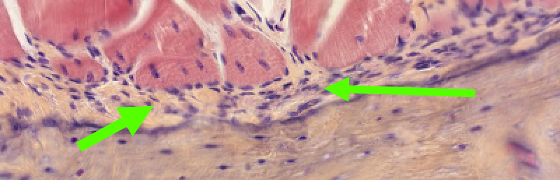
periosteum that is continuous with dense CT of tendon of skeletal muscl
14
Identify the cell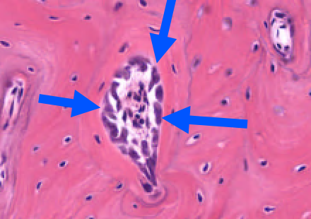
osteoblasts (secretes bone matrix), eventually a haversian canal will be left
15
Identify the structure 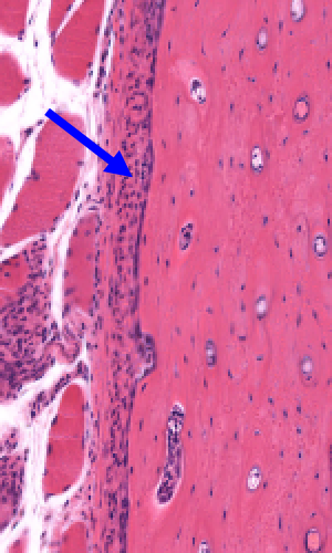
periosteum
16
Longitudinal of a long bone 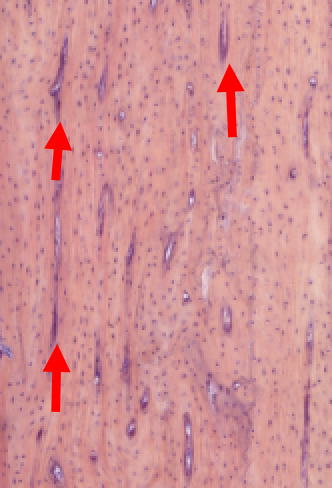
Haversian canals are parallel to bone surface
17
Identify the tissue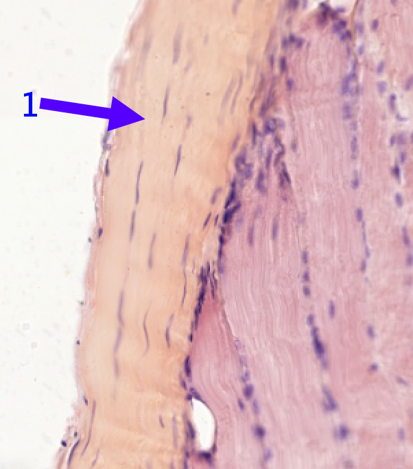
dense regular connective tissue
18
Identify the structure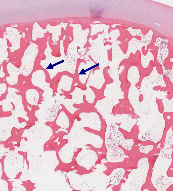
Spongy bone
19
Classify the tissue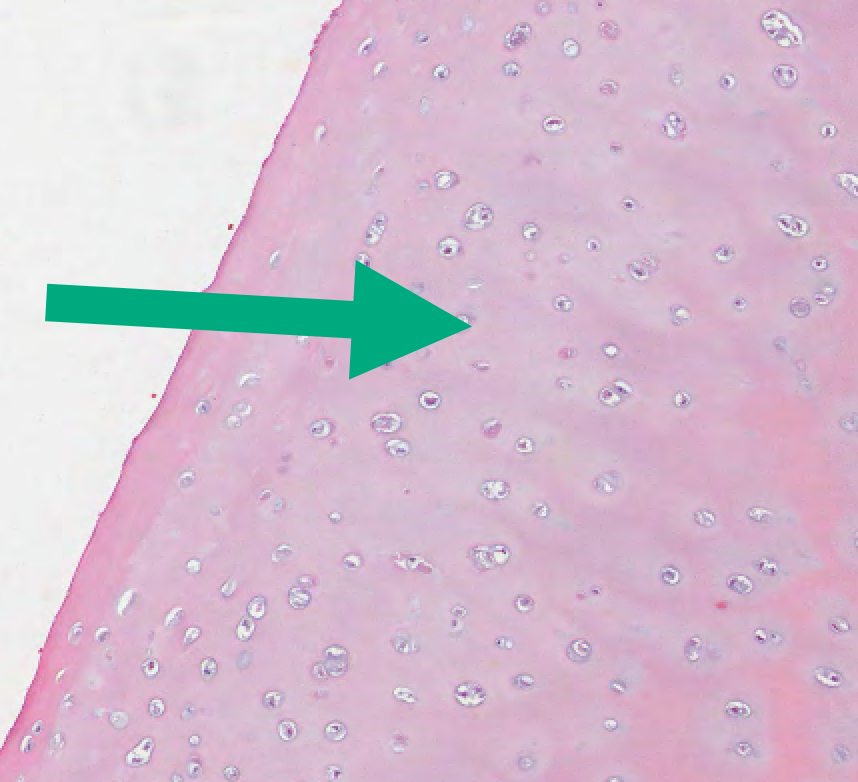
hyaline cartilage
20
Identify the cell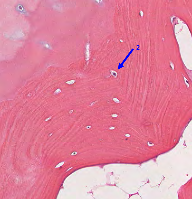
osteocyte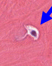
21
Identify the cell 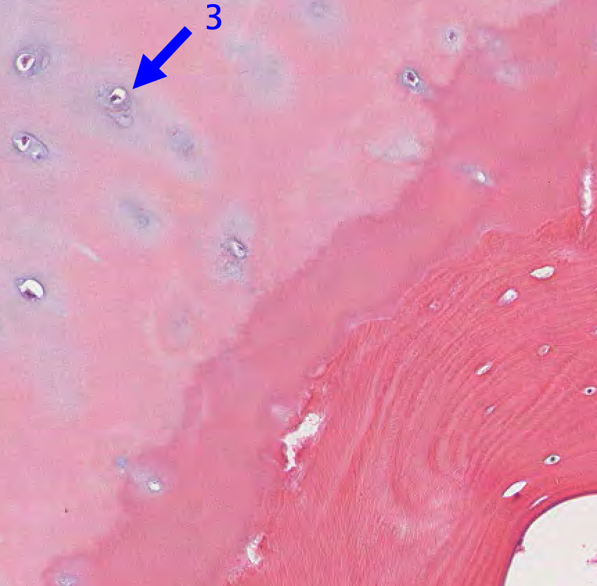
chondrocyte
22
Identify the cell 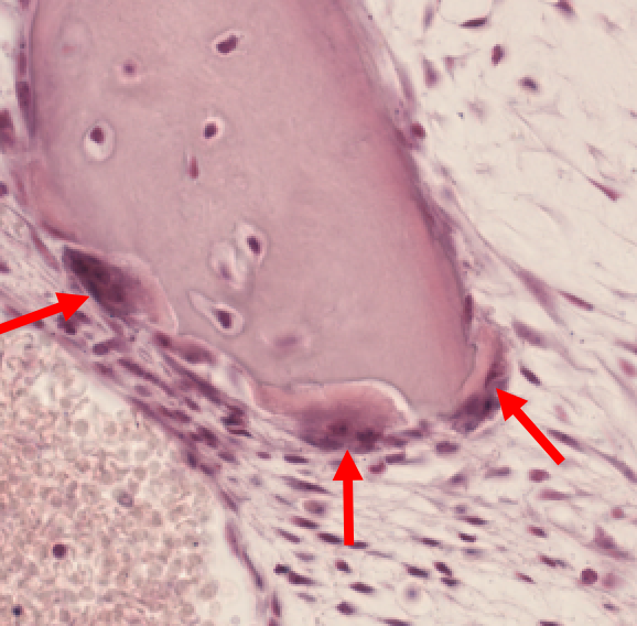
osteoclasts - multinucleated, phagocytic cells breaking down bone
23
Identify what is happening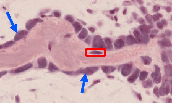
Osteoblasts lined up in rows against spongy bone. Newly formed osteocyte is indicated in red rectangle.
24
Identify the tissue 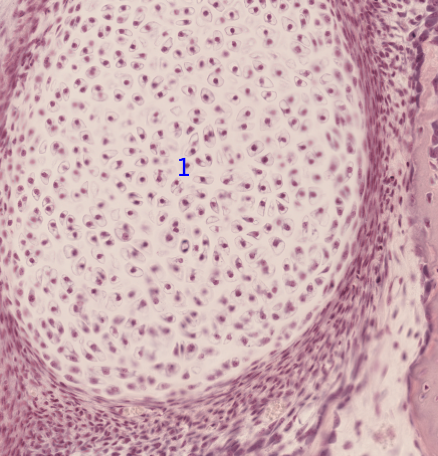
Hyaline
25
Identify what the blue arrows are pointing to 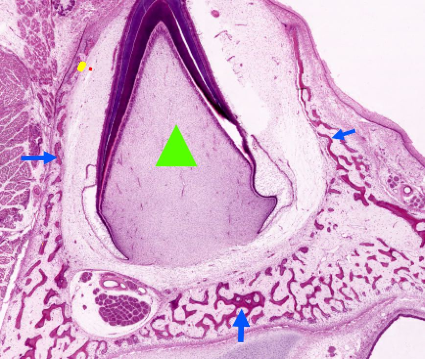
spongy bone surrounding a developing tooth
26
WHat is this?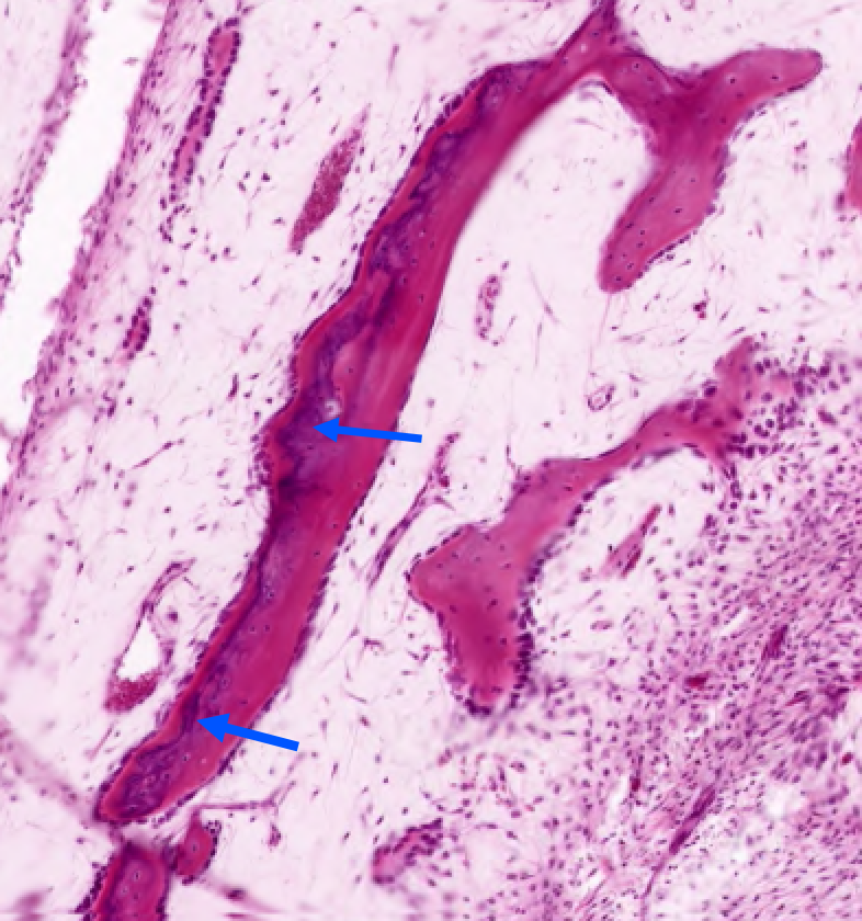
uneven staining in spongy bone
27
What is the arrow pointing to? 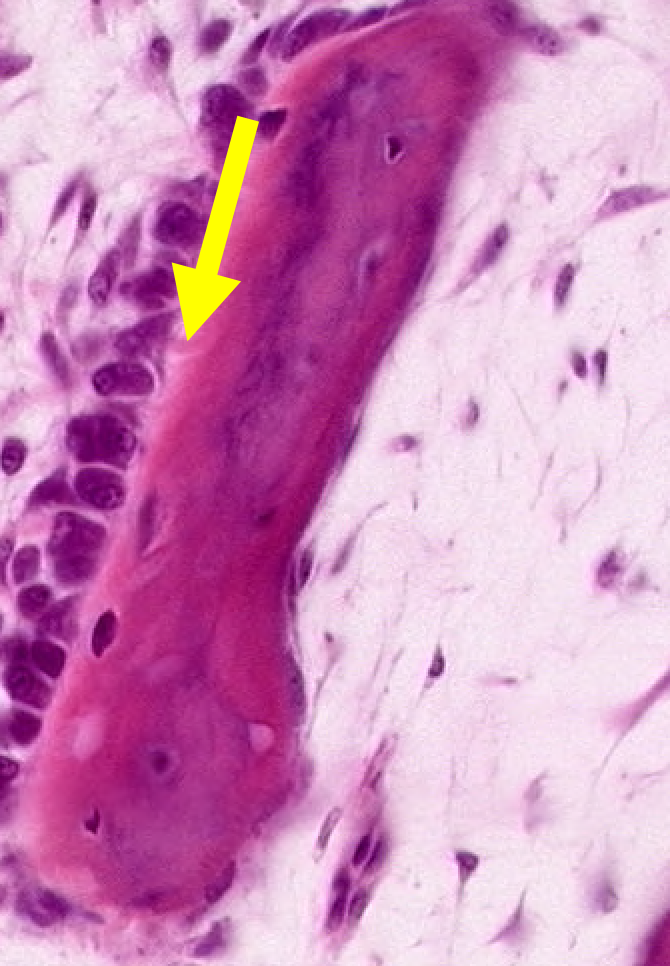
This is a unmineralized matrix called osteoid (appears as a light region between the osteoblasts and the red calcified bone matrix). This is secreted by osteoblasts
28
What does the H stand for?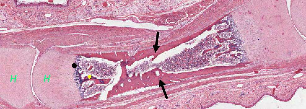
Hyaline cartilage. Explaining this picture: start with all cartilage and then begin to form bone bc more oxygen and blood vessels. The cartilage then begins to die (center).
29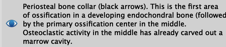
WHat are the black arrows pointing to?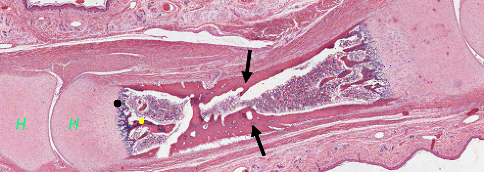
30
Classify the zones (numbers)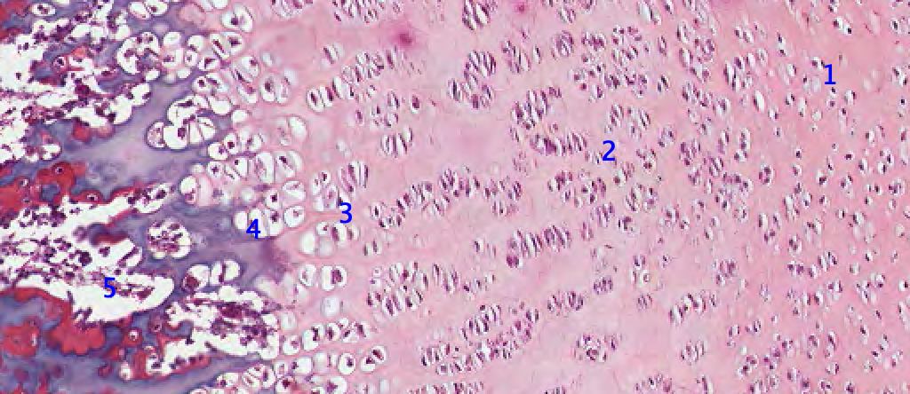
31
Identify the cell 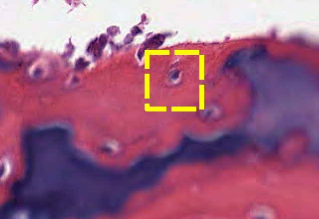
Osteocyte in red bone matrix
32
WHat is the arrow pointing to?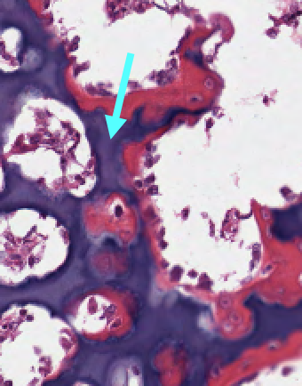
Calcified cartilage matrix
33
Identify the cell 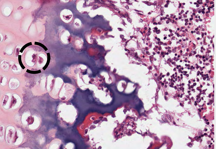
Hypertrophied chondrocytes in enlarged lacunae
34
Identify the cell 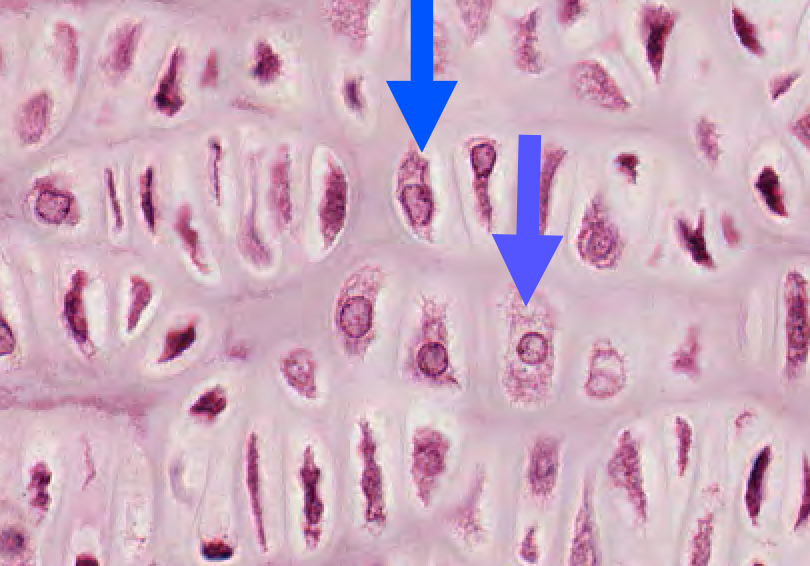
Hypertrophied chondrocytes and lacunae
35
What are the arrow pointing to?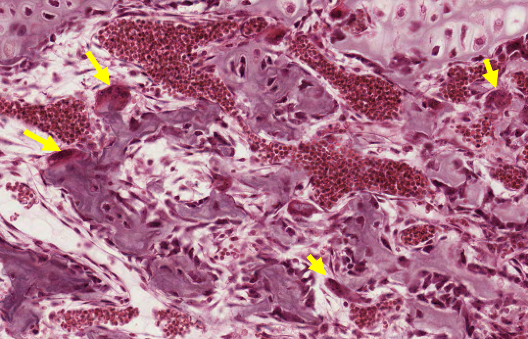
Osteoclasts in zone of resorption 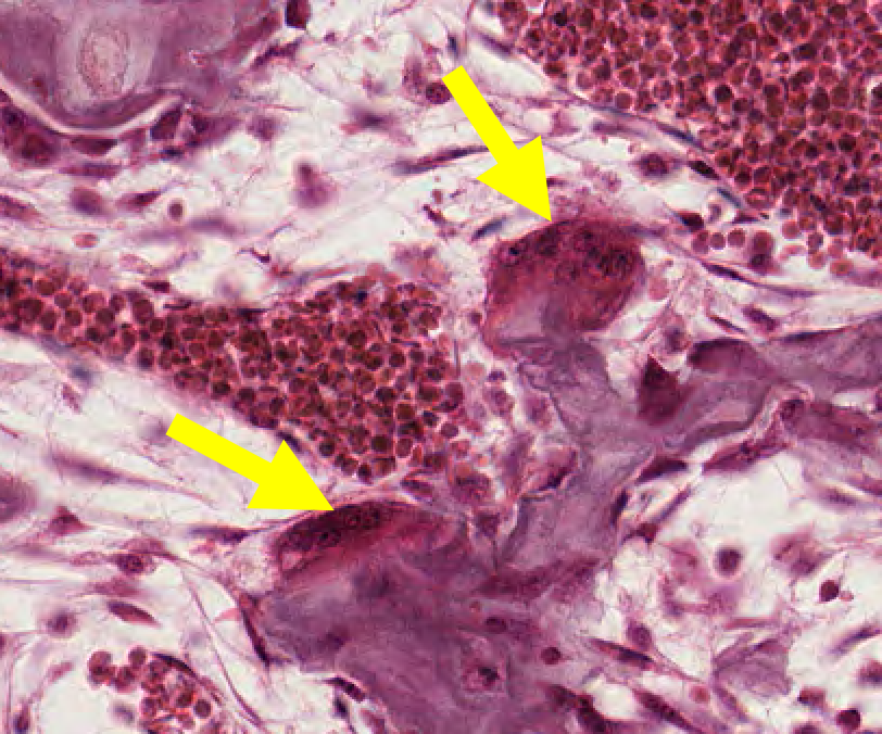
36
Identify the structure 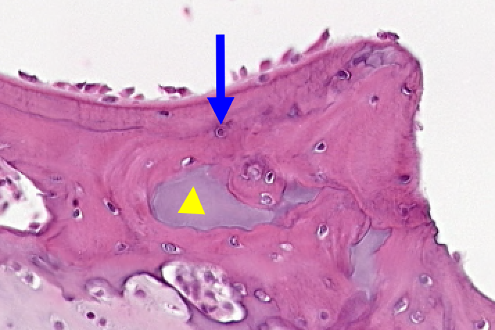
37
Identify what is in blue 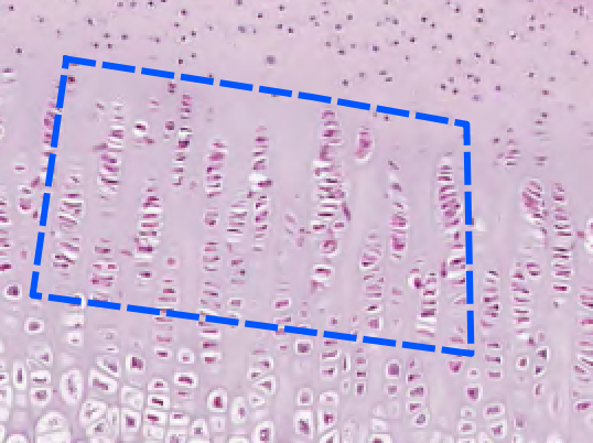
zone of proliferation
38
WHat is outlined?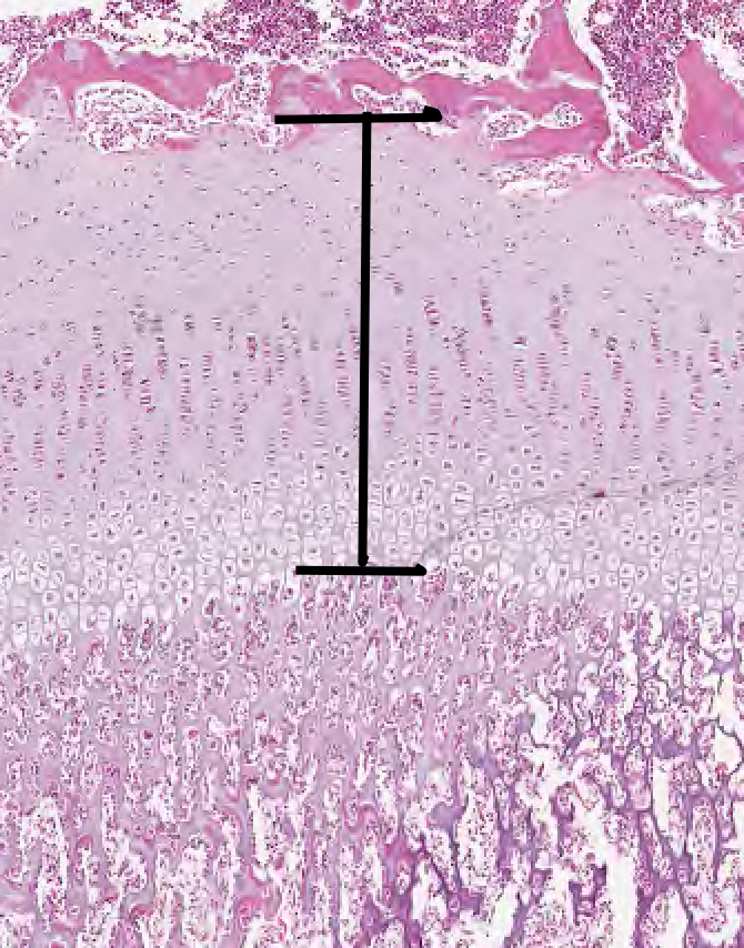
Growth of epiphyseal plate
39
WHat is shown?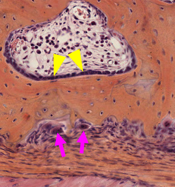
40
WHat is the arrow pointing to?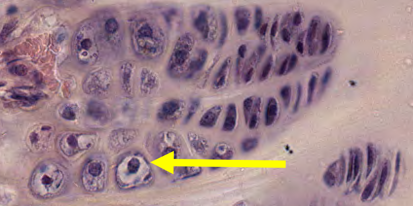
Hypertrophied chondrocyte
41
Classify the tissue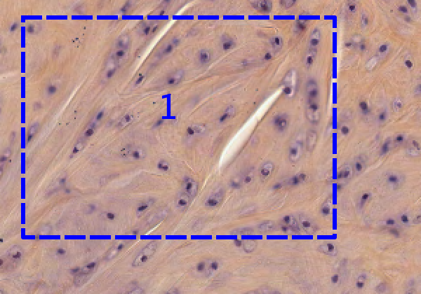
fibrocartilage
42
Identify the cell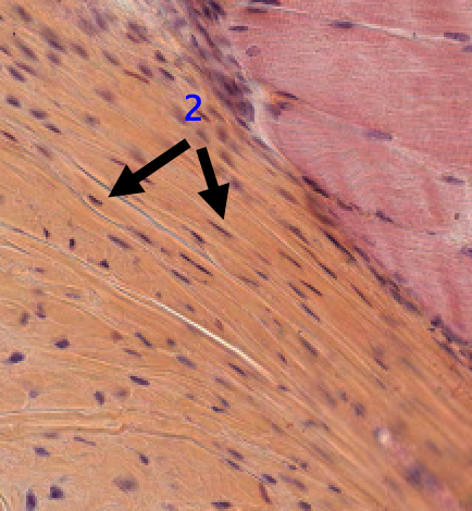
fibroblast
43
Identify the cell 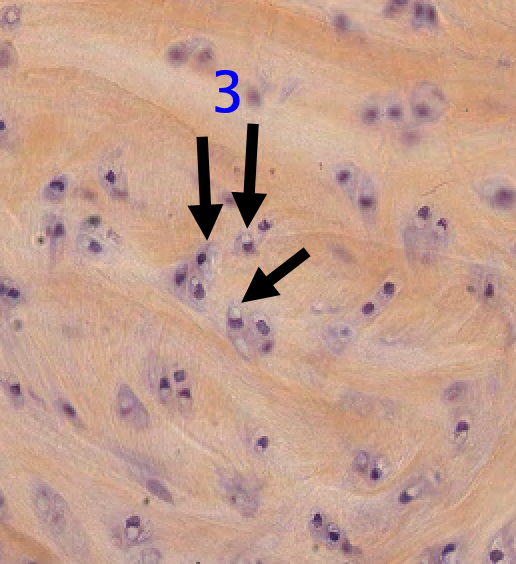
chondrocytes
-a cell which has secreted the matrix of cartilage and become embedded in it.
-a cell which has secreted the matrix of cartilage and become embedded in it.
44
Identify the cell 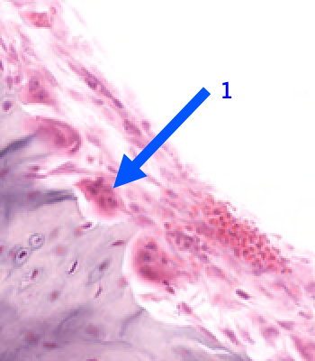
osteoclast
45
Classify the tissue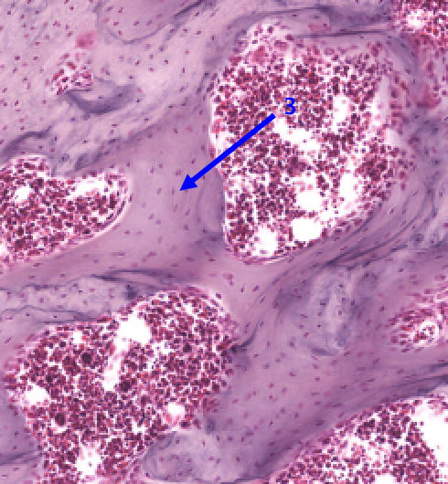
spongy bone
46
Lamellar or woven?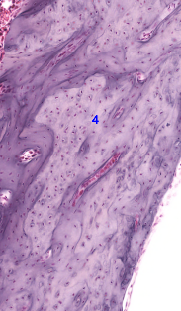
It is woven or primary bone
47
Classify the tissue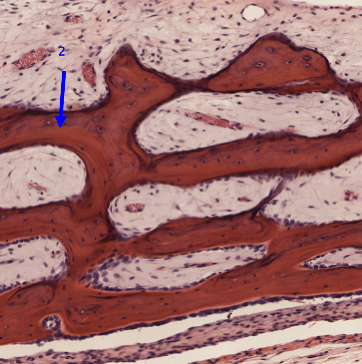
spongy
48
Identify the cells 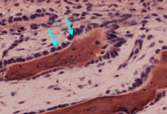
osteoblasts
49
Identify the fiber 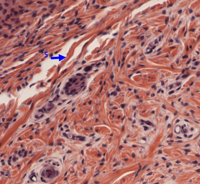
ollagen fiber (Type I)
50
Identify the lighter region of the tissue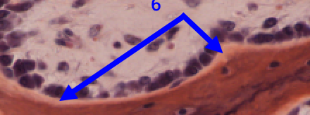
osteoid
51
Identify the zone of the growth plate 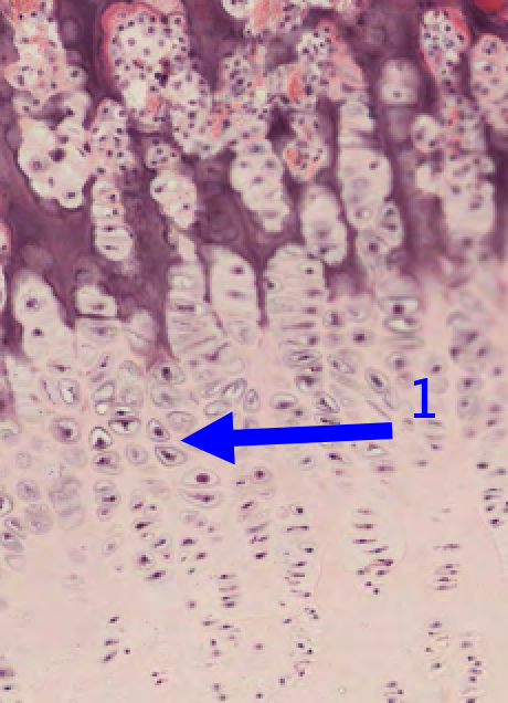
zone of hypertrophy
52
Identify the cells 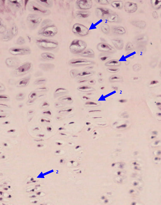
chondrocytes 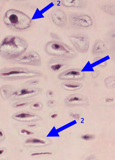
53
Identify the purple matrix or material 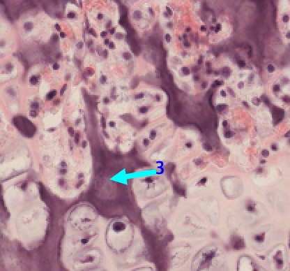
calcified cartilage matrix
54
Identify the cell 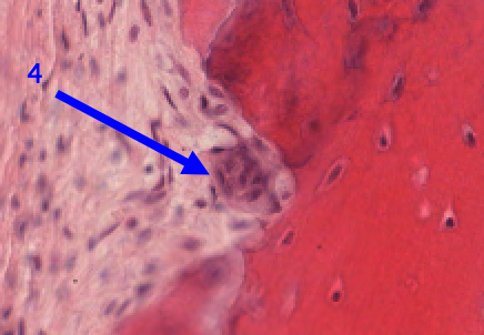
osteoclast
55
Identify the structure 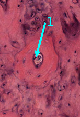
aversian canal in an osteon
56
Identify the structure 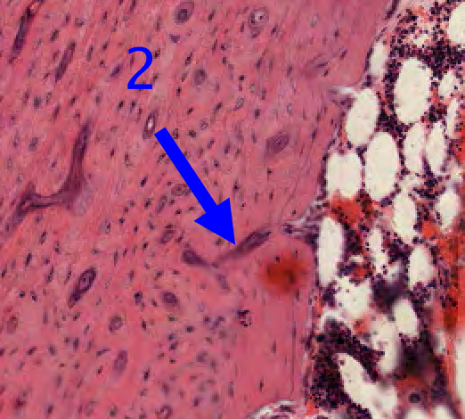
volkmann’s canal
57
Classify the tissue and is this a longitudinal or cross section? 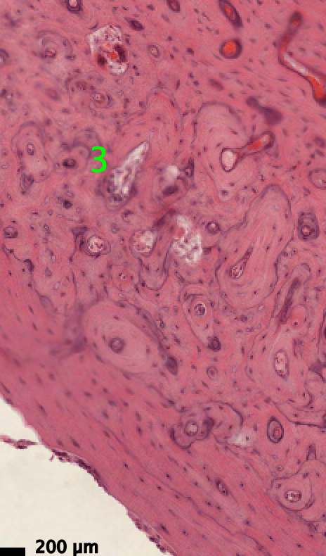
Compact bone in cross section 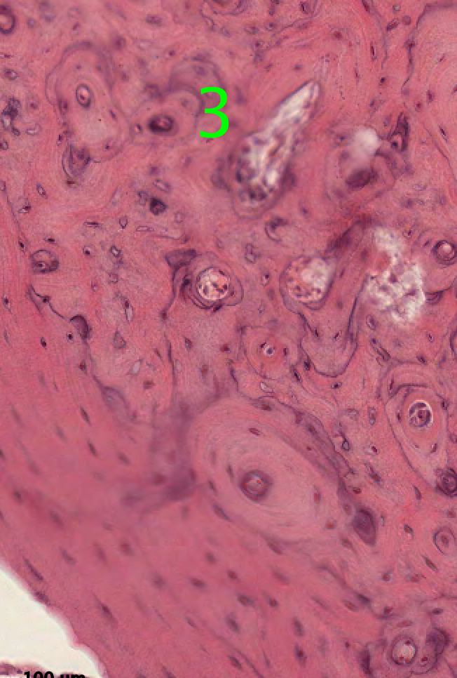
58
is this a longitudinal or cross section? 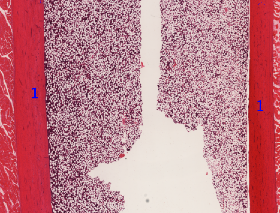
– longitudinal section of compact
bone
bone