Chapter 19
Study these flashcardsFree · spaced repetition · iPhone, Android, Mac & web
All cards
1
When the original flat embryonic disc folds into a tube, the becomes trapped inside forming the
epithelial lining of the
epithelial lining of the
When the original flat embryonic disc folds into a tube, the endoderm becomes trapped inside forming the
epithelial lining of the primitive gut tube.
epithelial lining of the primitive gut tube.
2
Mesoderm cells surround the tube and form and
Mesoderm cells surround the tube and form muscle and connective
tissue layers.
tissue layers.
3
.Neural crest cells migrate into the gut and differentiate into and cells.
.Neural crest cells migrate into the gut and differentiate into ganglion cells (Auerbach’s and
Meissner’s nerve plexus) and enteroendocrine cells.
Meissner’s nerve plexus) and enteroendocrine cells.
4
The primitive gut tube is divided into , , and , each supplied by a specific artery
and .
and .
The primitive gut tube is divided into FOREGUT, MIDGUT, and HINDGUT, each supplied by a specific artery
and autonomic nerve (see Table).
and autonomic nerve (see Table).
5
Foregut is from to
Foregut is from mouth to pharynx.
6
Foregut proper is , , and
Foregut proper is esophagus, stomach, and proximal duodenum
7
The midgut is the rest of the and the
The midgut is the rest of the small intestines and the proximal 2/3 of the transverse colon
8
The hindgut is the (splenic flexure) to (above the pectinate line)
The hindgut is the last 1/3 of the transverse colon (splenic flexure) to anal canal (above the pectinate line)
9
FOREGUT- gives rise to the , , , and .
FOREGUT- gives rise to the respiratory primordia, pancreas, liver, and gall bladder.
10
MIDGUT - connects to the which eventually becomes restricted to a which is
incorporated into the
incorporated into the
MIDGUT - connects to the yolk sac which eventually becomes restricted to a thin duct (vitelline duct) which is
incorporated into the umbilical cord.
incorporated into the umbilical cord.
11
The rapid growth and twisting of the results in a
temporary herniation of the developing intestines into the yolk sac.
temporary herniation of the developing intestines into the yolk sac.
The rapid growth and twisting of the midgut results in a
temporary herniation of the developing intestines into the yolk sac.
temporary herniation of the developing intestines into the yolk sac.
12
HINDGUT- enlargement at the end, the becomes subdivided into two passages, one
for food waste ( ) and the other for urine ( ).
for food waste ( ) and the other for urine ( ).
HINDGUT- enlargement at the end, the cloaca (sewer), becomes subdivided into two passages, one
for food waste (anorectal canal) and the other for urine (urethra and urinary bladder).
for food waste (anorectal canal) and the other for urine (urethra and urinary bladder).
13
Absence or a closure of an orifice or passage (like GI tube) in the body.
Atresia: Absence or a closure of an orifice or passage (like GI tube) in the body.
14
Abnormal passage between two hollow or tubular organs or between the surface and a tubular
organ
organ
Fistula: Abnormal passage between two hollow or tubular organs or between the surface and a tubular
organ
organ
15
Abnormal narrowing or contraction of a passage of the body
Stenosis Abnormal narrowing or contraction of a passage of the body
16
Abnormal passage between two hollow or tubular organs or between the surface and a tubular
organ
organ
Fistula: Abnormal passage between two hollow or tubular organs or between the surface and a tubular
organ
organ
17
The initially hollow gut tube becomes clogged with replicating epithelial cells around Weeks
or reopening of the tube occurs when the overgrowth cells die a few weeks later.
or reopening of the tube occurs when the overgrowth cells die a few weeks later.
The initially hollow gut tube becomes clogged with replicating epithelial cells around Weeks 6-7.
Recanalization or reopening of the tube occurs when the overgrowth cells die a few weeks later.
Recanalization or reopening of the tube occurs when the overgrowth cells die a few weeks later.
18
Failure to reform the lumen can result in or . This is especially common in two regions
of the foregut, the and .
of the foregut, the and .
Failure to reform the lumen can result in stenosis or atresia. This is especially common in two regions
of the foregut, the esophagus and duodenum.
of the foregut, the esophagus and duodenum.
19
The fetus continually swallows the , absorbs it into the body, and then adds fluid back into the
in the form of .
in the form of .
The fetus continually swallows the amniotic fluid, absorbs it into the body, and then adds fluid back into the
amnion in the form of fetal urine.
amnion in the form of fetal urine.
20
Increased amounts of can be indicative of a
(defective recanalization, twisting, compromised blood supply, tracheoesophageal
fistula, etc.) or the failure of the to develop (usually due to anencephaly).
(defective recanalization, twisting, compromised blood supply, tracheoesophageal
fistula, etc.) or the failure of the to develop (usually due to anencephaly).
Increased amounts of amniotic fluid (polyhydramnios) can be indicative of a
blockage in the GI system (defective recanalization, twisting, compromised blood supply, tracheoesophageal
fistula, etc.) or the failure of the swallow reflex to develop (usually due to anencephaly).
blockage in the GI system (defective recanalization, twisting, compromised blood supply, tracheoesophageal
fistula, etc.) or the failure of the swallow reflex to develop (usually due to anencephaly).
21
The foregut extends from the to the at the entrance of the bile duct. Buds arising from the
foregut give rise to the , , & .
foregut give rise to the , , & .
The foregut extends from the oral cavity to the duodenum at the entrance of the bile duct. Buds arising from the
foregut give rise to the respiratory primordium, liver, gall bladder & pancreas.
foregut give rise to the respiratory primordium, liver, gall bladder & pancreas.
22
The respiratory system develops as a bud that appears at in the ventral wall of the endoderm lined
foregut.
foregut.
The respiratory system develops as a bud that appears at Week 4 in the ventral wall of the endoderm lined
foregut.
foregut.
23
Growth of a septum separates the between the foregut (future ) from the bud (future
), and restricts their connection to a small opening at the .
), and restricts their connection to a small opening at the .
Growth of a septum separates the between the foregut (future esophagus) from the bud (future
trachea, bronchi and lung), and restricts their connection to a small opening at the larynx.
trachea, bronchi and lung), and restricts their connection to a small opening at the larynx.
24
Failure of the septum to develop properly during weeks can result in abnormal connections and/orblockage between the trachea and esophagus (ex. tracheoesophageal fistula with esophageal atresia).
Failure of the septum to develop properly during weeks 4-5 can result in abnormal connections and/orblockage between the trachea and esophagus (ex. tracheoesophageal fistula with esophageal atresia).
25
The most common defect (90%) is a between the . The
newborn infant and the gastric contents reflex into the lungs.
newborn infant and the gastric contents reflex into the lungs.
The most common defect (90%) is a fistula between the esophagus and distal third of the trachea. The
newborn infant gags after feeding and the gastric contents reflex into the lungs.
newborn infant gags after feeding and the gastric contents reflex into the lungs.
26
Prior to birth there is an increased volume of amniotic fluid (polyhydramnios). Amniotic fluid is normally
and , but in these infants the swallowed fluid is blocked
from entering the intestines.
and , but in these infants the swallowed fluid is blocked
from entering the intestines.
Prior to birth there is an increased volume of amniotic fluid (polyhydramnios). Amniotic fluid is normally
swallowed by the fetus and resorbed in the intestines, but in these infants the swallowed fluid is blocked
from entering the intestines.
swallowed by the fetus and resorbed in the intestines, but in these infants the swallowed fluid is blocked
from entering the intestines.
27
The respiratory bud branches repeatedly, forming and which continue to branch into
smaller and smaller channels, ending in the
smaller and smaller channels, ending in the
The respiratory bud branches repeatedly, forming bronchial and lungs buds which continue to branch into
smaller and smaller channels, ending in the alveoli (blind sacs).
smaller and smaller channels, ending in the alveoli (blind sacs).
28
The , , and of the gall bladder develop from buds that arise from the distal foregut (future )
The hepatocytes, bile ducts, and epithelial lining of the gall bladder develop from buds that arise from the distal foregut (future dudoenum)
29
Failure of the biliary ducts to develop or recanalize leads to or
Failure of the biliary ducts to develop or recanalize leads to biliary atresia or stenosis
30
Biliary atresia or stenosis is more likely to occur in . likely needed for survivial.
Biliary atresia or stenosis is more likely to occur in extrahepatic ducts . Liver transplants likely needed for survivial.
31
The pancreas develops as and buds, arising from oppoisite sides of the future duodenum. Rotation of the stomach and duodenum results in migration and fusion of the pancreatic buds dorsally to form the
The pancreas develops as dorsal and ventral buds, arising from oppoisite sides of the future duodenum. Rotation of the stomach and duodenum results in migration and fusion of the pancreatic buds dorsally to form the definitive pancreas.
32
The is formed when the bud splits and encircles the dudenum, constristing the lumen, leading to duodenal stenosis or atresia
The annular pancreas is formed when the ventral pancreatic bud splits and encircles the dudenum, constristing the lumen, leading to duodenal stenosis or atresia 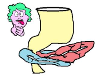
33
is a common malformation (1 in 150 males, 1 in 750 females) caused by hypertrophy of the smooth muscle of the pyloric sphincter. This severe stenosis blocks the movement of food and is associated with projectile vomiting in the infant.
Congenital Pyloric Stenosis is a common malformation (1 in 150 males, 1 in 750 females) caused by hypertrophy of the smooth muscle of the pyloric sphincter. This severe stenosis blocks the movement of food and is associated with projectile vomiting in the infant. 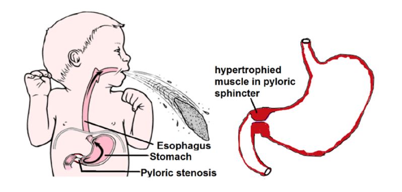
34
The midgut begins in the (after the entrance of the bile duct) and includes including the and
The midgut begins in the duodenum (after the entrance of the bile duct) and includes 2/3rds of the large
intestine including the cecum and appendix
intestine including the cecum and appendix
35
The midgut is connected to the yolk sac through the
at the site of the .
at the site of the .
The midgut is connected to the yolk sac through the vitelline duct
at the site of the future ileum.
at the site of the future ileum.
36
During the midgut grows and elongates so quickly that the developing intestines, no longer able to fit
inside the abdominal cavity, herniates into the and .
inside the abdominal cavity, herniates into the and .
During week 5 the midgut grows and elongates so quickly that the developing intestines, no longer able to fit
inside the abdominal cavity, herniates into the vitelline duct and yolk sac.
inside the abdominal cavity, herniates into the vitelline duct and yolk sac.
37
The unequal accelerated growth
also causes the herniated midgut to loop on itself forming the
also causes the herniated midgut to loop on itself forming the
The unequal accelerated growth
also causes the herniated midgut to loop on itself forming the primary intestinal loop
also causes the herniated midgut to loop on itself forming the primary intestinal loop
38
During weeks , the
gut returns or retracts to the now enlarged abdominal cavity.
gut returns or retracts to the now enlarged abdominal cavity.
During weeks 9-10, the
gut returns or retracts to the now enlarged abdominal cavity.
gut returns or retracts to the now enlarged abdominal cavity.
39
During the process of herniation and retraction, the midgut undergoes a massive
rotation.
rotation.
During the process of herniation and retraction, the midgut undergoes a massive 270º counter-clockwise
rotation.
rotation.
40
Initially the midgut with its developing artery (superior mesenteric artery) rotates
Initially the midgut with its developing artery (superior mesenteric artery) rotates 90º.
41
The final
rotation of occurs as the elongated gut is retracted back into the abdominal cavity.
rotation of occurs as the elongated gut is retracted back into the abdominal cavity.
The final
rotation of 180º occurs as the elongated gut is retracted back into the abdominal cavity.
rotation of 180º occurs as the elongated gut is retracted back into the abdominal cavity.
42
Failure of the gut to (ex. ) is a relatively common malformation. This condition is
asymptomatic unless the intestines become twisted and the becomes obstructed.
The infarcted region of the intestines can become
asymptomatic unless the intestines become twisted and the becomes obstructed.
The infarcted region of the intestines can become
Failure of the gut to rotate (ex.left-sided colon) is a relatively common malformation. This condition is
asymptomatic unless the intestines become twisted and the superior mesenteric artery becomes obstructed.
The infarcted region of the intestines can become gangrenous.
asymptomatic unless the intestines become twisted and the superior mesenteric artery becomes obstructed.
The infarcted region of the intestines can become gangrenous.
43
Omphalocoele: occurs when the midgut loops fail to return to the abdominal cavity during Weeks .
Infants are born with the intestines in an , covered by the amniotic membrane. This condition
is often associated with and and .
Infants are born with the intestines in an , covered by the amniotic membrane. This condition
is often associated with and and .
Omphalocoele: occurs when the midgut loops fail to return to the abdominal cavity during Weeks 6 to 10.
Infants are born with the intestines in an umbilical sac, covered by the amniotic membrane. This condition
is often associated with neural tube and cardiac defects and chromosomal abnormalities.
Infants are born with the intestines in an umbilical sac, covered by the amniotic membrane. This condition
is often associated with neural tube and cardiac defects and chromosomal abnormalities.
44
Gastroschisis: is caused by defective lateral folding of the embryo . The ventral abdominal wall
is weak and the viscera (intestines and neighboring organs) herniate directly into the a
(usually to the right of the umbilicus).This is often associated with malrotation.
is weak and the viscera (intestines and neighboring organs) herniate directly into the a
(usually to the right of the umbilicus).This is often associated with malrotation.
Gastroschisis: is caused by defective lateral folding of the embryo (weeks 3-4). The ventral abdominal wall
is weak and the viscera (intestines and neighboring organs) herniate directly into the amniotic cavity
(usually to the right of the umbilicus).This is often associated with malrotation.
is weak and the viscera (intestines and neighboring organs) herniate directly into the amniotic cavity
(usually to the right of the umbilicus).This is often associated with malrotation.
45
is the most common malformation of the small intestine.
Meckel’s diverticulum is the most common malformation of the small intestine.
46
Meckel’s diverticulum consists of an out pocketing
from the connected to the umbilicus by a or an .
It , , or . Meckel’s diverticulum is usually asymptomatic but can leadto inflammation and bleeding (if tissue secretes acid), obstruction and perforation
from the connected to the umbilicus by a or an .
It , , or . Meckel’s diverticulum is usually asymptomatic but can leadto inflammation and bleeding (if tissue secretes acid), obstruction and perforation
Meckel’s diverticulum consists of an out pocketing
from the ileum connected to the umbilicus by a fibrous cord or an open fistula.
It intestinal, stomach, or pancreatic tissue. Meckel’s diverticulum is usually asymptomatic but can leadto inflammation and bleeding (if tissue secretes acid), obstruction and perforation
from the ileum connected to the umbilicus by a fibrous cord or an open fistula.
It intestinal, stomach, or pancreatic tissue. Meckel’s diverticulum is usually asymptomatic but can leadto inflammation and bleeding (if tissue secretes acid), obstruction and perforation
47
The hindgut gives rise to the lining of the epithelium of the , , and upper part of
The hindgut gives rise to the lining of the epithelium of the distal one third of the trasnverse colon, desending sigmoid colon, and upper part of anal canal
48
The is a dilated terminal portion of the hindgut that becomes divided into the and urethra, urinary bladder, lower vagina
The cloaca is a dilated terminal portion of the hindgut that becomes divided into the anorectal canal and urogenital sinus urethra, urinary bladder, lower vagina
49
Failure of the urorectal septum to divide the properly can lead to abnormal openings
between the rectum, urethra, and vagina.
between the rectum, urethra, and vagina.
Failure of the urorectal septum to divide the CLOACA properly can lead to abnormal openings
between the rectum, urethra, and vagina.
between the rectum, urethra, and vagina.
50
or Hirschsprung’s disease is caused by defective neural crest migration of
(Auerbach’s and Meissner’s plexus) into a portion of the colon and rectum.
This leads to defective smooth muscle contraction and constriction of this portion of the bowel.
This is the most common cause of
(Auerbach’s and Meissner’s plexus) into a portion of the colon and rectum.
This leads to defective smooth muscle contraction and constriction of this portion of the bowel.
This is the most common cause of
Congenital Megacolon or Hirschsprung’s disease is caused by defective neural crest migration of
parasympathetic neurons (Auerbach’s and Meissner’s plexus) into a portion of the colon and rectum.
This leads to defective smooth muscle contraction and constriction of this portion of the bowel.
This is the most common cause of neonatal obstruction of the colon
parasympathetic neurons (Auerbach’s and Meissner’s plexus) into a portion of the colon and rectum.
This leads to defective smooth muscle contraction and constriction of this portion of the bowel.
This is the most common cause of neonatal obstruction of the colon
51
The blood supply of the foregut is the
The blood supply of the foregut is the celiac trunk
52
The blood supply of the midgut is the
The blood supply of the midgut is the superior mesenteric artery
53
The blood supply of the hindgut is the
The blood supply of the hindgut is the inferior mesenteric artery
54
The parasympathetic innervation of the foregut and midgut is the
The parasympathetic innervation of the foregut and midgut is the vagus nerve
55
The parasympathetic innervation of the hindgut is the pelvic
The parasympathetic innervation of the hindgut is the pelvic splanchnic nerves S2,S3,S4
56
Sympathetic of Foregut
Preganglion
Postganaglion
Preganglion
Postganaglion
Greater
Splanchnic
nerves
T5-T9
Celiac
ganglion
Splanchnic
nerves
T5-T9
Celiac
ganglion
57
Sympathetic of Midgut
Preganglion
Postganaglion
Preganglion
Postganaglion
Lesser
Splanchnic
nerves
T10-T11
Superior
mesenteric
ganglion
Splanchnic
nerves
T10-T11
Superior
mesenteric
ganglion
58
Sympathetic of Hindgut
Preganglion
Postganaglion
Preganglion
Postganaglion
Lumbar
Splanchnic
nerves
L1-— L 2
Inferior
mesenteric
ganglion
Splanchnic
nerves
L1-— L 2
Inferior
mesenteric
ganglion
59
3 Categories of things comming from ectoderm?
Surface ectoderm, neuroectoderm, neural crest
60
3 categories of things coming from meoderm?
Support tissues, cardiovascular, most GU
61
8 things from endoderm
Gut, buds from GI, gland, thyroid, lower GU, germ, pharyngeal pouches