Leg Study Tables
Study these flashcardsFree · spaced repetition · iPhone, Android, Mac & web
All cards
1
= between the thigh and knee
Femoral region = between the thigh and knee
2
= from the knee to the ankle
Leg = from the knee to the ankle
3
= from ankle to the toes
Foot = from ankle to the toes
4
What does the iliac crest correspond to?
L4/L5 Interspace
5
What is the ASIS?
Anterior Superior Iliac Spine (landmark on anterior surface)
6
What are the borders of the femoral triangle?
Lateral = Sartorius
Medial = Adductor Longus
Superior = Inguinal ligament
Medial = Adductor Longus
Superior = Inguinal ligament
7
What are the contents of the femoral triangle? (Lateral to Medial)
Femoral Nerve
Femoral Artery
Femoral Vein
Empty Space (femoral Canal)
Lymph Nodes
Femoral Artery
Femoral Vein
Empty Space (femoral Canal)
Lymph Nodes
8
= Sacrum and ilium and pubic syphysis
Greater / False pelvis= Sacrum and ilium and pubic syphysis
9
= the actual opening that is formed by the illiumsacrum and pubic
symphysis
symphysis
Lesser / true pelvis = the actual opening that is formed by the illiumsacrum and pubic
symphysis
symphysis
10
The patella is a seasmoid bone. Found within .
The patella is a seasmoid bone. Found within quadriceps fermoris.
11
Talus=
Talus= Ankle (forms the ankle joint)
12
Calcaneus =
Calcaneus = Heel (forms the heel)
13
Deep fascia encloses the leg and is called
Deep fascia encloses the leg and is called fascia lata
14
The opening to the is called the spahenous opening
The opening to the inguinal ligament is called the spahenous opening
15
What are the contents of the spahenous opening? What is the lower border and what is it filled with?
-Saphenous vein
-Branches of femoral artery
- Lymph vessels
Lower border is the falciform margin, it is filled with cribiform fascia
-Branches of femoral artery
- Lymph vessels
Lower border is the falciform margin, it is filled with cribiform fascia
16
What is the course of the great spahenous vein?
-Forms at the dorsal venous arch and ascends up the medial side of the leg
-Passes anteriorly to medial malleolus and posterior to medial condyle
-Passes anteriorly to medial malleolus and posterior to medial condyle
17
= between the thigh and knee
Femoral region = between the thigh and knee
18
Which two deep muscles of the gluteal region are innervated by the sacral plexus?
Superior gemellus and obturator
internus
internus
19
All of the muscles of the deep gluteal region contribute to which action?
Lateral rotation
20
What is the main flexor of the thigh? Where is it located?
-Iliopsoas is the main flexor of the thigh
-It is located in the anterior thigh
-It is located in the anterior thigh
21
All of the muscles of the anterior thigh are innervated by? Exception?
The femoral nerve (Psoas major, whose nerve supply is lumbar plexsus)
22
All of the muscles of the anterior thigh allow for what action?
Flexion of the thigh and extension of the leg
23
What is the other name for the sartorius muscle? What action does it allow?
-AKA tailors muscle, allows you to sit criss-cross
24
What is the function of the quadriceps muscles?
Extension of the leg
25
Quadratus Femoris has two
heads
= Asis
= upper brim of
acetabulum
heads
= Asis
= upper brim of
acetabulum
Quadratus Femoris has two
heads
Straight Head = Asis
Reflected head = upper brim of
acetabulum
heads
Straight Head = Asis
Reflected head = upper brim of
acetabulum
26
What is the action of the muscles in the medial thigh?
All contribute to adduction
27
Lumbosacral trunk (L4-L5) + Sacral plexus (S1-S4) =
Lumbosacral trunk (L4-L5) + Sacral plexus (S1-S4) = LUMBOSACRAL PLEXUS (L4-S4)
28
are responsible for forming the plexus
Ventral rami are responsible for forming the plexus
29
Femoral =
Femoral = Post. Division L2-L4
30
Obturator =
Obturator = Ant. Division L2-L4
31
= between the thigh and knee
Femoral region = between the thigh and knee
32
Inferior gluteal nerve =
Inferior gluteal nerve = L5-S2
33
Pudendal =
Pudendal = S2-S4
34
Sciatic=
○ Common fibular portion =
○ Tibial portion =
○ Common fibular portion =
○ Tibial portion =
Sciatic= L4-S3
○ Common fibular portion = L4-S2
○ Tibial portion = L4-S3
○ Common fibular portion = L4-S2
○ Tibial portion = L4-S3
35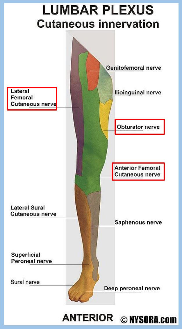
Draw cutaneous innervation of the leg
36
The internal iliac artery branches into which 3 arteries?
1. Superior gluteal a
2. Inferior gluteal a
3. Obturator a
2. Inferior gluteal a
3. Obturator a
37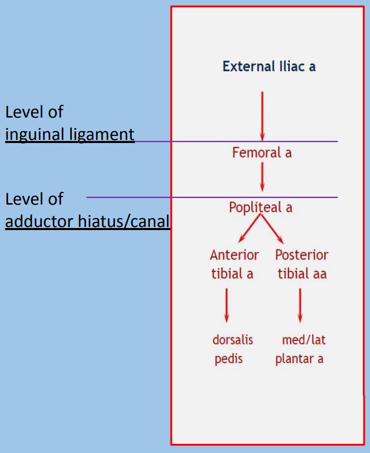
Draw the blood supply branch diagram
38
The hip joint is between the and the
The hip joint is between the head of the
femur and the acetabulum
femur and the acetabulum
39
The hip joint is a synovial .
The hip joint is a synovial ball-and-socket joint.
40
Acetabular cavity is deepened by
fibrocartilaginous rim called the
fibrocartilaginous rim called the
Acetabular cavity is deepened by
fibrocartilaginous rim called the
acetabular labrum
fibrocartilaginous rim called the
acetabular labrum
41
Blood supply to head of femur arises from
the
the
Blood supply to head of femur arises from
the medial circumflex artery
the medial circumflex artery
42
Fracture at the femoral neck can lead to of the head of the femur
Fracture at the femoral neck can lead to avascular necrosis of the head of the femur
43
What is the order of the nerves from superior to inferior?
Inferior gluteal nerves & vessels.
Sciatic nerve
Posterior cutaneous nerve of thigh
Nerve to quadratus femoris
Nerve to obturator internus
Pudendal N
Internal pudendal vessels
Sciatic nerve
Posterior cutaneous nerve of thigh
Nerve to quadratus femoris
Nerve to obturator internus
Pudendal N
Internal pudendal vessels
44
What is the Pes Anserinus?
Sartorius (anterior)
Gracilis (medial)
SemiTendinosus (posterior)
Femoral nerve
Obturator nerve
Tibial nerve [sciatic] || notes:SGT FOT
Gracilis (medial)
SemiTendinosus (posterior)
Femoral nerve
Obturator nerve
Tibial nerve [sciatic] || notes:SGT FOT
45
What is the common action of the posterior thigh?
EXTENSION of hip, FLEXION of knee
46
What is the blood supply of the posterior thigh?
PROFUNDA FEMORIS ARTERY
47
What is the blood supply of the posterior thigh?
PROFUNDA FEMORIS ARTERY
48
What is the common action of the superficial posterior compartment of the leg?
PLANTAR FLEXION.
49
What is the common innervation of the superifical posterior compartment of the leg?
TIBIAL N.
50
What is the common blood supply of the superficial posterior compartment of the leg?
POSTERIOR TIBIAL ARTERY
51
What is the common action of the deep posterior compartment of the leg?
Plantar flexion
52
What is the common innervation of the deep posterior compartment of the leg?
Tibial nerve
53
What is the blood supply of the deep posterior compartment of the leg?
POSTERIOR TIBIAL ARTERY
54
What is the second letter rule?
pEroneus longus
pEroneous brevis
pEroneous tertius
ALL EVERSION
tIbialis anterior
tIbialis posterior
ALL INVERSION
pEroneous brevis
pEroneous tertius
ALL EVERSION
tIbialis anterior
tIbialis posterior
ALL INVERSION
55
What is the common action of the anterior compartment of the leg?
Dorsiflexion
56
What is the common innervation of the anterior compartment of the leg?
Deep peroneal n.
57
What is the common blood supply of the anterior compartment of the leg?
Anterior tibial artery
58
What is the mnemonic for the anterior lower leg?
TEA DEPt
Tibialis anterior
Extensor hallucis longus
Anterior tibial artery
Deep peroneal nerve
Extensor digitorum longus
Peroneus tertius
Tibialis anterior
Extensor hallucis longus
Anterior tibial artery
Deep peroneal nerve
Extensor digitorum longus
Peroneus tertius
59
WHat is the common action of the lateral compartment of the leg?
PLANTAR FLEXION and EVERSION.
60
What is the common innervation of the lateral compartment of the leg?
SUPERFICIAL PERONEAL N.
61
All muscles in the anterior leg are supplied by?
deep peroneal nerve
62
Common fibular n ->
Common fibular n -> superficial and deep fibular n.
63
Sural n. = branches from
Sural n. = branches from tibial and common fibular n
64
Lateral sural cutaneous n. = branch from
Lateral sural cutaneous n. = branch from common fibular
65
Medial and lateral plantar n. =
Medial and lateral plantar n. = branch from tibial n.
66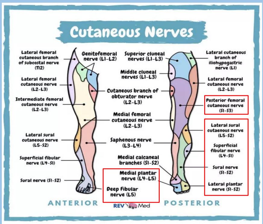
Draw out cutaneous innervations
67
What are the borders of the adductor canal?
Anterior: Sartorius and subartorial fascia
Lateral: vastus medialis
Medial: adductor longus and magnus
Lateral: vastus medialis
Medial: adductor longus and magnus
68
What are the contents of the adductor canal?
Femoral artery/vein
Saphenous nerve
Nerve to vastus medialis
Lymphatic vessels
Saphenous nerve
Nerve to vastus medialis
Lymphatic vessels
69
Where is the adductor hiatus? What happens here?
Located inside the tendon of the adductor
magnus, which opens in the popliteal fossa
Femoral artery → Popliteal Artery
Femoral Vein ←Popliteal Vein
Lymphatic vessels
magnus, which opens in the popliteal fossa
Femoral artery → Popliteal Artery
Femoral Vein ←Popliteal Vein
Lymphatic vessels
70
What are the boundaires of the popliteal fossa?
Superomedial: Semimembranosus and Semitendinosus
Superolateral: Biceps femoris
Inferior: heads of Gastrocnemius
Superolateral: Biceps femoris
Inferior: heads of Gastrocnemius
71
What are the contents of the popliteal fossa?
-Popliteal Artery
- Popliteal Vein
- Tibial Nerve
- Common Peroneal Nerve
Serve And Volley 2 Next Balls (medial to lateral)
Semimembranous Artery Vein 2 Nerves Biceps femoris
- Popliteal Vein
- Tibial Nerve
- Common Peroneal Nerve
Serve And Volley 2 Next Balls (medial to lateral)
Semimembranous Artery Vein 2 Nerves Biceps femoris
72
– inflammation and swelling of semimembranosus bursa
Damage to What Nerve
Symptoms:
Damage to What Nerve
Symptoms:
Baker’s Cyst – inflammation and swelling of semimembranosus bursa
Damage to What Nerve tibial nerve
Symptoms: Weak plantar flexion, Parathesia of foot and posterolateral leg
Damage to What Nerve tibial nerve
Symptoms: Weak plantar flexion, Parathesia of foot and posterolateral leg
73
– dilation of popliteal, popliteal dura is tough and
non-extensible
Damage to What Nerve:
Symptoms:
non-extensible
Damage to What Nerve:
Symptoms:
Popliteal aneurysm – dilation of popliteal, popliteal dura is tough and
non-extensible
Damage to What Nerve: Tibial
Symptoms: Weak plantar flexion, Parathesia of foot and posterolateral leg
non-extensible
Damage to What Nerve: Tibial
Symptoms: Weak plantar flexion, Parathesia of foot and posterolateral leg
74
Anterior tibial artery courses with the nerve to provide the pulse. It is
lateral to the
lateral to the
Anterior tibial artery courses with the Deep peroneal
(fibular) nerve to provide the dorsalis pedis pulse. It is
lateral to the extensor hallucis longus
(fibular) nerve to provide the dorsalis pedis pulse. It is
lateral to the extensor hallucis longus
75
Posterior tibial artery gives rise to the:
and arteries,
,
and the which runs with the nerve
and arteries,
,
and the which runs with the nerve
Posterior tibial artery gives rise to the:
lateral and medial plantar arteries,
plantar arch,
and the fibular artery which runs with the tibial nerve
lateral and medial plantar arteries,
plantar arch,
and the fibular artery which runs with the tibial nerve
76
TIBIAL SHAFT fractures can injure the anterior and
posterior tibial arteries and result in
posterior tibial arteries and result in
TIBIAL SHAFT fractures can injure the anterior and
posterior tibial arteries and result in COMPARTMENT
SYNDROME
posterior tibial arteries and result in COMPARTMENT
SYNDROME
77
Tibial collateral ligament (MCL)
• Attached to and
• Prevents displacement of tibia
• Tearing is caused by forces
• Attached to and
• Prevents displacement of tibia
• Tearing is caused by forces
Tibial collateral ligament (MCL)
• Attached to capsule and medial meniscus
• Prevents lateral displacement (valgus) of tibia
• Tearing is caused by lateral (valgus) forces
• Attached to capsule and medial meniscus
• Prevents lateral displacement (valgus) of tibia
• Tearing is caused by lateral (valgus) forces
78
Fibular Collateral Ligament (LCL)
• attached to lateral meniscus
• Prevents of tibia
• Tearing is caused by
• attached to lateral meniscus
• Prevents of tibia
• Tearing is caused by
Fibular Collateral Ligament (LCL)
• NOT attached to lateral meniscus
• Prevents medial displacement (varus) of tibia
• Tearing is caused by medial (varus) forces
• NOT attached to lateral meniscus
• Prevents medial displacement (varus) of tibia
• Tearing is caused by medial (varus) forces
79
Anterior Cruciate Ligament (ACL)
• Prevents of tibia
• Tearing allows the tibia to be easily
- sign
• Prevents of tibia
• Tearing allows the tibia to be easily
- sign
Anterior Cruciate Ligament (ACL)
• Prevents anterior displacement of tibia
• Tearing allows the tibia to be easily pulled forward
-anterior drawer sign
• Prevents anterior displacement of tibia
• Tearing allows the tibia to be easily pulled forward
-anterior drawer sign
80
Posterior Cruciate Ligament (PCL)
• Prevents of tibia
• Tearing allows the tibia to be easily
- sign
• Prevents of tibia
• Tearing allows the tibia to be easily
- sign
Posterior Cruciate Ligament (PCL)
• Prevents posterior displacement of tibia
• Tearing allows the tibia to be easily pulled posteriorly
- posterior drawer sign
• Prevents posterior displacement of tibia
• Tearing allows the tibia to be easily pulled posteriorly
- posterior drawer sign
81
Medial and lateral meniscus
• Serve as
• Serve as
Medial and lateral meniscus
• Serve as shock absorbers
• Serve as shock absorbers
82
What is the terrible triad?
Three most common injured
structures at knee are
• Tibial collateral ligament (MCL)
• Medial meniscus
• ACL
structures at knee are
• Tibial collateral ligament (MCL)
• Medial meniscus
• ACL
83
How is the terrible triad injured?
Results from a blow to lateral knee
with foot planted on ground
with foot planted on ground
84
What type of pain is seen with injury to the terrible triad?
Medical meniscus tear will cause pain
when leg is medially rotated at knee
when leg is medially rotated at knee
85
What is in the first layer of the foot?
Abductors
86
What is in the second layer of the foot?
Lumbricles
87
What is in the third layer of the foot?
Adductors
88
What is in the 4th layer of the foot?
interossi
89
What is the the most and only superifical muscle of the foot?
Extensor digitorum brevis
90
All of the muscles in the anterior leg do what action?
dorsiflexion of
the foot
the foot
91
All muscles in the anterior leg are supplied by?
deep peroneal nerve
92
What is seen with damage to superior gluteal nerve path?
-Weakness in abduction of hip
-Impaired gait (+) trendelenburg sign
- Gluteus medius and gluteus minimus are the
muscles affected
-Impaired gait (+) trendelenburg sign
- Gluteus medius and gluteus minimus are the
muscles affected
93
What is seen with damage to the inferior gluteal nerve path?
-Weakness in hip extension
- Difficulty rising from a chair and climbing stairs
- Gluteus maximus affected
- Difficulty rising from a chair and climbing stairs
- Gluteus maximus affected
94
What are the three symtpoms of damage to sciatic nerve path?
1. Weakened extension of thigh
2. Loss of knee flexion
3. Loss of all function below the knee
2. Loss of knee flexion
3. Loss of all function below the knee
95
Damage to sciatic nerve path is often seen when?
1. After hip replacement
2. Piriformis causing symptoms after spasm
2. Piriformis causing symptoms after spasm
96
What are the two symptoms of damage to the common fibular nerve?
1. Foot drop and loss of eversion
2. Loose sensation on lateral foot
2. Loose sensation on lateral foot
97
Fibular head fracture causes damage to which nerve?
Common fibular nerve
98
What are the two most common muscles to be strained?
1. Hamstring
2. Quadriceps
2. Quadriceps
99
- Increased pressure in a osseofacial compartment
- Increased pressure in a osseofacial compartment
Compartment syndrome
- Increased pressure in a osseofacial compartment
- Increased pressure in a osseofacial compartment
100
-Under the inguinal ligament the femoral artery
branches off to the
- Branches into the (watch the femur and AVN)
- Deep femoral then becomes the
(adductor hiatus)
- Splits into the anterior and posterior
branches off to the
- Branches into the (watch the femur and AVN)
- Deep femoral then becomes the
(adductor hiatus)
- Splits into the anterior and posterior
-Under the inguinal ligament the femoral artery
branches off to the deep femoral artery
- Branches into the medial/ lateral femoral
circumflex (watch the femur and AVN)
- Deep femoral then becomes the popiteal
(adductor hiatus)
- Splits into the anterior and posterior tibial
arteries
branches off to the deep femoral artery
- Branches into the medial/ lateral femoral
circumflex (watch the femur and AVN)
- Deep femoral then becomes the popiteal
(adductor hiatus)
- Splits into the anterior and posterior tibial
arteries
101
All veins of the lower extremity end up draining
into the
into the
All veins of the lower extremity end up draining
into the inferior vena cava
into the inferior vena cava
102
Deep veins
- Start with the and end with the
and eventually end in the
- Start with the and end with the
and eventually end in the
Deep veins
- Start with the plantar and end with the
common iliac and eventually end in the IVC
- Start with the plantar and end with the
common iliac and eventually end in the IVC
103
Superficial Vein
- Great saphenous veins ends and drains into the
(Dont forget about that saphenous
opening
- Great saphenous veins ends and drains into the
(Dont forget about that saphenous
opening
Superficial Vein
- Great saphenous veins ends and drains into the
femoral vein (Dont forget about that saphenous
opening
- Great saphenous veins ends and drains into the
femoral vein (Dont forget about that saphenous
opening
104
= supply medial compartment
of the thigh (think adductor muscles)
of the thigh (think adductor muscles)
Obturator artery = supply medial compartment
of the thigh (think adductor muscles)
of the thigh (think adductor muscles)
105
Femoral artery:
- = off femoral artery around
femur and prone to lacerations (Watch for
AVN)
- = supplies muscles in the
thigh with perforating branches
- = off femoral artery around
femur and prone to lacerations (Watch for
AVN)
- = supplies muscles in the
thigh with perforating branches
Femoral artery:
- Medial circumflex = off femoral artery around
femur and prone to lacerations (Watch for
AVN)
- Profunda femoris = supplies muscles in the
thigh with perforating branches
- Medial circumflex = off femoral artery around
femur and prone to lacerations (Watch for
AVN)
- Profunda femoris = supplies muscles in the
thigh with perforating branches
106
Popliteal artery:
- Supplies the knee and turns from femoral to
popliteal in the
- Supplies the knee and turns from femoral to
popliteal in the
Popliteal artery:
- Supplies the knee and turns from femoral to
popliteal in the adductor canal
- Supplies the knee and turns from femoral to
popliteal in the adductor canal
107
Anterior tibial artery:
- Travels with the down
the compartment of the leg
(Watch for those breaks)
- Travels with the down
the compartment of the leg
(Watch for those breaks)
Anterior tibial artery:
- Travels with the fibular nerve down
the anterior compartment of the leg
(Watch for those tibia breaks)
- Travels with the fibular nerve down
the anterior compartment of the leg
(Watch for those tibia breaks)
108
Posterior tibial artery:
- Courses with the in
posterior compartment
- Fibular artery supplies part of
leg
- Courses with the in
posterior compartment
- Fibular artery supplies part of
leg
Posterior tibial artery:
- Courses with the tibular nerve in
posterior compartment
- Fibular artery supplies lateral part of
leg
- Courses with the tibular nerve in
posterior compartment
- Fibular artery supplies lateral part of
leg
109
can lacerate
anterior or posterior arteries and
lead to compartment syndrome
anterior or posterior arteries and
lead to compartment syndrome
Tibial shaft fractures can lacerate
anterior or posterior arteries and
lead to compartment syndrome
anterior or posterior arteries and
lead to compartment syndrome
110
The femoral nerve is NOT in the
The femoral nerve is NOT in the femoral sheath
111
In the hip joint, the Acetabular labrum
goes on to form the
goes on to form the
In the hip joint, the Acetabular labrum
goes on to form the transverse
acetabular ligament
goes on to form the transverse
acetabular ligament
112
Most of the blood supply in the hip comes from
the (watch
out for breaks)
the (watch
out for breaks)
Most of the blood supply in the hip comes from
the medial circumflex artery (watch
out for femur breaks)
the medial circumflex artery (watch
out for femur breaks)
113
What are the three articulations of the knee?
1. Femoropatellar
2. Medial tibiofemoral
3. Lateral tibiofemoral
2. Medial tibiofemoral
3. Lateral tibiofemoral
114
Tibial Collateral Ligament =
- Firmly attached to
- Prevents of the tibia
- Firmly attached to
- Prevents of the tibia
Tibial Collateral Ligament =
- Firmly attached to medial meniscus
- Prevents lateral displacement of the tibia (abduction)
- Firmly attached to medial meniscus
- Prevents lateral displacement of the tibia (abduction)
115
Fibular collateral ligament =
- NOT attached to
- Prevents of the tibia
- NOT attached to
- Prevents of the tibia
Fibular collateral ligament =
- NOT attached to lateral meniscus
- Prevents medial displacement of the tibia
- NOT attached to lateral meniscus
- Prevents medial displacement of the tibia
116
Anterior cricuate ligament (ACL):
- Prevents displacement
- Tension highest with the knee
and resists
- sign (anterior movement)
- Prevents displacement
- Tension highest with the knee
and resists
- sign (anterior movement)
Anterior cricuate ligament (ACL):
- Prevents anterior displacement
- Tension highest with the knee extended
and resists hyperextension
- Positive drawer sign (anterior movement)
- Prevents anterior displacement
- Tension highest with the knee extended
and resists hyperextension
- Positive drawer sign (anterior movement)
117
Posterior curate ligament (PCL):
- Prevents displacement
- Tension highest when the knee is
- Positive test (posterior
movement)
- Prevents displacement
- Tension highest when the knee is
- Positive test (posterior
movement)
Posterior curate ligament (PCL):
- Prevents posterior displacement
- Tension highest when the knee is flexed
- Positive posterior drawer test (posterior
movement)
- Prevents posterior displacement
- Tension highest when the knee is flexed
- Positive posterior drawer test (posterior
movement)
118
Medial Meniscus:
- C-shaped and attach to
- C-shaped and attach to
Medial Meniscus:
- C-shaped and attach to tibial collateral ligament
(MCL)
- C-shaped and attach to tibial collateral ligament
(MCL)
119
Lateral Meniscus:
- Not attached to the
- Not attached to the
Lateral Meniscus:
- Not attached to the fibular collateral ligament
(LCL)
- Not attached to the fibular collateral ligament
(LCL)
120
Medial collateral/ deltoid ligaments are
- ligament
- ligament
- ligament
- ligament
- ligament
- ligament
- ligament
- ligament
Medial collateral/ deltoid ligaments are very strong
- Anterior tibotalar ligament
- Posterior tibotalar ligament
- Tibionavicular ligament
- Tibiocalcaneal ligament
- Anterior tibotalar ligament
- Posterior tibotalar ligament
- Tibionavicular ligament
- Tibiocalcaneal ligament
121
Lateral collateral ligaments Weak and prone to path
- ****Most damaged
-
-
- ****Most damaged
-
-
Lateral collateral ligaments Weak and prone to path
- Anterior talofibular ligament ****Most damaged
- Posterior talofibular ligament
- Calcaneofibular ligament
- Anterior talofibular ligament ****Most damaged
- Posterior talofibular ligament
- Calcaneofibular ligament
122
Anterior draw sign for the Ankle
- Will sign for ligament tears
- Will sign for ligament tears
Anterior draw sign for the Ankle
- Will see suction sign for ATF (anterior talofibular) ligament tears
- Will see suction sign for ATF (anterior talofibular) ligament tears
123
Watch out for lateral thigh pain as the and
nerve can be trapped under
the
nerve can be trapped under
the
Watch out for lateral thigh pain as the femoral and
lateral femoral cutaneous nerve can be trapped under
the inguinal ligament
lateral femoral cutaneous nerve can be trapped under
the inguinal ligament
124
Fibular head fractures and tight casts cause what damage?
Peroneal nerve damage (foot drop)
125
= pain and swelling the knee.
Watch for young children who are very active
Watch for young children who are very active
Osgood-Schlatter = pain and swelling below the knee.
Watch for young children who are very active
Watch for young children who are very active
126
What are the symptoms of tarsal tunnel pain?
Pain in plantar foot and heel
127
What are the contents of the tarsal tunnel?
- flexor posterior tendon
- flexor digitorum longus tendon
- posterior tibial artery
- posterior tibial vein
- tibial nerve
- flexor hallicus longus
Tom Dick And Very Nervous Harry
- flexor digitorum longus tendon
- posterior tibial artery
- posterior tibial vein
- tibial nerve
- flexor hallicus longus
Tom Dick And Very Nervous Harry