All cards
1
Glenohumoral joint- , , very unstable
Found Between the head of the and of scapula.
Found Between the head of the and of scapula.
Glenohumoral joint- ball and socket, multiaxial, synovial, very unstable
Found Between the head of the humerus and glenoid fossa of scapula.
Found Between the head of the humerus and glenoid fossa of scapula.
2
Ligaments of Glenohumoral joint
-coracoclavicular ligament that has two parts:Trapezoid and Conoid
o Acromioclavicular ligament
o Coracoacromial ligament
o Superior transverse scapular ligament
o Acromioclavicular ligament
o Coracoacromial ligament
o Superior transverse scapular ligament
3
Disocation: anterior (more common) due to blow . Posterior is due to
Disocation: anterior (more common) due to blow from back. Posterior is due to blow from front
4
• Sternoclavicular joint- between and
• Sternoclavicular joint- between sternum and clavicle.
5
Sternoclavicular joint has the ligament strengthens
Sternoclavicular joint has the ligament strengthens the joint capsule anteriorly and posteriorly
6
Stenoclavicular very strong. Rarely injured. If injury occurs there, usually results in
Stenoclavicular very strong. Rarely injured. If injury occurs there, usually results in clavicular fracture (fall on outstretched arm)
7
Acromioclavicular joint- between and
Dislocation is common- from hard fall on shoulder (
Dislocation is common- from hard fall on shoulder (
Acromioclavicular joint- between acromion of scapula and clavicle
Dislocation is common- SHOULDER SEPARATION- from hard fall on shoulder (contact sports)
Dislocation is common- SHOULDER SEPARATION- from hard fall on shoulder (contact sports)
8
Elbow joint- ulna goes into the of the humerus. Radius fits next to the and is connected to the ulna via the
Elbow joint- ulna goes into the trochlear notch of the humerus. Radius fits next to the capitulum of the humerus and is connected to the ulna via the annular ligament
9
will be most commonly torn rotator cuff muscle
Supraspinatous will be most commonly torn rotator cuff muscle
10
What is the function of the supraspinatous muscles?
Resting tone of these muscles act to pull the humeral head into the glenoid fossa
11
Axillary region: boundaries, contents.
Anterior: pectoralis major and minor
Posterior: subscapularis, teres major, and latissumus dorsi
Medial: serratus anterior, thoracic wall (ribs)
Lateral: intertubercular sulcus of humerus
Apex: lateral border of rib 1, superior border of scapula, and posterior border of clavicle
Floor: axillary fascia and skin
Contents: axillary vessels, cords and nerves of the brachial plexus, lymph nodes (pathway to breast and upper limb)
Posterior: subscapularis, teres major, and latissumus dorsi
Medial: serratus anterior, thoracic wall (ribs)
Lateral: intertubercular sulcus of humerus
Apex: lateral border of rib 1, superior border of scapula, and posterior border of clavicle
Floor: axillary fascia and skin
Contents: axillary vessels, cords and nerves of the brachial plexus, lymph nodes (pathway to breast and upper limb)
12
Proximal:
1)Superior thoracic artery
Deep:
1)Thoracoacromial artery
2)Lateral thoracic artery
Distal:
1)Subscapular artery
2)Anterior circumflex humeral artery
3)Posterior circumflex humeral artery
Parts of axillary artery
Proximal:
1)Superior thoracic artery
Deep:
1)Thoracoacromial artery
2)Lateral thoracic artery
Distal:
1)Subscapular artery
2)Anterior circumflex humeral artery
3)Posterior circumflex humeral artery
13
What separates axiallary artery into its three parts?
Pectoralis MINOR
14
Scaphoid is most and lunate is
Scaphoid is most often fractured and lunate is most commonly dislocated
15
•Colle: fracture of the with of the distal fragment and radial deviation of the hand at the wrist. Called
•Colle: fracture of the lower end of the radius with posterior displacement of the distal fragment and radial deviation of the hand at the wrist. Called dinner fork deformity
16
Smiths: same as except v deviation
Smiths: same as colle except volar (anterior) deviation
17
Bartons: dislocation of
Bartons: dislocation of distal radiocarpal joint
18
Scaphoid fraction: from fall of outstretched hand complications-
Scaphoid fraction: from fall of aBDucted outstretched hand complications- a vascular necrosis
19
Lunate fraction: from fall on Lunate dislocates
-complications- , a
-complications- , a
Lunate fraction: from fall on aDDucted hand. Lunate dislocates anteriorly
-complications- carpal tunnel syndrome, a vascular necrosis
-complications- carpal tunnel syndrome, a vascular necrosis
20
•Boxers: fracture neck of
•Boxers: fracture neck of fifth metacarpal
21
Bennet: fracture of base of
Bennet: fracture of base of first metacarpal
22
Upper Brachial Plexus Injury: (includes both and upper trunk)
Upper Brachial Plexus Injury: C5-6. (includes both anterior and posterior upper trunk)
23
Injury: Erb Duchene Paralysis- causes
Cause: and shoulder OR
Example of how this can happen:
Cause: and shoulder OR
Example of how this can happen:
Upper Brachial Plexsus Injury: Erb Duchene Paralysis- causes “waiters tip”
Cause: violent separation of the neck and shoulder OR over-stretch of infants neck during delivery
Example of how this can happen: contact sports (football)
Cause: violent separation of the neck and shoulder OR over-stretch of infants neck during delivery
Example of how this can happen: contact sports (football)
24
Symptoms of upper brachial plexux injury?
1. Loss of abduction, 2. external rotation at the shoulder, and 3. flexion of the elbow, 4. and
extension of the wrist occur
extension of the wrist occur
25
Nerves inmpacted by upper brachial plexsus injruy
(axillary n. [C5,6], suprascapular n. [C4,5,6]; radial
n. [C5,6,7,8])
n. [C5,6,7,8])
26
Muscles impacted by upper brachial plexusu injury
supraspinatus, infraspinatus, biceps, coracobrachialis, deltoid , teres minor
27
Middle Brachial Plexus Injury. C7 mostly.RARE
Cause: of the brachial plexus
Cause: of the brachial plexus
Middle Brachial Plexus Injury. C7 mostly.RARE
Cause: interscalene anesthetic of the brachial plexus
Cause: interscalene anesthetic of the brachial plexus
28
Impacts of middle brachial plexusu injury?
1. ADDuction, 2. ABduction, 3. extension, and 4. lateral rotation are impaired.
29
Lower Brachial Plexus Injury.
Lower Brachial Plexus Injury. C8-T1 .(effects lower trunk)
30
Djerine-Klumpke Paralysis-
Cause: shoulder dislocations, pulling baby out by arms during delivery, scalene syndrome, and apical tumor of the lung
Injury is similar to (ex: loss of flexion in the wrist, paralysis of these muscles Inability to due to involvement of the medial pectoral nerve.
Cause: shoulder dislocations, pulling baby out by arms during delivery, scalene syndrome, and apical tumor of the lung
Injury is similar to (ex: loss of flexion in the wrist, paralysis of these muscles Inability to due to involvement of the medial pectoral nerve.
Djerine-Klumpke Paralysis- Lower brachial plexsus injury
Cause: shoulder dislocations, pulling baby out by arms during delivery, scalene syndrome, and apical tumor of the lung
Injury is similar to ulnar nerve injury (ex: loss of flexion in the wrist, paralysis of these muscles (claw hand type). Inability to ADDuct due to involvement of the medial pectoral nerve.
Cause: shoulder dislocations, pulling baby out by arms during delivery, scalene syndrome, and apical tumor of the lung
Injury is similar to ulnar nerve injury (ex: loss of flexion in the wrist, paralysis of these muscles (claw hand type). Inability to ADDuct due to involvement of the medial pectoral nerve.
31
Radial nerve aka “
Causes
The injury occurs in the
Dislocation OR fracture to the of the humerus=
Causes
The injury occurs in the
Dislocation OR fracture to the of the humerus=
Radial nerve (C5-8), aka “Saturday night palsy” or “crunch palsy’.
Causes Wrist Drop.
The injury occurs in the axilla region
Dislocation at the shoulder joint OR fracture to the proximal part of the humerus= MIDSHAFT.
Causes Wrist Drop.
The injury occurs in the axilla region
Dislocation at the shoulder joint OR fracture to the proximal part of the humerus= MIDSHAFT.
32
Mortor Functions impacted by radial injury?
1. unable to extend at the forearm, wrist and finger
2. unopposed flexion of wrist occurs, known as wrist-drop.
2. unopposed flexion of wrist occurs, known as wrist-drop.
33
Sensory functions impacted by musculocutaneous injury?
1. All four cutaneous branches of the radial nerve are affected
2. sensory loss over the lateral and posterior arm, posterior
forearm, and dorsal surface of the lateral three and a half digits
2. sensory loss over the lateral and posterior arm, posterior
forearm, and dorsal surface of the lateral three and a half digits
34
Axillary nerve Injury
Cause: fracture to the
Result: affects and (c )
Recall: axillary n is content of so compression to the area affects it
Cause: fracture to the
Result: affects and (c )
Recall: axillary n is content of so compression to the area affects it
Axillary nerve Injury (C5,6)-
Cause: fracture to the surgical neck of the humerus
Result: affects deltoid and teres minor (cant do ABduction/ lateral rotation)
Recall: axillary n is content of quadrangular space so compression to the area affects it
Cause: fracture to the surgical neck of the humerus
Result: affects deltoid and teres minor (cant do ABduction/ lateral rotation)
Recall: axillary n is content of quadrangular space so compression to the area affects it
35
Lateral cord injury: ( )
Effect::cant flex , and sensory loss to the
Effect::cant flex , and sensory loss to the
Lateral cord injury: musculocutaneous nerve (C5,6,7)
Effect::cant flex forearm or shoulder, and sensory loss to the lateral forearm
Effect::cant flex forearm or shoulder, and sensory loss to the lateral forearm
36
Median Nerve Injury: . (inability to flex )
Causes:
1.Damage to
2.Laceration
3.The section above the elbow
4. of humerous
Also is injury to median n.
Causes:
1.Damage to
2.Laceration
3.The section above the elbow
4. of humerous
Also is injury to median n.
Median Nerve Injury: C6-T1. Hand of Benediction (inability to flex digits 1-3)
Causes:
1.Damage to axilla
2.Laceration at the wrist
3.The section above the elbow
4. Supracondylar fracture of humerous
Also carpal tunnel is injury to median n.
Causes:
1.Damage to axilla
2.Laceration at the wrist
3.The section above the elbow
4. Supracondylar fracture of humerous
Also carpal tunnel is injury to median n.
37
Midshaft Fracture
Radial nerve
38
Supracondylar fracture
Median nerve
39
Fracture of medial epicondyle
Ulnar nerve
40
Fracture at surgical neck
Axillary nerve
41
Ulnar Nerve Injury:
1.Occurs bc of trauma to the Causes (inability to extend fingers 4,5 at intephalangeal joints )
2.Section at the wrist - are paralyzed
1.Occurs bc of trauma to the Causes (inability to extend fingers 4,5 at intephalangeal joints )
2.Section at the wrist - are paralyzed
Ulnar Nerve Injury:
1.Occurs bc of trauma to the medial epicondyle. Causes Claw-hand . (inability to extend fingers 4,5 at intephalangeal joints )
2.Section at the wrist - hypothenar muscles are paralyzed
1.Occurs bc of trauma to the medial epicondyle. Causes Claw-hand . (inability to extend fingers 4,5 at intephalangeal joints )
2.Section at the wrist - hypothenar muscles are paralyzed
42
If radial groove is fractured-
If anterior to medial epicondyle is fractured-
If anterior to medial epicondyle is fractured-
If radial groove is fractured- radial n. affected
If anterior to medial epicondyle is fractured- ulnar n is affected
If anterior to medial epicondyle is fractured- ulnar n is affected
43
Anatomical snuff box: boundaries, contents
Medial: EPL
Lateral: APL and EPB
Proximal: styloid process of radius
Floor: scaphoid and trapezium
Roof: skin
Contents: radial artery, radial nerve, cephalic veins
Lateral: APL and EPB
Proximal: styloid process of radius
Floor: scaphoid and trapezium
Roof: skin
Contents: radial artery, radial nerve, cephalic veins
44
what nerve injury will lead to Claw hand, drop wrist, hand of Benediction?
• Claw hand: ulnar nerve
• Wrist drop: radial nerve
• Hand of benediction: median nerve
• Wrist drop: radial nerve
• Hand of benediction: median nerve
45
Lateral epicondylitis: pain and tenderness over the and the origin of the posterior muscles of the forearm via the common tendon
Lateral epicondylitis: Tennis elbow = pain and tenderness over the lateral epicondyle and the origin of the posterior muscles of the forearm via the common tendon (common extensor tendon).
46
Medial epicondylitis: = painful, from repetitive use of the superficial muscles of the anterior forearm. Pain is experienced on the Repetitive motion strains the common
Medial epicondylitis: Golfer’s elbow= painful, from repetitive use of the superficial muscles of the anterior forearm. Pain is experienced on the medial epicondyle. Repetitive motion strains the common flexor tendon
47
Cubital fossa: boundaries, contents…
Roof: superficial fascia, bicipital aponuerosis, cubcutaneous tissue
Lateral: brachioradialis
Medially: pronator teres
Upper limit: imaginary horizontal line between medial and lateral epicondyle of humerus
Floor or Base: brachialis and supinator
Apex: neck of radius
Lateral: brachioradialis
Medially: pronator teres
Upper limit: imaginary horizontal line between medial and lateral epicondyle of humerus
Floor or Base: brachialis and supinator
Apex: neck of radius
48
Contents of Cubital Fossa? What is NOT a content?
• radial nerve (can be damaged during fracture of later epicondyle of humerus)
o The deep branch pierces supinator muscle to get to posterior forearm
• Biceps tendon
• Brachial artery
• Median nerve
the ulnar nerve is NOT a content of the fossa, but its found posterior to the medial epicondyle of the humerus
o The deep branch pierces supinator muscle to get to posterior forearm
• Biceps tendon
• Brachial artery
• Median nerve
the ulnar nerve is NOT a content of the fossa, but its found posterior to the medial epicondyle of the humerus
49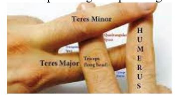
Draw out quadrangular space
50
Content of triangular interval
Radial artery nerve and brachial artery
51
Content of the quadrangular space?
axillary nerve, posterior circumflex humeral artery
52
Content of triangular space?
circumflex scapular artery and vein
53
Carpal tunnel boundaries and contents
• Anterior: flexor retinaculum
• Posterior: carpal bones
Contents: median nerve (lateral), tendons of FDS, FDP, FPL
• Posterior: carpal bones
Contents: median nerve (lateral), tendons of FDS, FDP, FPL
54
when you tap the flexor retinaculum to check for tingling sensation
Tinel sign: when you tap the flexor retinaculum to check for tingling sensation
55
Hands back to back
Phalens sign: Hands back to back
56
Injury to long thoracic nerve causes problem with serratus anterior muscle —>
Injury to long thoracic nerve causes problem with serratus anterior muscle —> Winged scapula
57
What is the bicep reflex?
C5-C6
58
What is the tricep reflex?
C7-8
59
Anterior upper arm: innervated by
Anterior upper arm: flexors, innervated by musculocutaneous (C5-7)
60
What are the anterior upper arm flexors?
•biceps brachii
• Brachialis
-inserts into choronoid process of the ulna
•Corachobrachialis (doesnt flex the elbow. It aDDucts the shoulder)
• Brachialis
-inserts into choronoid process of the ulna
•Corachobrachialis (doesnt flex the elbow. It aDDucts the shoulder)
61
Posterior upper arm: innervated by radial nerve:
Posterior upper arm: extensors, innervated by radial nerve:
62
What is the muscle in the upper arm?
•triceps brachii
63
Anterior forarm: of the wrist and digits, innervated by the except for 2 muscle ( and , which are innervated by ulnar nerve).
Anterior forarm: flexion of the wrist and digits, innervated by the median nerve except for 2 muscle (flexor carpi ulnaris and flexor digitorum profundus, which are innervated by ulnar nerve).
64
Deep muscles of anterior forarm: supplied by anterior interosseous nerve (
Deep muscles of anterior forarm: supplied by anterior interosseous nerve (branch of median nerve)
65
Posterior forearm: of the wrist and digits, innervated by
Posterior forearm: extension of the wrist and digits, innervated by radial nerve and its branch the posterior interosseous nerve
66
Superfical and deep muscles of forarm?
• superficial:
o Anconeus
o Brachioradialis (does flexion of the elbow and supination)
o Extensor carpi radialis longus
o Extensor carpi radialis brevis
o Extensor digitorum
o Extensor digiti minimi
o Extensor carpi ulnaris
• Deep:
o Abductor policis longus
o Extensor policis longus
o Extensor policis brevis
o Supinator
o Extensor indices
o Anconeus
o Brachioradialis (does flexion of the elbow and supination)
o Extensor carpi radialis longus
o Extensor carpi radialis brevis
o Extensor digitorum
o Extensor digiti minimi
o Extensor carpi ulnaris
• Deep:
o Abductor policis longus
o Extensor policis longus
o Extensor policis brevis
o Supinator
o Extensor indices
67
Thenar muscles: all innervated by except for 2 (part of flexor policis brevis and adductor policis- which are supplied by the
Thenar muscles: all innervated by median nerve except for 2 (part of flexor policis brevis and adductor policis- which are supplied by the ulnar nerve)
68
Thenar muscles:
o Abductor policis brevis
o Flexor policis brevis
o Opponens policis
o Adductor policis
o Flexor policis brevis
o Opponens policis
o Adductor policis
69
• Hypothenar:
• Hypothenar: innervated by ulnar nerve
70
Hypothenar muscles
o Abductor digiti minimi
o Flexor digiti mini brevis
o Opponens digiti minimi
o Flexor digiti mini brevis
o Opponens digiti minimi
71
• Lumbricles: 4 lumbricals. 1-2 innervated by 3-4 is innervation by
• Lumbricles: 4 lumbricals. 1-2 innervated by median nerve, 3-4 is innervation by ulnar nerve
72
• Interosseous muscles: (4 , 3
o All supplied by the
o All supplied by the
• Interosseous muscles: (4 dorsal-abduct, 3 palmar-adduct)
o All supplied by the ulnar nerve
o All supplied by the ulnar nerve
73
HALF LOAF : THESE ARE BY MEDIAN AND NOT ULNAR
1. half of lumbricles
2. abductor pollicus brevis
3. opponens pollicus
4. Lumbricles 1 and 2
2. abductor pollicus brevis
3. opponens pollicus
4. Lumbricles 1 and 2