Large Intestine
Study these flashcardsFree · spaced repetition · iPhone, Android, Mac & web
All cards
1
= small masses of fat on the
colon
colon
Appendices epiploicae (omental appendices) = small masses of fat on the
colon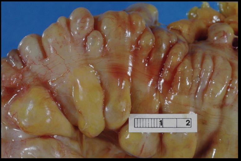
colon
2
= 3 bands of longitudinal muscle
Teniae coli = 3 bands of longitudinal muscle 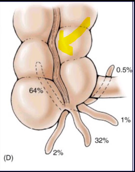
3
= sac-like bulges
Haustra = sac-like bulges
4
Inferior to ileocolic junction (iliac fossa) =
Inferior to ileocolic junction (iliac fossa) = appendix
5
The holds the appendix in place
The mesoappendix holds the appendix in place
6
The proper hepatic artery splits into the and
The proper hepatic artery splits into the right and left heaptic artery
7
Inguinal ligament and external inguinal ring both come from
Inguinal ligament and external inguinal ring both come from external oblique muscle
8
Posterior relationships of Cecum =
muscle, vessels and
nerves of the
muscle, vessels and
nerves of the
Posterior relationships of Cecum = iliopsoas
muscle, vessels and
nerves of the iliac
fossa
muscle, vessels and
nerves of the iliac
fossa
9
***TQ Opening of the cecum is the inferior and
***TQ Opening of the cecum is the inferior ileocecal valve and appendix
10
****TQ The blood supply of the cecum is the?
Ileocolic artery (SMA)
11
The appendix is located at point: 1.5 to 2 in on the line from to
The appendix is located at McBurney's point: 1.5 to 2 in on the line from ASIS to ubilicius 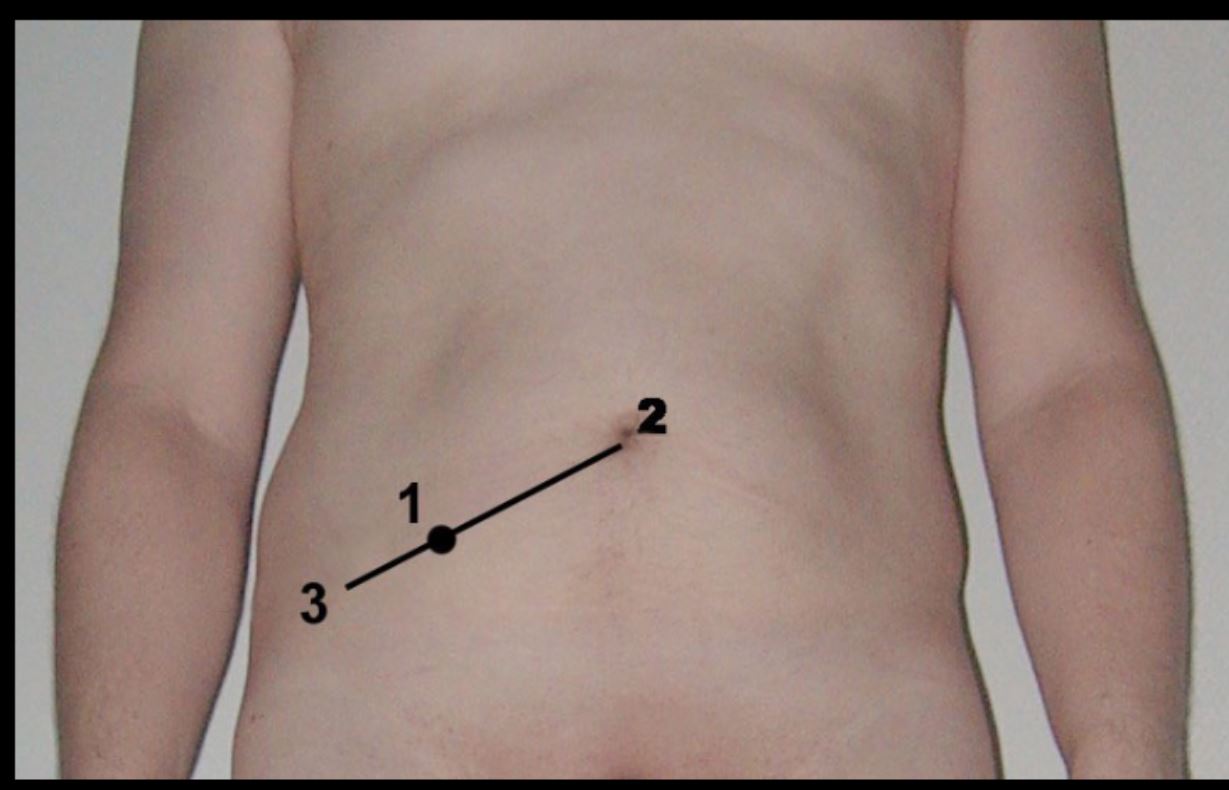
12
The apendix lacks a and
The apendix lacks a teniae coli and haustra
13
The asending colon is
The asending colon is retroperitoneal
14
Transverse Colon = hangs like a “loop”transversely to the
diaphragm and it extends to the (splenic
flexure).
diaphragm and it extends to the (splenic
flexure).
Transverse Colon = hangs like a “loop”transversely to the
diaphragm and it extends to the left colic flexure (splenic
flexure).
diaphragm and it extends to the left colic flexure (splenic
flexure).
15
The transverse colon is more on the right side, with the
lying deep to ribs and and being
overlapped by the
lying deep to ribs and and being
overlapped by the
The transverse colon is more posterior on the right side, with the
right colic flexure lying deep to ribs 9 and 10 and being
overlapped by the liver.
right colic flexure lying deep to ribs 9 and 10 and being
overlapped by the liver.
16
The transverse colon is
The transverse colon is intraperitoneal.
17
The left colic flexure is more , more , and than the right colic flexure. It is anterior to the left
The left colic flexure is more superior, more acute, and less
mobile than the right colic flexure. It is anterior to the left
kidney
mobile than the right colic flexure. It is anterior to the left
kidney
18
The left colic flexure is supported by the and
The left colic flexure is supported by the Phrenicolic ligament and Transverse mesocolon
19
Descending = on the left side, the portion up to
the
•It is
the
•It is
Descending = on the left side, the portion up to
the pelvic rim.
•It is retroperitoneal.
the pelvic rim.
•It is retroperitoneal.
20
Sigmoid = “S”-shaped loop of variable length
(about 40 cm) located between the descending
and the . It extends from the pelvic
rim to the sacrum where it forms the rectum.
•It is
(about 40 cm) located between the descending
and the . It extends from the pelvic
rim to the sacrum where it forms the rectum.
•It is
Sigmoid = “S”-shaped loop of variable length
(about 40 cm) located between the descending
colon and the rectum. It extends from the pelvic
rim to the sacrum where it forms the rectum.
•It is intraperitoneal.
(about 40 cm) located between the descending
colon and the rectum. It extends from the pelvic
rim to the sacrum where it forms the rectum.
•It is intraperitoneal.
21
From the to the is supplied by by the SMA (Superior Mesenteric Asrtery)
From the 2nd part of the
duodenum to the left colic
flexure is supplied by by the SMA (Superior Mesenteric Asrtery)
duodenum to the left colic
flexure is supplied by by the SMA (Superior Mesenteric Asrtery)
22
4 branches of superior mesenteric artery
•Jejunal and ileal
branches
•Ileocolic artery to the
ascending colon, cecum,
appendix
•Right colic artery to the
right colic flexure
•Middle colic artery
supplies the transverse
colon
branches
•Ileocolic artery to the
ascending colon, cecum,
appendix
•Right colic artery to the
right colic flexure
•Middle colic artery
supplies the transverse
colon
23
From the left colic
flexure to the rectum
by the and its
branches
flexure to the rectum
by the and its
branches
From the left colic
flexure to the rectum
by the IMA and its
branches
flexure to the rectum
by the IMA and its
branches
24
What are the 4 branches of the inferior mesenteric artery?
•Left colic: to the left
colic flexure
•Descending branch of
IMA
•Sigmoidal artery
•Superior rectal artery
AKA superior
hemorrhoidal artery
(end artery of the
IMA)
colic flexure
•Descending branch of
IMA
•Sigmoidal artery
•Superior rectal artery
AKA superior
hemorrhoidal artery
(end artery of the
IMA)
25
TQ The left colic artery (IMA) and the middle colic artery
(SMA) anastomose to form the It is where all the arteries join
together
(SMA) anastomose to form the It is where all the arteries join
together
TQ The left colic artery (IMA) and the middle colic artery
(SMA) anastomose to form the marginal artery. It is where all the arteries join
together
(SMA) anastomose to form the marginal artery. It is where all the arteries join
together
26
The are small branches from the marginal
artery to the colon.
artery to the colon.
The vasa recta are small branches from the marginal
artery to the colon.
artery to the colon.
27
TQ The IMA and SMA anastamose to form the
TQ The IMA and SMA anastamose to form the marginal artery
28
What innervates the large intestine?
1. The right scaral plexus
2. Right pelvic splanchinic nerves
2. Right pelvic splanchinic nerves