Abdomen Wall
Study these flashcardsFree · spaced repetition · iPhone, Android, Mac & web
All cards
1
The abdomen is the part of the trunk that lies between the
and the .
and the .
The abdomen is the part of the trunk that lies between the thorax
and the pelvis.
and the pelvis.
2
The abdominal cavity is located between the and
the
the
The abdominal cavity is located between the thoracic diaphragm and
the pelvic rim (linea terminalis)
the pelvic rim (linea terminalis)
3
The diaphragm forms the roof of the
The diaphragm forms the roof of the abdominal cavity.
4
The cavity is continuous with the pelvic cavity; therefore it has
The cavity is continuous with the pelvic cavity; therefore it has no
floor.
floor.
5
******The abdominal cavity extends superiorly into the osteocartilaginous
thoracic cage to about the in the supine position;
consequently, parts of the liver, stomach, and spleen are protected by
the thoracic cage.
thoracic cage to about the in the supine position;
consequently, parts of the liver, stomach, and spleen are protected by
the thoracic cage.
******The abdominal cavity extends superiorly into the osteocartilaginous
thoracic cage to about the 5th intercostal space in the supine position;
consequently, parts of the liver, stomach, and spleen are protected by
the thoracic cage.
thoracic cage to about the 5th intercostal space in the supine position;
consequently, parts of the liver, stomach, and spleen are protected by
the thoracic cage.
6
The abdominal wall extends from the osteocartilagenous to the
The abdominal wall extends from the osteocartilagenous thoracic cage to the pelvis
7
Anterior Abdominal Wall
-Superiorly by the and
-Inferiorly by a line connecting each .
-Laterally by a .
-Superiorly by the and
-Inferiorly by a line connecting each .
-Laterally by a .
Anterior Abdominal Wall
-Superiorly by the right and left costal margins.
-Inferiorly by a line connecting each ASIS to the pubis.
-Laterally by a vertical line through ASIS.
-Superiorly by the right and left costal margins.
-Inferiorly by a line connecting each ASIS to the pubis.
-Laterally by a vertical line through ASIS.
8
Umbilicus:
intervertebral disc
intervertebral disc
Umbilicus: L3-L4
intervertebral disc
intervertebral disc
9
median fibrous
white line divides the
abdomen into right and
left sides. Indicated by a
vertical groove from the
xiphoid process to the
pubis
white line divides the
abdomen into right and
left sides. Indicated by a
vertical groove from the
xiphoid process to the
pubis
Line Alba: median fibrous
white line divides the
abdomen into right and
left sides. Indicated by a
vertical groove from the
xiphoid process to the
pubis
white line divides the
abdomen into right and
left sides. Indicated by a
vertical groove from the
xiphoid process to the
pubis
10
*****Linea Semilunaris: curved
line or groove (convex
laterally). It extends from
the to
the pubis. It is located from the .
Represents the lateral
border of the
line or groove (convex
laterally). It extends from
the to
the pubis. It is located from the .
Represents the lateral
border of the
*****Linea Semilunaris: curved
line or groove (convex
laterally). It extends from
the 9th costal cartilage to
the pubis. It is located 5-8
cm from the median plane.
Represents the lateral
border of the rectus
abdominis muscle
line or groove (convex
laterally). It extends from
the 9th costal cartilage to
the pubis. It is located 5-8
cm from the median plane.
Represents the lateral
border of the rectus
abdominis muscle
11
:
transverse lines over the
rectus abdominis muscle
transverse lines over the
rectus abdominis muscle
Tendinous Intersections:
transverse lines over the
rectus abdominis muscle
transverse lines over the
rectus abdominis muscle
12
: division
between the anterior
abdominal wall and
anterior thigh region
between the anterior
abdominal wall and
anterior thigh region
Inguinal Groove: division
between the anterior
abdominal wall and
anterior thigh region
between the anterior
abdominal wall and
anterior thigh region
13
****** : firm
resistance felt at the inferior
end of the linea alba at the
anterior aspect of the
median plane
resistance felt at the inferior
end of the linea alba at the
anterior aspect of the
median plane
******Pubic Symphysis: firm
resistance felt at the inferior
end of the linea alba at the
anterior aspect of the
median plane
resistance felt at the inferior
end of the linea alba at the
anterior aspect of the
median plane
14
Pubic crest and tubercle:
bony landmark located . The attaches there
bony landmark located . The attaches there
Pubic crest and tubercle:
bony landmark located 2.5
cm laterally of the pubic
symphysis. The inguinal
ligament attaches there
bony landmark located 2.5
cm laterally of the pubic
symphysis. The inguinal
ligament attaches there
15
The abdomen is the part of the trunk that lies between the
and the .
and the .
The abdomen is the part of the trunk that lies between the thorax
and the pelvis.
and the pelvis.
16
slight
depression in the
epigastric region just
inferior to the xiphoid
process. Particularly
noticeable in the supine
position
depression in the
epigastric region just
inferior to the xiphoid
process. Particularly
noticeable in the supine
position
Epigastric Fossa: slight
depression in the
epigastric region just
inferior to the xiphoid
process. Particularly
noticeable in the supine
position
depression in the
epigastric region just
inferior to the xiphoid
process. Particularly
noticeable in the supine
position
17
*****Subcostal Plane: joins the inferior margins of the passing through .
*****Subcostal Plane: joins the inferior margins of the 10th costal
cartilage passing through L2-L3.
cartilage passing through L2-L3.
18
*****Transtubercular Plane (TTP): through the iliac tubercles.
Usually passes through the body of
Usually passes through the body of
*****Transtubercular Plane (TTP): through the iliac tubercles.
Usually passes through the body of L5.
Usually passes through the body of L5.
19
*****Transpyloric Plane (TPP): This plane is usually midway
between the and the . It passes
through the inferior borders of . Key plane: many abdominal
viscera related to it.
between the and the . It passes
through the inferior borders of . Key plane: many abdominal
viscera related to it.
*****Transpyloric Plane (TPP): This plane is usually midway
between the xiphosternal joint and the umbilicus. It passes
through the inferior borders of L1. Key plane: many abdominal
viscera related to it.
between the xiphosternal joint and the umbilicus. It passes
through the inferior borders of L1. Key plane: many abdominal
viscera related to it.
20
*****Transumbilical Plane (TUP): through the . Indicates
the level of intervertebral disc between .
the level of intervertebral disc between .
*****Transumbilical Plane (TUP): through the umbilicus. Indicates
the level of intervertebral disc between L3-L4.
the level of intervertebral disc between L3-L4.
21
TRANSPYLORIC PLANE (
TRANSPYLORIC PLANE (L1)
22
SUBCOSTAL PLANE (
SUBCOSTAL PLANE (10TH costal cartilage-L2/L3)
23
TRANSUMBILICAL PLANE (
TRANSUMBILICAL PLANE (L3-L4)
24
INTERTUBERCULAR PLANE (
INTERTUBERCULAR PLANE (L5)
25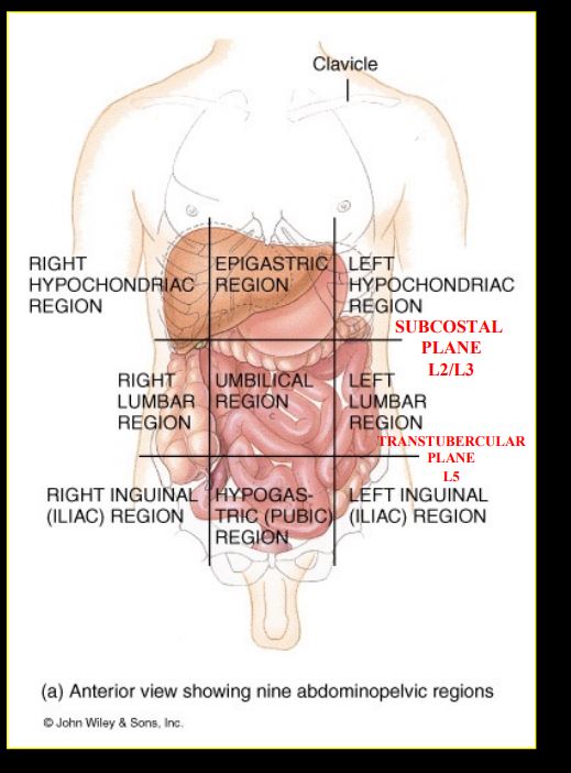
Image occlusion: name the hidden labels
26
The fascia of the anterior abdominal wall is broken into and
The fascia of the anterior abdominal wall is broken into superifical and deep
27
The fatty superifical fascia is called and the membranous deep superifical fascia is called . There is also a that covers the superifical muscles.
The fatty superifical fascia is called camper's fascia and the membranous deep superifical fascia is called scarpa's fasica . There is also a deep fasica that covers the superifical muscles. 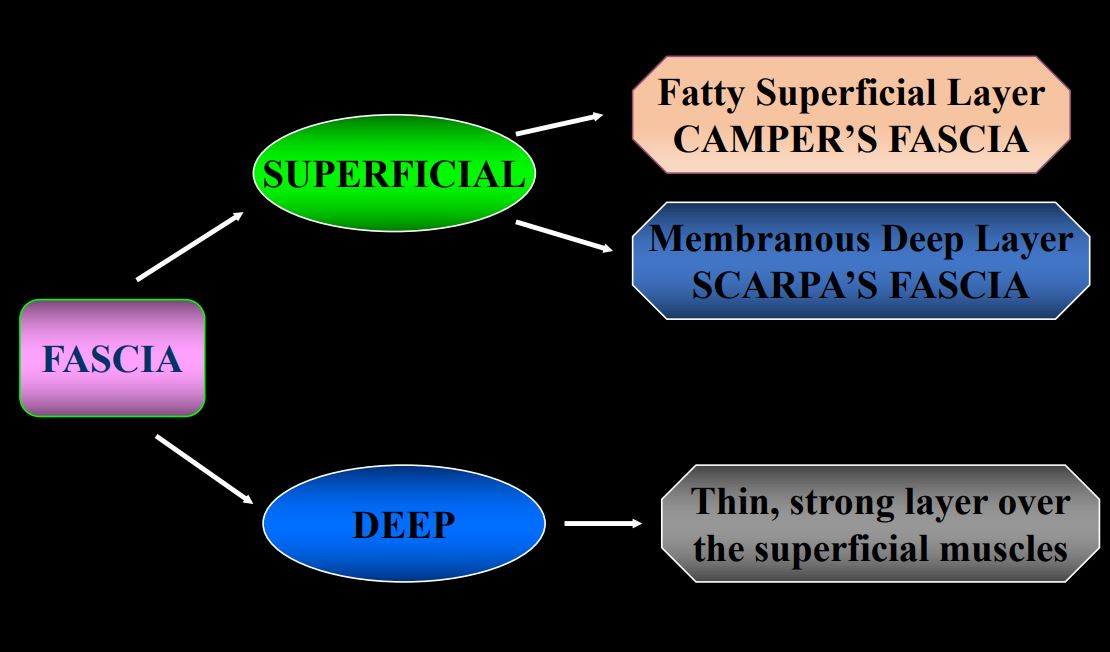
28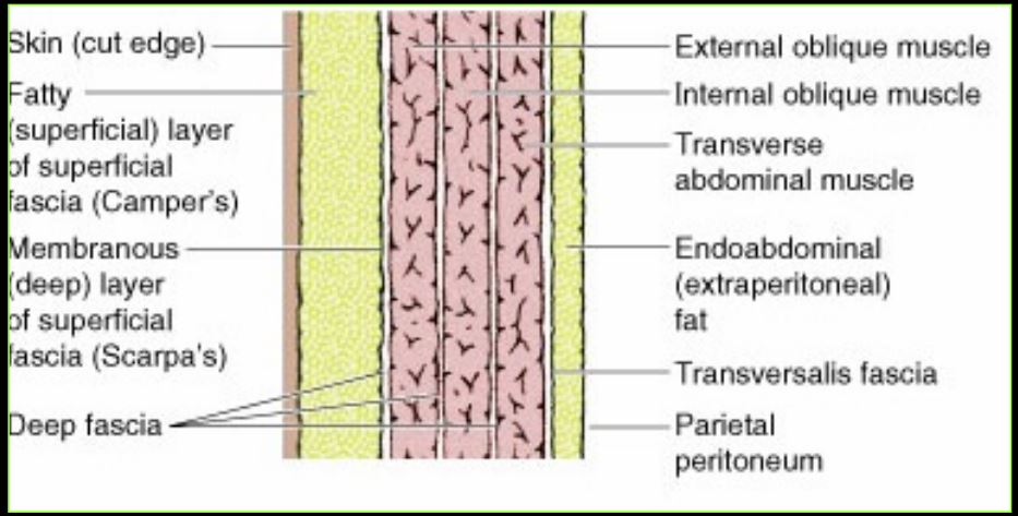
Image occlusion: name the hidden labels
29
The lower 1/3 of the rectus
sheath (between the ASIS
and the pubic symphysis) is
formed by:
sheath (between the ASIS
and the pubic symphysis) is
formed by:
- aponeuroses of all three
muscles form the anterior wall
of the rectus sheath.
-FASCIA TRANSVERSALIS
muscles form the anterior wall
of the rectus sheath.
-FASCIA TRANSVERSALIS
30
The membranous deep layer is also continuous with the superficial
fascia of the perineum or , and with that investing the
scrotum, penis, and the labia majora
fascia of the perineum or , and with that investing the
scrotum, penis, and the labia majora
The membranous deep layer is also continuous with the superficial
fascia of the perineum or Colle’s fascia, and with that investing the
scrotum, penis, and the labia majora
fascia of the perineum or Colle’s fascia, and with that investing the
scrotum, penis, and the labia majora
31
********SUPERFICIAL VESSELS IN THE SUPERFICIAL FASCIA
( )
( )
********SUPERFICIAL VESSELS IN THE SUPERFICIAL FASCIA
(Superficial Epigastric arteries [femoral artery] and veins)
(Superficial Epigastric arteries [femoral artery] and veins)
32
4 pairs muscles
1. External oblique
2. Internal oblique
3. Tranversus Abdominis
4. Rectus abdominis
2. Internal oblique
3. Tranversus Abdominis
4. Rectus abdominis
33
Nerve and action of external oblique
N: Inferior 6 thoracic nerve + subcostal nerve
A: Compress and support abdominal viscera, flex/ rotate the trunk
A: Compress and support abdominal viscera, flex/ rotate the trunk
34
-The fibers of the external oblique muscle run and
-As the fibers pass medially, they become This aponeurosis ends in the
-As the fibers pass medially, they become This aponeurosis ends in the
-The fibers of the external oblique muscle run inferoanteriorly and medially.
-As the fibers pass medially, they become aponeurotic. This aponeurosis ends in the linea alba.
-As the fibers pass medially, they become aponeurotic. This aponeurosis ends in the linea alba.
35
Inferiorly, the aponeurosis of the EOM folds back on itself to form the
(Poupart’s Ligament) which extends from the
to the .
(Poupart’s Ligament) which extends from the
to the .
Inferiorly, the aponeurosis of the EOM folds back on itself to form the
INGUINAL LIGAMENT (Poupart’s Ligament) which extends from the
ASIS to the pubic tubercle.
INGUINAL LIGAMENT (Poupart’s Ligament) which extends from the
ASIS to the pubic tubercle.
36
Nerve and action of the internal oblique muscle
N: Inferior 6 thoracic nerve
A: Compress and support the abdominal visera, flex and rotate the trunk
A: Compress and support the abdominal visera, flex and rotate the trunk
37
*****• In the male the spermatic cord passes through the fibers from which the is formed.
• The and, it is formed in part by the
IOM.
• The and, it is formed in part by the
IOM.
*****• In the male the spermatic cord passes through the internal oblique
muscle fibers from which the cremasteric muscle is formed.
• The inguinal canal passes through it and, it is formed in part by the
IOM.
muscle fibers from which the cremasteric muscle is formed.
• The inguinal canal passes through it and, it is formed in part by the
IOM.
38
What is the nerve and action of the transversus abdominis muscle?
N: Inferior 6 thoracic nerves
AL Compress and support abdominal visera
AL Compress and support abdominal visera
39
What is the conjoint tendon?
Insertion of IOM and
transversus abdominis onto the pubic symphysis
transversus abdominis onto the pubic symphysis
40
What is the nerve supply and action of the rectus abdominis muscle?
N: Inferior six thoracic nerves
A: Flexes the trunk and compresses the abdominal viscera
A: Flexes the trunk and compresses the abdominal viscera
41
The rectus sheath is formed by the aponeurosis of the ,
and as they insert into the
linea alba.
and as they insert into the
linea alba.
The rectus sheath is formed by the aponeurosis of the EOM, IOM
and transversus abdominis as they insert into the
linea alba.
and transversus abdominis as they insert into the
linea alba.
42
The internal oblique muscle aponeurosis splits into two
layers, with the anterior portion
going the rectus
abdominis, and the posterior
part going the rectus.
layers, with the anterior portion
going the rectus
abdominis, and the posterior
part going the rectus.
The internal oblique muscle aponeurosis splits into two
layers, with the anterior portion
going ABOVE the rectus
abdominis, and the posterior
part going BELOW the rectus.
layers, with the anterior portion
going ABOVE the rectus
abdominis, and the posterior
part going BELOW the rectus.
43
The upper 2/3 of the rectus sheath is made by the , the , and the
The upper 2/3 of the rectus sheath is made by the EOM aponeursis, the IOM aponeurosis, and the transversus abdominis
44
Superior to the acurate line the rectus sheath goes , , , and then
Superior to the acurate line the rectus sheath goes external oblique, internal oblique *SPLITS*, ***rectus abdominus, internal oblique again *SPLITS*and then transversus abdominis
45
Inferior to the acurate line, the rectus sheath goes , , , , and then
Inferior to the acurate line, the rectus sheath goes external oblique, internal oblique, transversus abdominis, **** rectus abdominus, and then transversalis fascia
46
The lower 1/3 of the rectus sheath is formed by the and the
The lower 1/3 of the rectus sheath is formed by the aponeurosis of all three muscles of anterior wall and the fascia transversalis (posterior)
47
UPPER 2/3 OF RECTUS
SHEATH:
Between and
SHEATH:
Between and
UPPER 2/3 OF RECTUS
SHEATH:
Between ASIS and Costal
margins
SHEATH:
Between ASIS and Costal
margins
48
LOWER 1/3 OF RECTUS
SHEATH:
Between and
SHEATH:
Between and
LOWER 1/3 OF RECTUS
SHEATH:
Between ASIS and Pubic
symphysis
SHEATH:
Between ASIS and Pubic
symphysis
49
The inferior limit of the posterior wall of the rectus sheath is marked
by a crescentic border called the which is located
midway between the umbilicus and the pubis.
by a crescentic border called the which is located
midway between the umbilicus and the pubis.
The inferior limit of the posterior wall of the rectus sheath is marked
by a crescentic border called the ARCUATE LINE which is located
midway between the umbilicus and the pubis.
by a crescentic border called the ARCUATE LINE which is located
midway between the umbilicus and the pubis.
50
The internal spermatic fascia of the spermatic cord is derived from the
The internal spermatic fascia of the spermatic cord is derived from the fascia transversalis
51
Layers of penetration abdomen
• Skin
• Superficial fascia: Camper’s
• Scarpa’s
• Deep fascia
• EOM and aponeurosis
• IOM and aponeurosis
• Transversus Abdominis
• Fascia transversalis
• Extraperitoneal fat
• Peritoneum: • Parietal • Peritoneal cavity • Visceral • Abdominal viscera
• Superficial fascia: Camper’s
• Scarpa’s
• Deep fascia
• EOM and aponeurosis
• IOM and aponeurosis
• Transversus Abdominis
• Fascia transversalis
• Extraperitoneal fat
• Peritoneum: • Parietal • Peritoneal cavity • Visceral • Abdominal viscera
52
Thoraco-abdominal nerves
T7-T11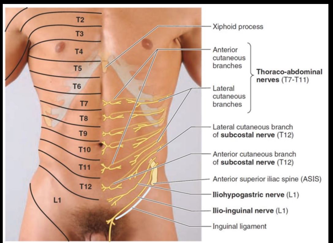
53
Lateral cutaneous branch of subcostal nerve
T12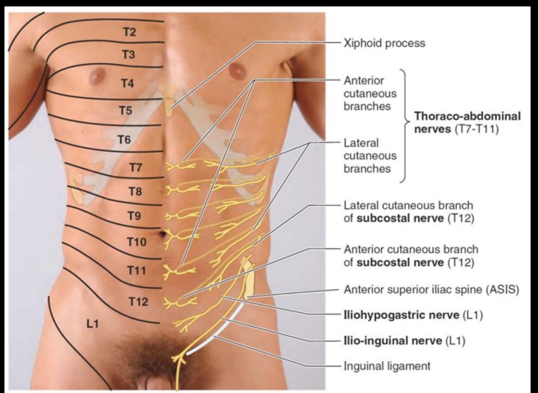
54
Anterior cuteaneous branch of subcostal nerve
T12 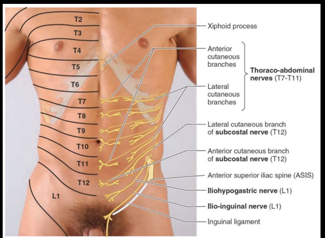
55
Iliohypogastric nerve
L1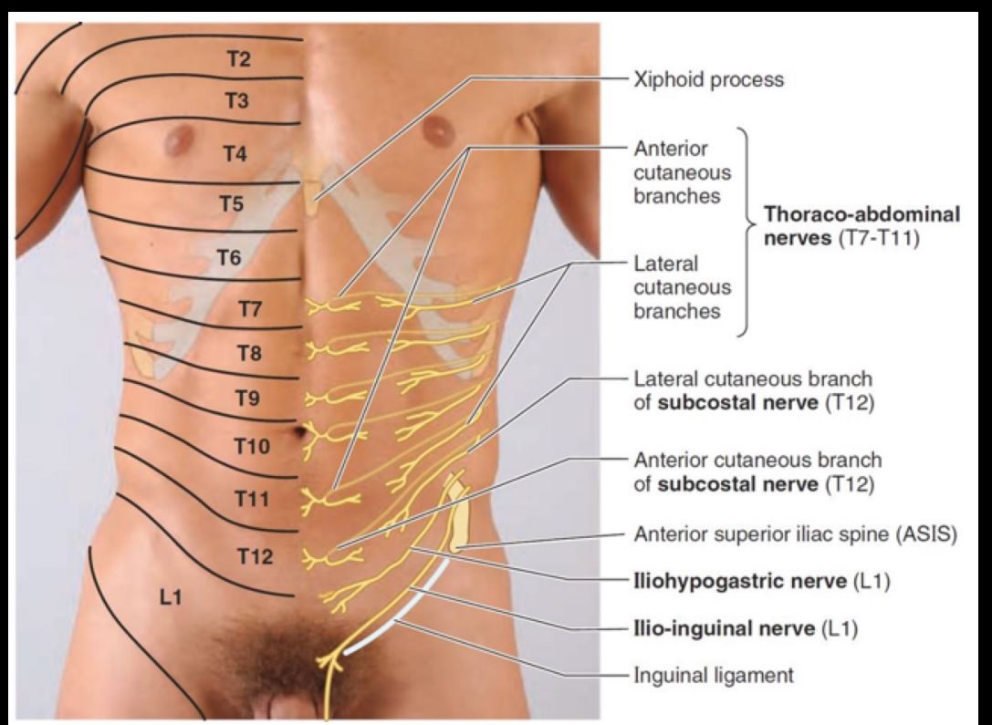
56
Ilioinguinal nerve
L1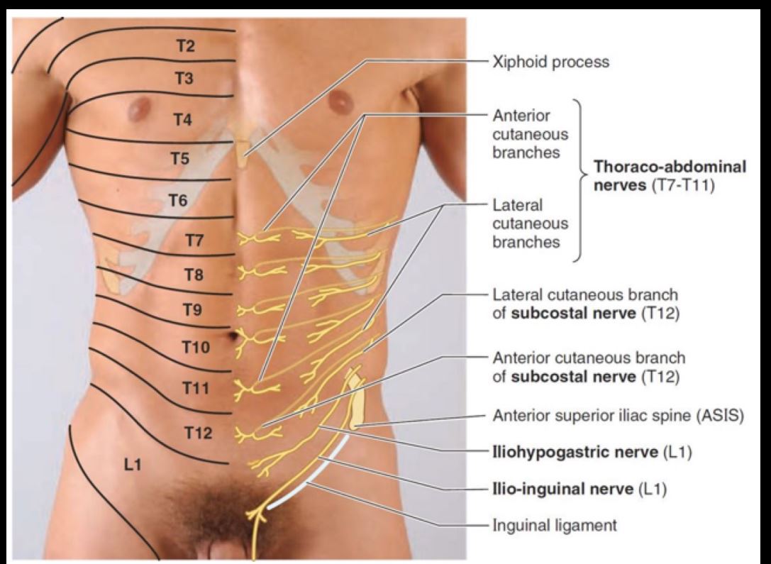
57
*****Iliohypogastric nerve (L1) = skin over upper , skin over
*****Iliohypogastric nerve (L1) = skin over upper inguinal
region (groin), skin over iliac crest.
region (groin), skin over iliac crest.
58
******lioinguinal nerve (L1) = skin over the . Also, the skin of the lower , , , or .
******lioinguinal nerve (L1) = skin over the superomedial
aspect of the thigh. Also, the skin of the lower inguinal
region, mons pubis, anterior scrotum, or labia majora.
aspect of the thigh. Also, the skin of the lower inguinal
region, mons pubis, anterior scrotum, or labia majora.
59
Superior epigastric artery (
Superior epigastric artery (Internal thoracic)
60
Inferior epigastric artery (
Inferior epigastric artery (External iliac artery)
61
Lumbar arteries (
Lumbar arteries (abdominal aorta)
62
Deep circumflex iliac (
Deep circumflex iliac (External iliac artery)
63
Superficial epigastric artery (
Superficial epigastric artery (femoral artery)
64
The inferior epigastric artery runs superiorly on the
and enters the to gain
the rectus sheath.
and enters the to gain
the rectus sheath.
The inferior epigastric artery runs superiorly on the
transversalis fascia and enters the arcuate line to gain
the rectus sheath.
transversalis fascia and enters the arcuate line to gain
the rectus sheath.
65
********The superior epigastric artery enters the rectus sheath
superiorly just inferior to the
superiorly just inferior to the
********The superior epigastric artery enters the rectus sheath
superiorly just inferior to the 7th costal cartilage.
superiorly just inferior to the 7th costal cartilage.
66
Superior to the umbilicus
Large median fold =
which goes
to the liver and
diaphragm. Its free edge
is the containing the
remnant of the umbilical
vein (obliterated)
Large median fold =
which goes
to the liver and
diaphragm. Its free edge
is the containing the
remnant of the umbilical
vein (obliterated)
Superior to the umbilicus
Large median fold =
FALCIFORM
LIGAMENT which goes
to the liver and
diaphragm. Its free edge
is the LIGAMENTUM
TERES containing the
remnant of the umbilical
vein (obliterated)
Large median fold =
FALCIFORM
LIGAMENT which goes
to the liver and
diaphragm. Its free edge
is the LIGAMENTUM
TERES containing the
remnant of the umbilical
vein (obliterated)
67
Inferior to the Umbilicus:
– Median Umbilical Fold =
– Medial Umbilical Fold =
– Lateral Umbilical Fold =
– Median Umbilical Fold =
– Medial Umbilical Fold =
– Lateral Umbilical Fold =
Inferior to the Umbilicus:
– Median Umbilical Fold =
urachus
– Medial Umbilical Fold =
obliterated umbilical
artery
– Lateral Umbilical Fold =
inferior epigastric artery
– Median Umbilical Fold =
urachus
– Medial Umbilical Fold =
obliterated umbilical
artery
– Lateral Umbilical Fold =
inferior epigastric artery