All cards
1
Bone is a tissue that acts as a support tissue and as a reservoir for
Bone is a calcified tissue that acts as a support tissue and as a reservoir for minerals.
2
The extracellular matrix of bone contains extracellular MATRIX that contains , and
The extracellular matrix of bone contains extracellular MATRIX that contains Type I collagen fibers, ground substance and minerals.
3
Whta are the two parts of the organic phase of bone?
1. Type I collagen fibers
2. Ground substance
2. Ground substance
4
Bone has what type of matrix (acidophilic or basophilic)? Why?
Acidophilic due to the high amount of calcium
5
What is the ground substance of bone made up of?
proteoglycans, GAGs, glycoproteins (osteonectin, osteocalcin, sialoprotein)
6
What is the inorganic phase of bone?
mineral salts in the form of hydroxyapatite (calcium phosphate crystals).
7
Bone is studied in or bone preparations
Bone is studied in decalcified or dried bone preparations
8
The matrix contains trapped in
The matrix contains osteocytes trapped in lacunae
9
Bone cells are interconnected by thin channels called
Bone cells are interconnected by thin channels called canaliculi
10
What is the function of the canaliculi?
Carries nutrients and gases to nearby blood vessels
11
Bone is highly (vascular or avascular)?
highly vascular, containing many blood vessels.
12
Bone matrix is constantly being
Bone matrix is constantly being remodeled
13
What are the two histological classifications of bone?
1. Spongy (cancellous, trabecular)
2. COmpact (dense, corticle)
2. COmpact (dense, corticle)
14
What is spongy bone made of?
Small pieces of bone, usually by bone marrow cavity
15
What is compact bone made of?
Large peices of bone with enclosed channels with blood vessels
16
Each ”gross” bone consists of a mixture of and .
Each ”gross” bone consists of a mixture of compact and spongy bone.
17
Bone is a and an
Bone is a tissue and an 'organ”
18
All exposed surfaces of bone must be covered or it will be destroyed
All exposed surfaces of bone must be covered by a lining of cells or tissues or it will be destroyed
19
What are the 4 parts of the bone "life cycle"
1. Osteoprogenitor cells
2. osteoblasts
3. MAKE BONE
4. Osteocytes
2. osteoblasts
3. MAKE BONE
4. Osteocytes
20
What are osteoprogenitor cells?
Osteogenic stem cells
21
Where are osteoprogenitor cells? What do they do? What do they become?
-They are mesenchymal stem cell hiding in the periosteum and endostum
-They can undergo mitosis
-They can differentiate into osteoblasts
-They can undergo mitosis
-They can differentiate into osteoblasts
22
What are osteoblasts?
-They are bone producers
23
What color do osteoblasts stain in H and E?
Blue (they are basphilic cells with round euchromatic nucleus)
24
How are osteoblasts arranged?
. lined up in one-cell thick rows on surfaces of bone (appositional growth)
25
What do osteoblasts secrete?
secrete organic unmineralized matrix called osteoid (collagen &
ground substance) and matrix vesicles that initiate bone mineralization.
ground substance) and matrix vesicles that initiate bone mineralization.
26
Can osteoblasts undergo mitosis?
No, they are terminally differeniyaed
27
When do osteoblasts become osteocytes?
When trapped in the mineralizing matrix, they become osteocytes
28
What are ostecytes?
They are trapped in lacunae with long, thin cell processes extendinng into canalculi
29
How do osteocytes communicate with each other?
gap junctions at the tips of their processes
30
What role do osteocytes play in the bone matrix?
They maintain the bone matrix (with some synthetic and resportive ability)
31
Can osteocytes divide?
No, they are terminally differentiated
32
What is an osteoclast?
It is a bone resorbing cell
33
How are osteoclasts formed?
Monocytes become osteoclasts and osteoclasts destroy bone
34
How do osteoclasts stain in H and E?
They have an acidic cytoplasm because they are abundant in mitochondria
35
What is a Howship's lacunae?
Any of the tiny depressions, pits, or irregular grooves in bone that is being resorbed by osteoclasts
36
Describe the structure of the cell membrane of osteoclasts?
Membrane is highly folded into a ruffled border
37
What do osteoclasts contain?
Lysosomes, collagenase, and other lytic enzymes
38
When osteoclasts break down bone, what do they release into the blood?
Calcium and phosphate
39
, the functional unit of compact bone, consists of
concentric lamellae (layers) of bone matrix and osteocytes surrounding a canal
with blood vessels
concentric lamellae (layers) of bone matrix and osteocytes surrounding a canal
with blood vessels
Haversian systems or osteons, the functional unit of compact bone, consists of
concentric lamellae (layers) of bone matrix and osteocytes surrounding a canal
with blood vessels
concentric lamellae (layers) of bone matrix and osteocytes surrounding a canal
with blood vessels
40
How are osteons aligned in the Haversian system (osteon)? How are they linked?
Chains of 10-20 osteocytes in 4-10 lamellae are linked by canaliculi.
41
How does oxygen and nutrient diffusion work in the Haversian system?
Oxygen and nutrients from the central
blood vessel, diffuses through the canaliculi to reach all the osteocytes in the osteon.
blood vessel, diffuses through the canaliculi to reach all the osteocytes in the osteon.
42
Why is the osteon strong?
Lamellae have a slightly different angle of collagen
fibers
fibers
43
What are haversian canals lined with?
endosteum
44
What is a Volmann's canal?
-transverse canals aligned perpendicular to the long axis of bone
-bring blood vessels from the periosteum to the Haversian canals, marrow
cavity and then back out.
-bring blood vessels from the periosteum to the Haversian canals, marrow
cavity and then back out.
45
What are Volkmann's canals lined with?
Endosteum
46
What are intersitial lamellae?
- remnants of partially resorbed osteons formed during bone
remodeling
remodeling
47
What are circumferential lamellae?
The first few lamellae of bone near the periosteum or the
marrow cavity. T
marrow cavity. T
48
What is the blood supply of cicrumferential lamellae?
They are supplied with blood from vessels in the periosteum or marrow
49
Endosteum lines structures including , & canals.
Endosteum lines internal structures including bone marrow, Haversian & Volkmann’s canals.
50
Periosteum lines the of bone. Collagen fibers ( ) in the periosteum insert
directly into the .
directly into the .
Periosteum lines the outside of bone. Collagen fibers (Sharpey’s fibers) in the periosteum insert
directly into the bone matrix.
directly into the bone matrix.
51
Both the endosteum and periosteum are
Both the endosteum and periosteum are osteogenic.
52
(cancellous, trabecular) consists of small finger-like pieces of bone, usually
associated with the marrow cavity.
associated with the marrow cavity.
Spongy bone (cancellous, trabecular) consists of small finger-like pieces of bone, usually
associated with the marrow cavity.
associated with the marrow cavity.
53
Blood vessels in the surrounding spaces between the
trabeculae of spongy bone supply and to nearby osteocytes.
trabeculae of spongy bone supply and to nearby osteocytes.
Blood vessels in the surrounding spaces between the
trabeculae of spongy bone supply oxygen and nutrients to nearby osteocytes.
trabeculae of spongy bone supply oxygen and nutrients to nearby osteocytes.
54
(dense, cortical) consists of thick pieces of bone with osteons.
Compact bone (dense, cortical) consists of thick pieces of bone with osteons.
55
Blood vessels in the supply oxygen and nutrients to osteocytes in the / system
Blood vessels in the Haversian canals supply oxygen and nutrients to osteocytes in the osteon/Haversian system
56
What are the three features of COMPACT bone?
1. Volmann's and Harversian canals
2. Osteons
3. Circumferential and interstitial lamellae
2. Osteons
3. Circumferential and interstitial lamellae
57
Bone develops in , a loosely organized embryonic connective tissue.
Bone develops in mesenchyme, a loosely organized embryonic connective tissue.
58
Mesodermgives rise to most of the mesenchyme in the embryo except for the region
where most of the mesenchyme is derived from
where most of the mesenchyme is derived from
Mesodermgives rise to most of the mesenchyme in the embryo except for the head and neck region
where most of the mesenchyme is derived from neural crest ectoderm.
where most of the mesenchyme is derived from neural crest ectoderm.
59
Bone forms by two processes: or
Bone forms by two processes: intramembranous or endochondral ossification
60
What happens during INTRAMEMBRANOUS ossification?
Bones develops directly from the mesenchyme
61
What is developed from intramembranous ossification? 4 structures
All head related = Flat bones of face and skill (vault of cranium), manible, maxilla, and clavicle
62
What happens during endochrondral ossifcation?
-Temporary cartilage model is formed in mesenchyme
-Continuously growing cartilage model is progressive replaced by bone
-Continuously growing cartilage model is progressive replaced by bone
63
What are general three steps of endochondral ossifcation?
Mesenchyme to cartilage model (chondroblasts/cytes) to bone (osteoblasts)
64
What is developed from endochondral ossfication?
Extremities, vertebrae, pelvis, and bones at base of skull (NO HEAD)
65
The terms, endochondral and intramembranous ossification refer ONLY to the
and
and
The terms, endochondral and intramembranous ossification refer ONLY to the mechanism
and environment by which bone is initially formed.
and environment by which bone is initially formed.
66
What 4 things are similar between the ossification processes? (Endochondral and intramembranous)
1. Both give rise to same histological types of bone
2. Both processes have primary, immature bone made of collagen first that is poorly arranged
3. Bone remodeling makes all of immature WOVEN bone to strong LAMELLAR bone
4. Spongy bone is then filled in
2. Both processes have primary, immature bone made of collagen first that is poorly arranged
3. Bone remodeling makes all of immature WOVEN bone to strong LAMELLAR bone
4. Spongy bone is then filled in
67
What are the 4 steps of intramembranous ossifcation?
1. Mesenchyme differeniate to osteoblasts
2. Osteoblasts secrete unmineralized bone matrix (osteoid) to form primary woven bone. Osteoblast get trapped in lacunae, making osteocytes
3. Osteoblasts differeniate and secrete osteoid at surface of developing bone (appositional growth) forming spicules
4. Bone is continuously remodeled through antagonistic actions of osteoblastsm
5. The periosteum, endosteum, and marrow develop
2. Osteoblasts secrete unmineralized bone matrix (osteoid) to form primary woven bone. Osteoblast get trapped in lacunae, making osteocytes
3. Osteoblasts differeniate and secrete osteoid at surface of developing bone (appositional growth) forming spicules
4. Bone is continuously remodeled through antagonistic actions of osteoblastsm
5. The periosteum, endosteum, and marrow develop
68
What occurs during bone remodeling in intramembranous ossification?
1. Primary bone is replaced with stronger, secondary lamellar bone
2. Compact bone forms by filling in spaces of spongy
3. Proper poertions of bone is maintained by adding new layers
2. Compact bone forms by filling in spaces of spongy
3. Proper poertions of bone is maintained by adding new layers
69
What are the 1-7 steps of the endochondrial ossification model?
1. Mesenchyme cells condense, differenitae into chondroblasts, and form HYALINE CARTILAGE MODEL
2. Blood vessels develop into a connective tissue. Increased oxygen causes conversion of PERICHONDRIUM TO PERIOSTEUM.
3. Oxgen and nutrients cannot diffuse through bone matrix of PERIOSTEAL COLLAR to cartilage within. This causes the chrondrocytes to hyperthrphy, calcify, and chondrocytes die
4. PERIOSTEAL bud breaks the bone collar in center shaft, which brings in vessels, blood cells, osteoclasts, and osteoprogenitaor cells
5. BECOMES PRIMARY OSSIFICATION CENTER.
6. OSTEOBLASTS = differentiate and deposit bone on pieces of calcifed cartilage
7. OSTEOCLASTS = reabsorb the calified cartilage and newly formed bone FORMS THE MARROW CAVITY
2. Blood vessels develop into a connective tissue. Increased oxygen causes conversion of PERICHONDRIUM TO PERIOSTEUM.
3. Oxgen and nutrients cannot diffuse through bone matrix of PERIOSTEAL COLLAR to cartilage within. This causes the chrondrocytes to hyperthrphy, calcify, and chondrocytes die
4. PERIOSTEAL bud breaks the bone collar in center shaft, which brings in vessels, blood cells, osteoclasts, and osteoprogenitaor cells
5. BECOMES PRIMARY OSSIFICATION CENTER.
6. OSTEOBLASTS = differentiate and deposit bone on pieces of calcifed cartilage
7. OSTEOCLASTS = reabsorb the calified cartilage and newly formed bone FORMS THE MARROW CAVITY
70
What are the 8- steps of the endochondrial ossification model?
8. Growth in length occurs by chondrocyte division (interstital growth)
9. Growth in width occurs by appositional growth by osteoblasts that differenitae from the periosteum. Osteoclasts enlarge marrow cavity.
10. Secondary ossification center forms at end of bone restricting cartilage growth to growth/epiphyseal plate (occurs POSTNATALLY) m
11. Growth in length ends with cartilage in the growth epiphyseal plate is destroyed
12. Bones can increase in WIDTH throughout life from differentiation of osteoblasts in periosteum
9. Growth in width occurs by appositional growth by osteoblasts that differenitae from the periosteum. Osteoclasts enlarge marrow cavity.
10. Secondary ossification center forms at end of bone restricting cartilage growth to growth/epiphyseal plate (occurs POSTNATALLY) m
11. Growth in length ends with cartilage in the growth epiphyseal plate is destroyed
12. Bones can increase in WIDTH throughout life from differentiation of osteoblasts in periosteum
71
What are the 5 zones of bone? (proximal to more distal)
1. Resting cartilage (young healthy cartilage)
2. Proliferating cartilage (condrocytes divide interstitally, proudce matrix, increase length, arrange in columns)
3. Hypertrophying cartilage (chondrocytes and lacunae enlarge - apoptosis
4. Calcifying cartilage (insoluble calcium salts in cartilage matrix
5. Resorption (osteoclasts eat clacified matrix)
2. Proliferating cartilage (condrocytes divide interstitally, proudce matrix, increase length, arrange in columns)
3. Hypertrophying cartilage (chondrocytes and lacunae enlarge - apoptosis
4. Calcifying cartilage (insoluble calcium salts in cartilage matrix
5. Resorption (osteoclasts eat clacified matrix)
72
Maintenance of the appropriate WIDTH of bone during development (and life) occurs by
of osteoblasts and osteoclasts.
of osteoblasts and osteoclasts.
Maintenance of the appropriate WIDTH of bone during development (and life) occurs by
antagonistic actions of osteoblasts and osteoclasts.
antagonistic actions of osteoblasts and osteoclasts.
73
Bone matrix is added to the outside of the bone (appositional growth) by osteoblasts that
differentiate from the
differentiate from the
Bone matrix is added to the outside of the bone (appositional growth) by osteoblasts that
differentiate from the periosteum.
differentiate from the periosteum.
74
Bone matrix is resorbed from the inside by osteoclasts in the
Bone matrix is resorbed from the inside by osteoclasts in the marrow cavity
75
What two changes can cause bone disease?
1. Number or activity of osteoclasts and osteoblasts
2. Poor bone matrix construction by issues with osteoid or matrix or mineralization
2. Poor bone matrix construction by issues with osteoid or matrix or mineralization
76
Spongy bone has a high to area allowing more space for osteoblasts and
clasts.
clasts.
Spongy bone has a high surface to volume area allowing more space for osteoblasts and
clasts.
clasts.
77
Are disordered related to rate of bone turnover rate more likely to impact spongy bone or compact bone?
Spongy bone
78
What are the two major hormones that regulate calcium levels?
(1) Calcitonin - from tyroid gland, lowers blood calcium levels, inhibits osteoclast
(2) Parathyroid hormone - from parathyroid gland, raise blood calcium, increases osteoclast
(2) Parathyroid hormone - from parathyroid gland, raise blood calcium, increases osteoclast
79
causes excessive osteoclast activity and bone resorption.
Hyperparathyroidism causes excessive osteoclast activity and bone resorption.
80
Osteoporosis – is a disease in which there is a reduction in due to an
rate of bone resorption. Bone appears histologically . Decreased bone is
especially visible in bone. Fractures are common especially in
vertebrae.
rate of bone resorption. Bone appears histologically . Decreased bone is
especially visible in bone. Fractures are common especially in
vertebrae.
Osteoporosis – is a disease in which there is a reduction in total bone mass due to an
increased rate of bone resorption. Bone appears histologically normal. Decreased bone is
especially visible in spongy or trabecular bone. Fractures are common especially in
vertebrae.
increased rate of bone resorption. Bone appears histologically normal. Decreased bone is
especially visible in spongy or trabecular bone. Fractures are common especially in
vertebrae.
81
Vitamin D (and Calcium) deficiency – results in of newly
formed bone matrix resulting in an accumulation of osteoid, the unmineralized organic
components of bone matrix. Vitamin D is necessary for .
formed bone matrix resulting in an accumulation of osteoid, the unmineralized organic
components of bone matrix. Vitamin D is necessary for .
Vitamin D (and Calcium) deficiency – results in inadequate mineralization of newly
formed bone matrix resulting in an accumulation of osteoid, the unmineralized organic
components of bone matrix. Vitamin D is necessary for calcium absorption.
formed bone matrix resulting in an accumulation of osteoid, the unmineralized organic
components of bone matrix. Vitamin D is necessary for calcium absorption.
82
Osteomalacia – in leads to inadequate mineralization during
bone remodeling. Bones are soft and easily fracture.
bone remodeling. Bones are soft and easily fracture.
Osteomalacia – low vitamin D in adults leads to inadequate mineralization during
bone remodeling. Bones are soft and easily fracture.
bone remodeling. Bones are soft and easily fracture.
83
Rickets – in , leads to inadequate mineralization during bone
growth and remodeling as well as disrupts the growth plates. Cartilage does not calcify,
and bone does not mineralize adequately in the growth plate. Growth is and
with weight baring the softer bones of the legs
growth and remodeling as well as disrupts the growth plates. Cartilage does not calcify,
and bone does not mineralize adequately in the growth plate. Growth is and
with weight baring the softer bones of the legs
Rickets – low vitamin D in children, leads to inadequate mineralization during bone
growth and remodeling as well as disrupts the growth plates. Cartilage does not calcify,
and bone does not mineralize adequately in the growth plate. Growth is stunted and
with weight baring the softer bones of the legs become bowed.
growth and remodeling as well as disrupts the growth plates. Cartilage does not calcify,
and bone does not mineralize adequately in the growth plate. Growth is stunted and
with weight baring the softer bones of the legs become bowed.
84
Growth hormone (GH), secreted from the anterior pituitary, stimulates
, including that of and .
, including that of and .
Growth hormone (GH), secreted from the anterior pituitary, stimulates
general tissue growth, including that of cartilage and bone.
general tissue growth, including that of cartilage and bone.
85
Acromegaly - in increases . Causes excess growth from the periosteum that produces thicker bones and coarse facial features
Acromegaly - excess GH in adults increases bone formation. Causes excess growth from the periosteum that produces thicker bones and coarse facial features
86
Gigantism - in results in increased and extended bone growth. Results in bones that are thicker and wider and longer due to increased cartilage proliferation at
Gigantism - excess GH in children results in increased and extended bone growth. Results in bones that are thicker and wider and longer due to increased cartilage proliferation at growth plate
87
Pituitary dawrfism - in reduces overall growth in all tissues, including cartilage and bone. AND are effected. Causes decreased sature, but normal proportions.
Pituitary dawrfism - decreased GH in children reduces overall growth in all tissues, including cartilage and bone. Endochondral AND intermembranous ossification are effected. Causes decreased sature, but normal proportions.
88
Achondroplasia - the most common form of . Is a genetiic disease where is inhibited. Bones formed by are affected. Individuals will have short extremeities, bowed legs, and swayed back. appears large compared to .
Achondroplasia - the most common form of drawfism. Is a genetiic disease where chondrocyte division is inhibited. Bones formed by endochondral ossification are affected. Individuals will have short extremeities, bowed legs, and swayed back. Head appears large compared to short limbs.
89
Osteogenesis imperfecta (brittle bone disease), is a group of hereditary disorders with
defective synthesis of . Tissues with , like bone, are
severely affected. Osteoblasts secrete the into the matrix, with the
result that bones are weak and easily fractured.
defective synthesis of . Tissues with , like bone, are
severely affected. Osteoblasts secrete the into the matrix, with the
result that bones are weak and easily fractured.
Osteogenesis imperfecta (brittle bone disease), is a group of hereditary disorders with
defective synthesis of Type I collagen. Tissues with high Type I collagen, like bone, are
severely affected. Osteoblasts secrete the defective collagen into the matrix, with the
result that bones are weak and easily fractured.
defective synthesis of Type I collagen. Tissues with high Type I collagen, like bone, are
severely affected. Osteoblasts secrete the defective collagen into the matrix, with the
result that bones are weak and easily fractured.
90
What are the four steps of fracture repair?
1. large hematoma from clotted blood forms a fibrin scaffold. The damaged cells and tissue are removed and new vessels, inflammatory cells, fibroblasts, and chrondrogenic and osteogenic cells appear
2. SOFT CALLUS containing cartilage and CT connects ends of the fracture bones
3. BONY CALLUS with parts of primary woven bone replace soft cartilaginous callus
4. Bone remodeling replaces woven bone with strong lamellar bone
2. SOFT CALLUS containing cartilage and CT connects ends of the fracture bones
3. BONY CALLUS with parts of primary woven bone replace soft cartilaginous callus
4. Bone remodeling replaces woven bone with strong lamellar bone
91
Glance at table and just loosely familiarize.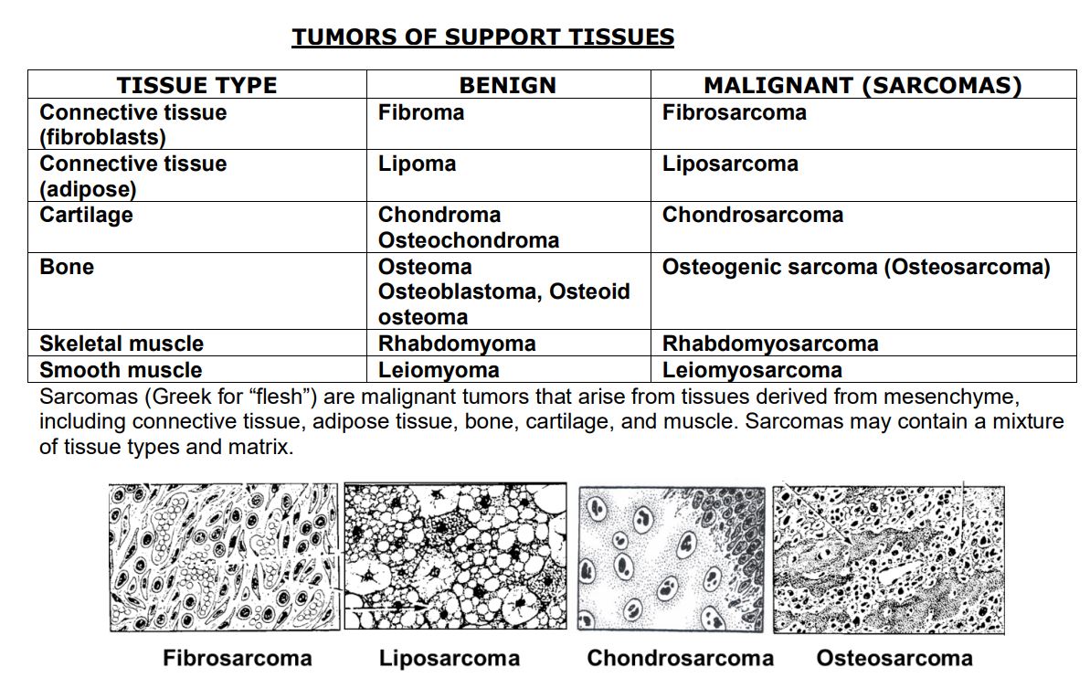
92
Growth in length ends when all chondrocytes are
Growth in length ends when all chondrocytes are destroyed