Chapter 17
Study these flashcardsFree · spaced repetition · iPhone, Android, Mac & web
All cards
1
Three major paired salivary glands are the , , and
Three major paired salivary glands are the parotid, submandibular, and sublingual
2
The major paired salivary glands and the minor glands in the submucosa cavity make , , , , and .
The major paired salivary glands and the minor glands in the submucosa cavity make saliva, serous/muscous, water, electrolytes, and antibodies.
3
What are the two types of specialized ducts that are within lobes?
1. Intercalcated duct
2. Striated duct
2. Striated duct
4
Intercalcated ducts are lined with or
Intercalcated ducts are lined with simple cubodial or squamous
5
Intercalcated ducts add
Intercalcated ducts add biocarbonate ions to secretion
6
Straited ducts are lined by with
Straited ducts are lined by acidophilic simple columnar epithelia with basal striations
7
The straitions of the striated duct are caused by in a highly cell membrane
The straitions of the striated duct are caused by parallel rows of mitrochondria in a highly folded cell membrane
8
In a striated duct, the nuceli are pushed towards the
In a striated duct, the nuceli are pushed towards the lumen
9
The striated duct is involved in and re-absorption of and addition of to secretion.
The striated duct is involved in ion transport and re-absorption of Na+ and addition of K+ to secretion.
10
Excretory ducts are indentified because they are ducts that are surrounded by increasing amounts of .
Excretory ducts are indentified because they are progressively larger ducts that are surrounded by increasing amounts of connective tissue.
11
Excretory ducts are lined by to
Excretory ducts are lined by simple columnar to stratified squamous
12
are the main duct that enters into the oral cavity
Excretory ducts are the main duct that enters into the oral cavity
13
Parotid glands are
Parotid glands are pure serous 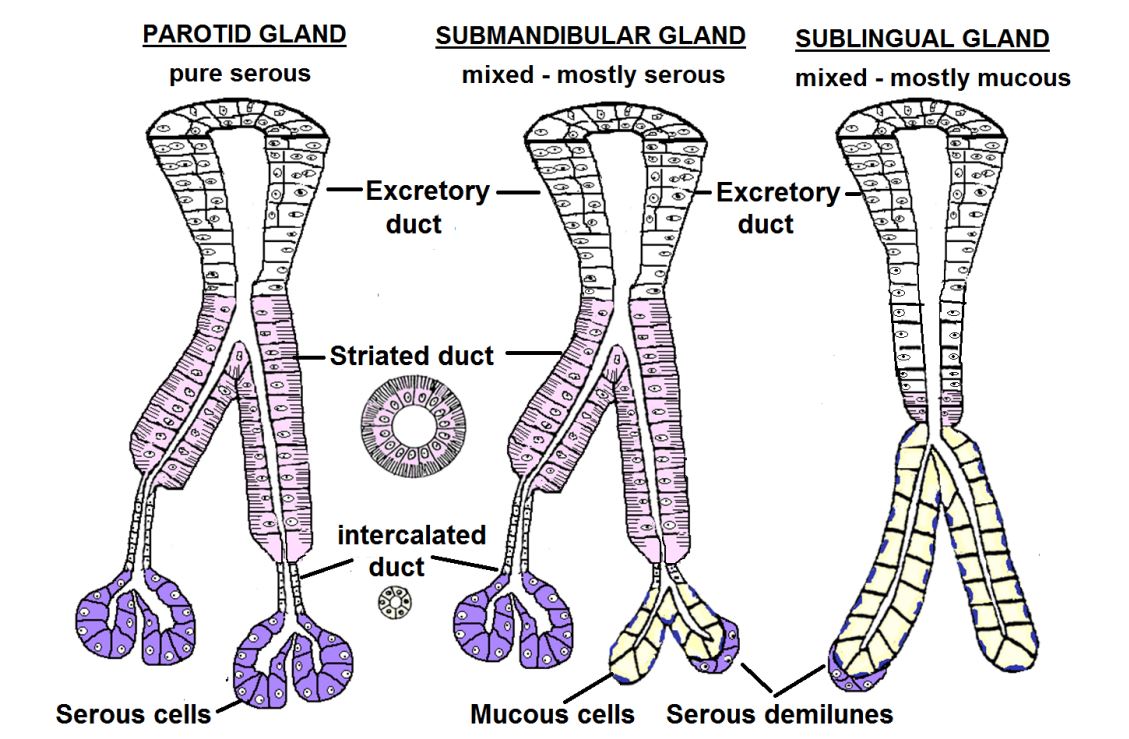
14
Submandibular glands are
Submandibular glands are mixed-mostly serous 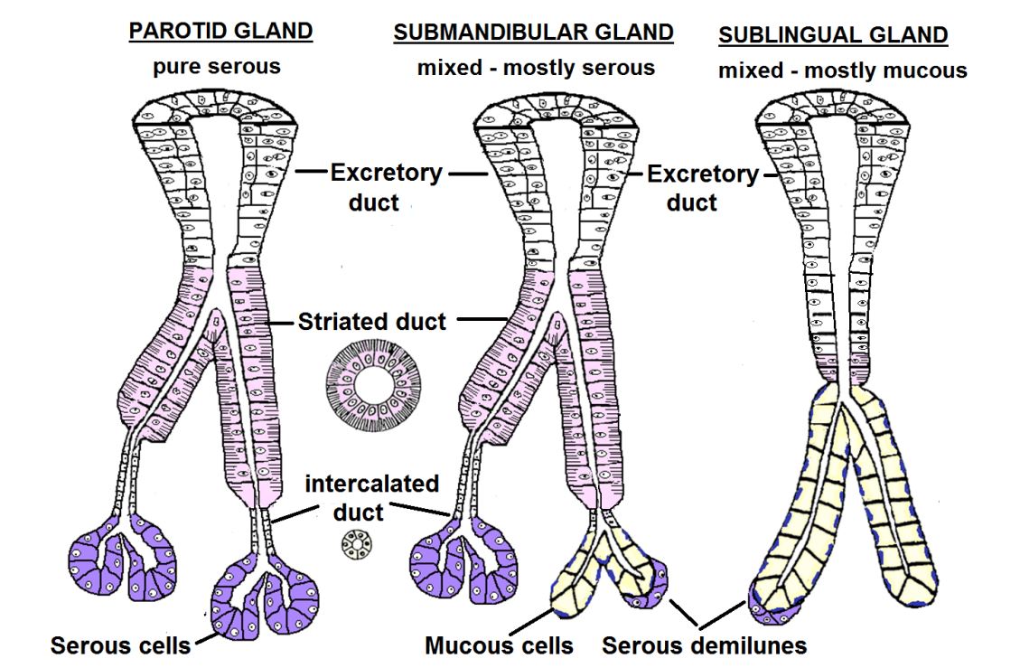
15
Sublingual glands are
Sublingual glands are mixed-mostly mucous 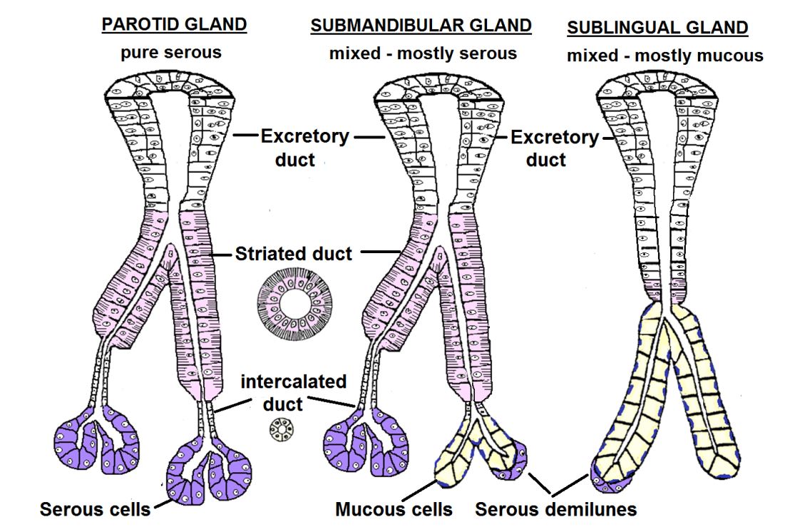
16
Parotid glands and submandibular glands have and
The sublingual gland has NO and
The pancreas has many and NO
have excretory ducts
The sublingual gland has NO and
The pancreas has many and NO
have excretory ducts
Parotid glands and submandibular glands have many intercalcated ducts and striated ducts
The sublingual gland has NO intercalcated and few striated ducts
The pancreas has many intercalcated and NO striated ducts.
All have excretory ducts
The sublingual gland has NO intercalcated and few striated ducts
The pancreas has many intercalcated and NO striated ducts.
All have excretory ducts
17
A pancreas is a gland
A pancreas is a pure serous gland
18
Straited ducts are only in
Straited ducts are only in salivary glands
19
Pleomorphic adenoma, a benign tumor of the derived from a mixture of
, , and
, , and
Pleomorphic adenoma, a benign tumor of the salivary gland derived from a mixture of
secretory, duct, and myoepithelial cells
secretory, duct, and myoepithelial cells
20
The pancrea is considered to have two glands in one. The exocrine gland ( ) and endocrine gland ( ).
The pancrea is considered to have two glands in one. The exocrine gland (serous gland) and endocrine gland (islet of langerhans).
21
The pancreas is a gland (shape of gland)
The pancreas is a compound alveolar gland (shape of gland)
22
The two parts of the exocrine pancreas are the and
The two parts of the exocrine pancreas are the serous cells and ducts
23
Acinar cells, a protein secreting cell, have a nucleus, cytoplasm
(due mostly to ) and visible secretory ( ) granules that contain numerous digestive
enzymes including , , , , and .
(due mostly to ) and visible secretory ( ) granules that contain numerous digestive
enzymes including , , , , and .
Acinar cells, a protein secreting cell, have a euchromatic nucleus, basophilic cytoplasm
(due mostly to RER) and visible secretory (zymogen) granules that contain numerous digestive
enzymes including trypsinogen, chymotrypsinogen, amylase, lipase, and deoxyribonuclease.
(due mostly to RER) and visible secretory (zymogen) granules that contain numerous digestive
enzymes including trypsinogen, chymotrypsinogen, amylase, lipase, and deoxyribonuclease.
24
Enzymes of acinar or zymogen cells are inactive proenzyme until they reach the
Enzymes of acinar or zymogen cells are inactive proenzyme until they reach the duodenum
25
The pancreas has intercalcated ducts that are and have . Secrete and water to make the secretion . cells are the first duct cells, found in the middle of the
The pancreas has intercalcated ducts that are simple cuboidal and have clear cells. Secrete bicarbonate and water to make the secretion alkaline . Centroacinar cells are the first duct cells, found in the middle of the secretory acinus
26
The excretory ducts of the pancreas are usually , eventually empties into . The last duct is lined by the same epithelium as the
The excretory ducts of the pancreas are usually simple columnar, eventually empties into duodenum. The last duct is lined by the same epithelium as the surface it empties onto.
27
In the salivary glands
the last duct is lined by a , while the pancreatic
duct is lined by , similar to the duodenum.
the last duct is lined by a , while the pancreatic
duct is lined by , similar to the duodenum.
In the salivary glands
the last duct is lined by a stratified squamous epithelium (oral cavity lining), while the pancreatic
duct is lined by simple columnar epithelium, similar to the duodenum.
the last duct is lined by a stratified squamous epithelium (oral cavity lining), while the pancreatic
duct is lined by simple columnar epithelium, similar to the duodenum.
28
The two diseases of the exocrine portion of the pancreas are and .
The two diseases of the exocrine portion of the pancreas are acute necrotizing pancretitis and pancreatic carcinoma.
29
ISLETS OF LANGERHANS: Islands of light staining cells scattered among the and . Separated from the other cells by a
ISLETS OF LANGERHANS: Islands of light staining cells scattered among the acinar (serous cells) and ducts. Separated from the other cells by a delicate CT
30
cell (70%) – secrete insulin, lowers blood glucose
Beta cell (70%) – secrete insulin, lowers blood glucose
31
cell (20%) - secretes glucagon, raises blood glucose
Alpha cell (20%) - secretes glucagon, raises blood glucose
32
cell – secretes somatostatin
Delta cell – secretes somatostatin
33
cell – secretes pancreatic polypeptide
PP cell – secretes pancreatic polypeptide
34
There are many to easily pick up and carry away the hormones. These are common in endocrine tissues and organs.
There are many fenestrated capillaries to easily pick up and carry away the hormones. These are common in endocrine tissues and organs.
35
What is the major disease of the endocrine portion of the pancreas?
Type 1 diabetes mellitus
36
Compared to salivary glands, the pancreas has
Compared to salivary glands, the pancreas has less connective tissue
37
Striated ducts are only in
Striated ducts are only in salivary glands
38
Undermicroscope, pancreas looks like it only has few ducts, but it is actually filled with ducts that are
Undermicroscope, pancreas looks like it only has few ducts, but it is actually filled with ducts that are small intercalcated
39
The three functions of the liver are , , and .
The three functions of the liver are exocine, modification and filtration of blood, and hematopoietic function of the embryo.
40
The exocine function of the liver involve secretion of into ducts which eventually empty into the
The exocine function of the liver involve secretion of BILE into ducts which eventually empty into the duodenum
41
Bile contains , that emulsify fats in the small intestine and waste products: such as ) a toxic breakdown product of eliminated in feces
Bile contains cholesterol, bile salts that emulsify fats in the small intestine and waste products: such as bilirubin (conjugated) a toxic breakdown product of hemoglobin eliminated in feces
42
The liver is responsible for secretion of , including albumen, blood clotting factors (fibrinogen and
prothrombin).
prothrombin).
The liver is responsible for secretion of plasma proteins, including albumen, blood clotting factors (fibrinogen and
prothrombin).
prothrombin).
43
The liver removes debris from the
The liver removes debris from the breakdown of RBC
44
The liver had a artery and vein
The liver had a heptatic artery and portal vein
45
The hepatic artery brings blood
The hepatic artery brings oxygenated blood
46
The brings blood from capillaries in the , , and
The hepatic portal vein brings blood from capillaries in the intestines (food), pancreas (hormones), and spleen (RBC breakdown products)
47
The blood of the liver flows from the to the
The blood of the liver flows from the portal traid to the central vein
48
The hepatocytes are the or functional cells and they are arranged in
The hepatocytes are the parenchymal or functional cells and they are arranged in plates
49
The portal canal has edges of , contains and
The portal canal has edges of lobule, contains portal triad and lympathic vessels
50
Bile is kept separated and flows from to the , to the
flow of blood.
flow of blood.
Bile is kept separated and flows from bile canaliculi to the portal triad, opposite to the
flow of blood.
flow of blood.
51
Hepatocytes secrete bile into , tiny channels formed by tight junctions between
hepatocytes
hepatocytes
Hepatocytes secrete bile into canaliculi, tiny channels formed by tight junctions between
hepatocytes
hepatocytes
52
Bile flows along the canaliculi to the edge of the lobule where they empty into true
epithelial lined ducts, the
epithelial lined ducts, the
Bile flows along the canaliculi to the edge of the lobule where they empty into true
epithelial lined ducts, the bile ducts.
epithelial lined ducts, the bile ducts.
53
Bile ducts are lined by or in the larger ducts,
.
.
Bile ducts are lined by simple cuboidal or in the larger ducts,
simple columnar epithelium.
simple columnar epithelium.
54
Flow of bile goes from to to to to
Flow of bile goes from hepatocytes to bile canaliculi to bile ductules to bile ducts to hepatic duct
55
Branches of the hepatic portal vein and artery in the CT at the edge of the lobule, empty their
blood into the which are lined by AND
blood into the which are lined by AND
Branches of the hepatic portal vein and artery in the CT at the edge of the lobule, empty their
blood into the liver sinusoids which are lined by endothelial AND Kupffer cells (macrophages)
blood into the liver sinusoids which are lined by endothelial AND Kupffer cells (macrophages)
56
Blood from the hepatic artery and hepatic portal vein drain into which drain into the which drain into the , which drain into the
Blood from the hepatic artery and hepatic portal vein drain into sinusoids which drain into the central vein which drain into the hepatic vein, which drain into the vena cava
57
Blood flows from the to to the
Blood flows from the portal triad to sinusoids to the central vein.
58
Heaptocytes with nuclei
Heaptocytes acidophilic cuboidal epithelial cells with euchromatic nuclei
59
Hepatocytes are exoposed to blood in space
Hepatocytes are exoposed to blood in perisinusodial space
60
Bile is secreted into small pockets ( ) formed by between adjacent hepatocytes.
Bile canaliculi empty into
Bile canaliculi empty into
Bile is secreted into small pockets (bile canaliculi) formed by tight
junctions (zonula occludens) between adjacent hepatocytes.
Bile canaliculi empty into bile ductules
junctions (zonula occludens) between adjacent hepatocytes.
Bile canaliculi empty into bile ductules
61
Hepatocytes divide if stimulated
Hepatocytes can divide if stimulated
62
The sinusoidal capillaries of the liver are
with in the endothelial cells and large gaps
between cells. The basal lamina is
with in the endothelial cells and large gaps
between cells. The basal lamina is
The sinusoidal capillaries of the liver are discontinuous
with fenestrations in the endothelial cells and large gaps
between cells. The basal lamina is incomplete or absent.
with fenestrations in the endothelial cells and large gaps
between cells. The basal lamina is incomplete or absent.
63
Unlike sinusoids in other organs, the hepatic sinusoids
are lined not only by endothelial cells but also by the
phagocytic,
are lined not only by endothelial cells but also by the
phagocytic,
Unlike sinusoids in other organs, the hepatic sinusoids
are lined not only by endothelial cells but also by the
phagocytic, Kupffer cell.
are lined not only by endothelial cells but also by the
phagocytic, Kupffer cell.
64
KUPFFER CELLS
- in the liver, derived from that flow in
- in the liver, derived from that flow in
KUPFFER CELLS
-macrophages in the liver, derived from monocytes that flow in sinusoid
-macrophages in the liver, derived from monocytes that flow in sinusoid
65
PERISINUSOIDAL SPACE ( )
- Space between the and the .
- Blood fluids readily pass through the endothelial fenestrations and gaps into this space
- contains only
- Space between the and the .
- Blood fluids readily pass through the endothelial fenestrations and gaps into this space
- contains only
PERISINUSOIDAL SPACE (SPACE OF DISSE)
- Space between the hepatocytes and the sinusoids.
- Blood fluids readily pass through the endothelial fenestrations and gaps into this space
- contains only small amounts of CT (usually reticular fibers)
- Space between the hepatocytes and the sinusoids.
- Blood fluids readily pass through the endothelial fenestrations and gaps into this space
- contains only small amounts of CT (usually reticular fibers)
66
Hepatic stellate cells ( cells) – are a small population of cells hidden in the
space. They store in lipid droplets. With inflammation, the stellate
cell differentiates into a cell that secretes excess and
(reticular) collagen into the perisinusoidal space, resulting in .
space. They store in lipid droplets. With inflammation, the stellate
cell differentiates into a cell that secretes excess and
(reticular) collagen into the perisinusoidal space, resulting in .
Hepatic stellate cells (Ito cells) – are a small population of cells hidden in the
perisinusoidal space. They store Vitamin A in lipid droplets. With inflammation, the stellate
cell differentiates into a myofibroblast cell that secretes excess Type I and Type III
(reticular) collagen into the perisinusoidal space, resulting in liver scarring or cirrhosis.
perisinusoidal space. They store Vitamin A in lipid droplets. With inflammation, the stellate
cell differentiates into a myofibroblast cell that secretes excess Type I and Type III
(reticular) collagen into the perisinusoidal space, resulting in liver scarring or cirrhosis.
67
The 3 types of liver lobules are , , and
The 3 types of liver lobules are classic, liver acinus, and portal lobule
68
In the liver acinus, is the best supplied and is the worst supplied.
In the liver acinus, zone 1 is the best supplied and zone 3 is the worst supplied.
69
The portal lobule emphasizes with the in the center
The portal lobule emphasizes bile secretion (exocrine) with the bile duct in the center
70
In the venous portal system, capillaries go to which go to
In the venous portal system, capillaries go to veins which go to capillaries
71
Increased connective tissue in the liver can through the liver. This leads to increased
portal venous pressure termed .
portal venous pressure termed .
Increased connective tissue in the liver can impede blood flow through the liver. This leads to increased
portal venous pressure termed portal hypertension.
portal venous pressure termed portal hypertension.
72
Anastomoses open up between the portal
circulation and the systemic venous system, resulting in large, dilated veins called (prone to rupture)
circulation and the systemic venous system, resulting in large, dilated veins called (prone to rupture)
Anastomoses open up between the portal
circulation and the systemic venous system, resulting in large, dilated veins called varices. (prone to rupture)
circulation and the systemic venous system, resulting in large, dilated veins called varices. (prone to rupture)
73
(side of heart?) can lead to back up of blood flow through the liver. The central veins and the sinusoids are with blood.
Right side heart failure (side of heart?) can lead to back up of blood flow through the liver. The central veins and the sinusoids are distended with blood.
74
Cirrhosis – is a chronically damaged liver characterized by widespread (fibrosis). Hepatocytes containing and necrosis are commonly present. The is the major source of excess collagen.
Cirrhosis – is a chronically damaged liver characterized by widespread CT scarring (fibrosis). Hepatocytes containing fat vacuoles (FATTY change) and necrosis are commonly present. The Stellate (Ito) cell is the major source of excess collagen.
75
/ is
Persistent congestive heart failure impedes the venous outflow from the liver, producing a chronic
passive congestion of that organ
Persistent congestive heart failure impedes the venous outflow from the liver, producing a chronic
passive congestion of that organ
Chronic Passive Venous Congestion / Nutmeg liver is
Persistent congestive heart failure impedes the venous outflow from the liver, producing a chronic
passive congestion of that organ
Persistent congestive heart failure impedes the venous outflow from the liver, producing a chronic
passive congestion of that organ
76
Pattern of destruction of the
liver radiating from the central vein is referred to as
liver radiating from the central vein is referred to as
Pattern of destruction of the
liver radiating from the central vein is referred to as Centrilobular necrosis.
liver radiating from the central vein is referred to as Centrilobular necrosis.
77
Necrotic fat cells have , intensely with faint outline AKA ( ), and nucleus
Necrotic fat cells have fatty vacuoles, intensely acidophilic cytoplasm with faint outline AKA (coagulation necrosis), and pynotic nucleus
78
Metastases to the are common.
Metastases to the liver are common.
79
Liver cancer- primary cancers of the liver can arise from the (hepatocellular carcinoma)
or cells (cholangiocarcinoma).
or cells (cholangiocarcinoma).
Liver cancer- primary cancers of the liver can arise from the hepatocytes (hepatocellular carcinoma)
or bile duct cells (cholangiocarcinoma).
or bile duct cells (cholangiocarcinoma).
80
Hepatoblastoma – rare, cancer of children, associated with secretion of
Hepatoblastoma – rare, cancer of children, associated with secretion of alpha fetoprotein.
81
The gall bladder is lined by with and . There are
or glands in the mucosa.
or glands in the mucosa.
The gall bladder is lined by simple columnar epithelium with tight
junctions and microvilli. There are no goblet cells
or glands in the mucosa.
junctions and microvilli. There are no goblet cells
or glands in the mucosa.
82
Avoid getting confused! The gall bladder LACKS , ,
Avoid getting confused! The gall bladder LACKS goblet cells, glands, discrete muscle layers
83
Obstruction of the biliary tract is commonly caused by .
Accumulation of bile within the liver causes increasing levels of in the blood,
resulting in .
Accumulation of bile within the liver causes increasing levels of in the blood,
resulting in .
Obstruction of the biliary tract is commonly caused by gallstones.
Accumulation of bile within the liver causes increasing levels of bilirubin in the blood,
resulting in jaundice (yellow color of the skin).
Accumulation of bile within the liver causes increasing levels of bilirubin in the blood,
resulting in jaundice (yellow color of the skin).
84
The epithelial cells of the liver (hepatocytes, bile ducts), gall bladder, and pancreas are
derived from the of the distal portion of the foregut.
derived from the of the distal portion of the foregut.
The epithelial cells of the liver (hepatocytes, bile ducts), gall bladder, and pancreas are
derived from the endoderm of the distal portion of the foregut.
derived from the endoderm of the distal portion of the foregut.
85
or is an important malformation of the liver.
Biliary atresia or hypoplasia is an important malformation of the liver.
86
The endothelial cells of the blood vessels including the sinusoids, Kupffer cells
(macrophages), and CT are derived from surrounding
(macrophages), and CT are derived from surrounding
The endothelial cells of the blood vessels including the sinusoids, Kupffer cells
(macrophages), and CT are derived from surrounding mesoderm.
(macrophages), and CT are derived from surrounding mesoderm.
87
The liver is an important embryonic site of . Red and white blood cells first
arise in the wall of the of the early embryo, followed by the liver ( month).
arise in the wall of the of the early embryo, followed by the liver ( month).
The liver is an important embryonic site of hematopoiesis. Red and white blood cells first
arise in the wall of the yolk sac of the early embryo, followed by the liver (2nd month).
arise in the wall of the yolk sac of the early embryo, followed by the liver (2nd month).
88
The
remains as the major site of hematopoiesis in the embryo and fetus (with some help by
the spleen). At the time of birth has replaced the liver as the major site.
However, in certain diseases in the adult (ex. anemia), the liver is stimulated to again
produce and
remains as the major site of hematopoiesis in the embryo and fetus (with some help by
the spleen). At the time of birth has replaced the liver as the major site.
However, in certain diseases in the adult (ex. anemia), the liver is stimulated to again
produce and
The
liver remains as the major site of hematopoiesis in the embryo and fetus (with some help by
the spleen). At the time of birth bone marrow has replaced the liver as the major site.
However, in certain diseases in the adult (ex. anemia), the liver is stimulated to again
produce red and white blood cells.
liver remains as the major site of hematopoiesis in the embryo and fetus (with some help by
the spleen). At the time of birth bone marrow has replaced the liver as the major site.
However, in certain diseases in the adult (ex. anemia), the liver is stimulated to again
produce red and white blood cells.