THINGS TO KNOW EXAM 3
Study these flashcardsFree · spaced repetition · iPhone, Android, Mac & web
All cards
1
ASIS- more
PSIS- more
PSIS- more
ASIS- more lateral
PSIS- more medial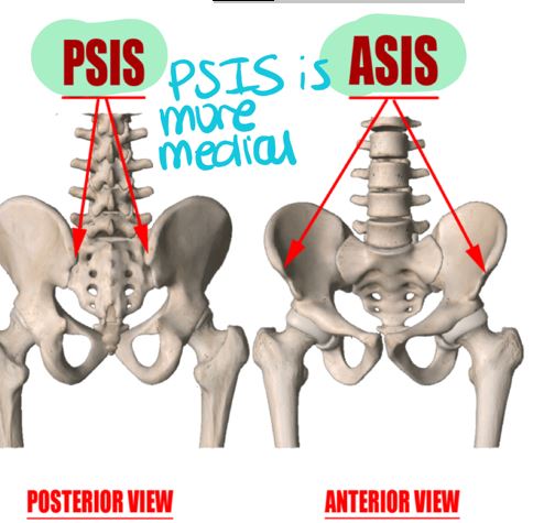
PSIS- more medial
2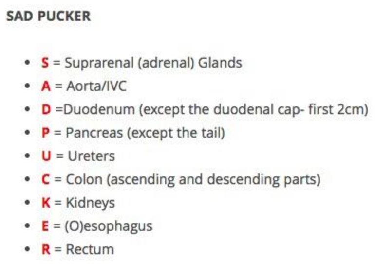
What are the retroperitoneal organs?
3
Pubic symphysis- [projects anteriorly]- firm resistance found at the end of in aspect of plane
Pubic symphysis- [projects anteriorly]- firm resistance found at the end of linea alba in ANTERIOR aspect of median plane
4
Pubic tubercle- bony landmark cm laterally of the . Where the attaches / insert
Pubic tubercle- bony landmark 2.5 cm laterally of the pubic symphysis. Where the inguinal ligament attaches / insert
5
Iliac crest- extends from . The iliac tubercle can be palpated cm posterior to
Iliac crest- extends posteriorly from ASIS. The iliac tubercle can be palpated 6 cm posterior to ASIS
6
Xyphoid process- inferior border of sternum found at
Xyphoid process- inferior border of sternum found at T10-11
7
Linea semilunaris-curved line ( laterally) that extends from the to the . It is located cm from the median plane. Represents the lateral border of the muscle
Linea semilunaris-curved line (convex laterally) that extends from the 9th costal cartilage to the pubis. It is located 5-8 cm from the median plane. Represents the lateral border of the rectus abdominis muscle
8
Linea alba- that’s - divides abdomen into right and left sides. Goes from the process to
Linea alba-white line that’s midline- divides abdomen into right and left sides. Goes from the xiphoid process to pubis
9
Inguinal ligament- folded over onto itself
Inguinal ligament- EOM folded over onto itself inferiorly
10
Tendinous intersections- over rectus (six pack)
Tendinous intersections-transverse lines over rectus abdominis muscle (six pack)
11
Gallbladder is found on the in the of the of the liver. You palpate the .
Gallbladder is found on the transpyloric plane (L1) in the quadrate lobe of the right lobe of the liver. You palpate the RUQ subcostal region.
12
Stab wound to the right 5th-10th rib?
Liver
13
Stab wound between T11-L2 posterolateral?
Kidney
14
Stab wound between T7-T11 posterolateral, left?
Spleen
15
Midline Stab Wound (7 layers)
1. Skin
2. Superficial fascia
3. Deep facia
4. Linea alba
5. Fascia transversalis
6. Extraperitoneal fat
7. Parietal peritoneum
2. Superficial fascia
3. Deep facia
4. Linea alba
5. Fascia transversalis
6. Extraperitoneal fat
7. Parietal peritoneum
16
Midaxillary Stab Wound (9 layers)
1. Skin
2. Superifical Fascia (Camper then Scarpa's)
3. Deep fascia
4. External oblique m.
5. Internal oblique m.
6. Transversus abdominis m.
7. Facia transversalis
8. Extraperitoneal fat
9. Parietal peritoneum
2. Superifical Fascia (Camper then Scarpa's)
3. Deep fascia
4. External oblique m.
5. Internal oblique m.
6. Transversus abdominis m.
7. Facia transversalis
8. Extraperitoneal fat
9. Parietal peritoneum
17
Paramidline/Midclavicular (superior to arcuate line (RUQ/LUQ) - 11 layers
1. Skin
2. Superifical fascia (Camper the Scarpa's
3. Deep fascia
4. External oblique m. apneurosis
5. Ant internal oblique muscle apneurosis
6. Rectus abdominis
7. Post internal oblique muscle apneurosis
8. Transversus abdominus muscle apneurosis
9. Fascia transversalis
10. Extraperitoneal fat
11. Parietal peritoneum
2. Superifical fascia (Camper the Scarpa's
3. Deep fascia
4. External oblique m. apneurosis
5. Ant internal oblique muscle apneurosis
6. Rectus abdominis
7. Post internal oblique muscle apneurosis
8. Transversus abdominus muscle apneurosis
9. Fascia transversalis
10. Extraperitoneal fat
11. Parietal peritoneum
18
Paramidline/Midclavicular (inferior to arcuate line (RLQ/LLQ) - 10 layers
1. Skin
2. Superifical fascia (Camper the Scarpa's
3. Deep fascia
4. External oblique m. apneurosis
5. Internal oblique m. aponeurosis
6. Transversus abdominus aponeurosis
7. Rectus abdominus m.
8. Fascia transversalis
9. Extraperitoneal fat
10. Parietal peritoneum
2. Superifical fascia (Camper the Scarpa's
3. Deep fascia
4. External oblique m. apneurosis
5. Internal oblique m. aponeurosis
6. Transversus abdominus aponeurosis
7. Rectus abdominus m.
8. Fascia transversalis
9. Extraperitoneal fat
10. Parietal peritoneum
19
Subcostal plane- Vertebral level : Joins the inferior margins of the
Subcostal plane- Vertebral level L2-3: Joins the inferior margins of the 10th costal cartilage 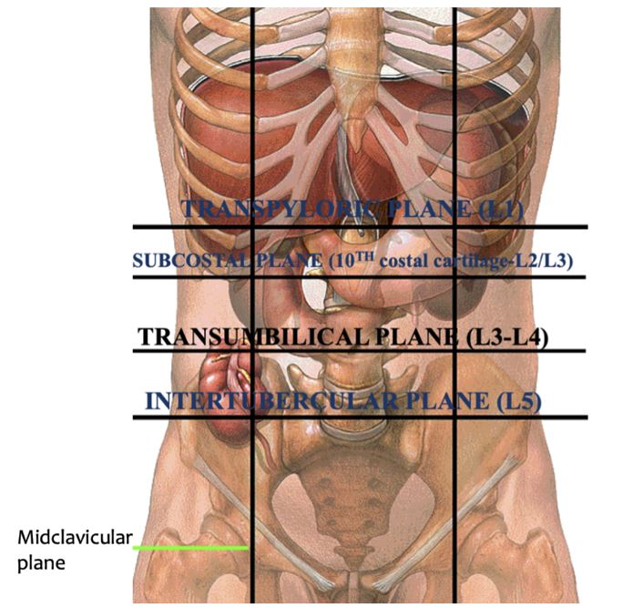
20
HIGH YIELD
Transpyloric Plane
-Found at vertebral level
- costal cartilage
-Many organs are found there- , , , , and , , ,
Transpyloric Plane
-Found at vertebral level
- costal cartilage
-Many organs are found there- , , , , and , , ,
HIGH YIELD
Transpyloric Plane
-Found at vertebral level L1
-Ninth costal cartilage
-Many organs are found there- gallbladder, liver, pancreas, pyloris of stomach, colon- right and left colic flexure and transverse colon, portal vein begins, kidney, spleen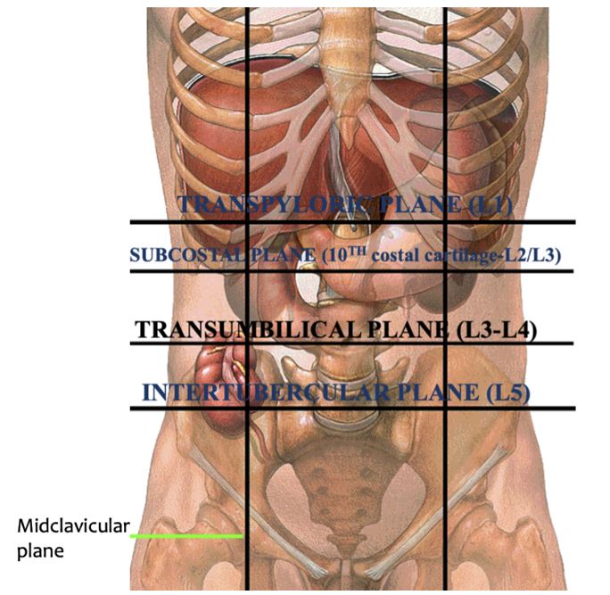
Transpyloric Plane
-Found at vertebral level L1
-Ninth costal cartilage
-Many organs are found there- gallbladder, liver, pancreas, pyloris of stomach, colon- right and left colic flexure and transverse colon, portal vein begins, kidney, spleen
21
Transumbilical plane- vertebral level
Transumbilical plane- vertebral level L3-4 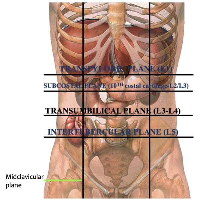
22
Transtubercular plane- Vertebral level . Through the
Transtubercular plane- Vertebral level L5. Through the iliac tubercles 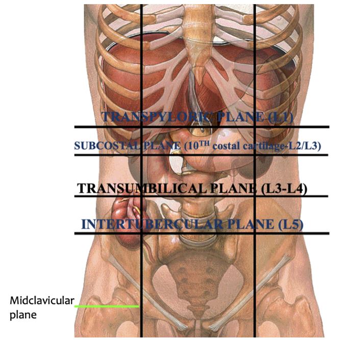
23
Caval opening (T )
Contents:
Contents:
Caval opening (T8)
Contents: IVC, phrenic nerve, lymphatic vessels
Contents: IVC, phrenic nerve, lymphatic vessels
24
Esophageal opening (T )
Contents:
Contents:
Esophageal opening (T10)
Contents: Esophogous, right and left vagus nerves, Esophageal branches of left gastric artery and vein
Contents: Esophogous, right and left vagus nerves, Esophageal branches of left gastric artery and vein
25
Aortic opening (T )
Contents:
Contents:
Aortic opening (T12)
Contents: thoracic aorta, thoracic duct, greater splanchnic nerves, azygos vein
Contents: thoracic aorta, thoracic duct, greater splanchnic nerves, azygos vein
26
What is the blood supply of the abdominal muscle?
1. Superior epigastric a.
2. Inferior epigastric a.
3. Lumbar a.
4. Deep circumflex a.
5. Iliac a.
6. Superficial epigastric a.
2. Inferior epigastric a.
3. Lumbar a.
4. Deep circumflex a.
5. Iliac a.
6. Superficial epigastric a.
27
What is the origin of the EOM?
Outer surface of ribs 5-12
28
What is the insertion of the EOM? (3)
***linea alba, pubic tubercle, anterior ½ of iliac crest
29
What is the nerve supply of the abdominal muscles? EOM has what extra supply?
-inferior 6 thoracic nerve (T7-T12)
[thoracoabdominal]
EOM ONLY Subcostal nerve (T12)
[thoracoabdominal]
EOM ONLY Subcostal nerve (T12)
30
What is the action of the EOM?
-Compress and support abdominal visera
-Flexion and rotation of trunk
-Flexion and rotation of trunk
31
What 3 structures does the EOM form?
1. Inguinal ligament (apnuerosis)
2. Superficial inguinal ring
3. External spermatic fascia
2. Superficial inguinal ring
3. External spermatic fascia
32
What is the orientation of the fibers of the EOM?
Inferior, medial, and anterior (Hand in pockets)
33
What is the origin of the IOM? (3)
thoracolumbar fascia, anterior 2/3 of iliac crest, lateral ½ of inguinal ligament
34
What is the insertion of the IOM? (3)
inferior border of ribs 10-12, ***linea alba, pubis (via the conjoint tendon)
35
What is the action of the IOM?
-Compress and support the abdominal visera
-Flexion and rotation of the trunk
-Flexion and rotation of the trunk
36
What stucture does the IOM form? What structure does it insert on?
1. Cremaster muscle
2. Conjoint tendon
2. Conjoint tendon
37
What is the orientation of the fibers of the IOM?
Superior, medial, anterior
"Hands on boobs"
"Hands on boobs"
38
What is the origin of the transversis abdominus? (3)
1. Thoracolumbar fascia
2. Internal surface of ribs 7-11
3. Lateral 1/3 of inguinal ligament
2. Internal surface of ribs 7-11
3. Lateral 1/3 of inguinal ligament
39
What is teh insertion of the transversis abdominus?
1. Linea alba ***
2. Pubis (via conjoint tendon)
2. Pubis (via conjoint tendon)
40
What structure does the transverisis abdominius insert on?
Conjoint tendon
41
What is the orientation of the fibers of the transversis abdominus?
Horizontal
42
Which two sturctures insert on the conjoint tendon?
The IOM and transversis abdominus
43
What is the origin of the rectus abdominus?
Pubic symphysis
44
ASIS- more
PSIS- more
PSIS- more
ASIS- more lateral
PSIS- more medial
PSIS- more medial
45
What is the function of the rectus abdominus? (2)
1. Compress abdominal visera
2. Flex the trunk
2. Flex the trunk
46
What is teh orientation of the ribers of the rectus abdominus?
Vertical
47
Where is an esophageal hernia most likely to occur?
Esophageal hiatus of diaphram (T10)
48
Hiatal hernias- abdominal organs slip through the into the thoracic cavity. May result in
Hiatal hernias- abdominal organs slip through the diaphragm into the thoracic cavity. May result in GERD
49
Rectus abdominis muscle sheath- made up of the aponeurosis of the , and as they insert into the .
Rectus abdominis muscle sheath- made up of the aponeurosis of the External oblique muscle, internal oblique muscle and transversus abdominis muscle as they insert into the linea alba.
50
Superior epigastric artery [comes from ]
Enters the rectus sheath superior LH just inferior the the
Enters the rectus sheath superior LH just inferior the the
Superior epigastric artery [comes from internal thoracic]
Enters the rectus sheath superior LH just inferior the the 7th costal cartilage
Enters the rectus sheath superior LH just inferior the the 7th costal cartilage
51
***HY
Inferior epigastric artery [comes from ]
Runs superior LH on the transversalis fascia and enters the to gain the
Inferior epigastric artery [comes from ]
Runs superior LH on the transversalis fascia and enters the to gain the
***HY
Inferior epigastric artery [comes from external iliac]
Runs superior LH on the transversalis fascia and enters the arcuate line to gain the rectus sheath
Inferior epigastric artery [comes from external iliac]
Runs superior LH on the transversalis fascia and enters the arcuate line to gain the rectus sheath
52
Superficial epigastric artery [ ]
Superficial epigastric artery [comes from branch of femoral]
53
Above the arcuate line (upper 2/3) is between the and the
Above the arcuate line (upper 2/3) is between the costal margins and the ASIS
54
Below the arcuate line (lower 1/3) is between the and
Below the arcuate line (lower 1/3) is between the ASIS and pubic symphysis
55
What are the two approaches to paracentesis?
1. RLQ and LLQ (lateral to rectus sheet!!)
2. 2 cm below ubilicus with empty bladder.
2. 2 cm below ubilicus with empty bladder.
56
Below the ubilicus paracentesis approach (6 layers)
1. Skin
2. Lina alba
3. Fascia transversalis
4. Extraperitoneal fat
5. Parietal peritoneum
6. Peritoneal cavity
2. Lina alba
3. Fascia transversalis
4. Extraperitoneal fat
5. Parietal peritoneum
6. Peritoneal cavity
57
RLQ or LLQ paracentesis approach (9 layers)
1. Skin
2. Superifical fascia
3. External oblique m.
4. Internal oblique m.
5. Transversus abdominis
6. Fascia transversalis
7. Extraperitoneal fat
8. Parietal peritoneum
9. Peritoneal cavity
2. Superifical fascia
3. External oblique m.
4. Internal oblique m.
5. Transversus abdominis
6. Fascia transversalis
7. Extraperitoneal fat
8. Parietal peritoneum
9. Peritoneal cavity
58
Superior to the umbilicus theres is the large median fold AKA
Superior to the umbilicus theres is the large median fold AKA falciform ligament
59
The large median fold goes from ligament to diaphragm. It has a free edge called the which has
The large median fold goes from ligament to diaphragm. It has a free edge called the liagmentum teres which has obliterated umbilical vein
60
Inferior to the ubilicus:
Median Umbilical Fold=
Median Umbilical Fold=
Inferior to the ubilicus:
Median Umbilical Fold= urachus
Median Umbilical Fold= urachus
61
Inferior to the umbilicus:
Medial Umbilical Fold=
Medial Umbilical Fold=
Inferior to the umbilicus:
Medial Umbilical Fold= obliterated umbilical ARTERY
Medial Umbilical Fold= obliterated umbilical ARTERY
62
Inferior to the umbilicus:
Lateral Umbilical Fold= *****ONLY ONE THAT IS STILL USED
Lateral Umbilical Fold= *****ONLY ONE THAT IS STILL USED
Inferior to the umbilicus:
Lateral Umbilical Fold= inferior epigastric artery (patent) *****ONLY ONE THAT IS STILL USED
Lateral Umbilical Fold= inferior epigastric artery (patent) *****ONLY ONE THAT IS STILL USED
63
What is the site where injuinal hernias pass through called?
Hesselbach’s/inguinal triangle
64
What is the inferior, lateral, and medial border of Hasselbach's/inguinal triangle?
Inferior: Inguinal ligament
Lateral: Inferior epigastric vessels
Medial: Lateral border of the rectus abdominus muscle
Lateral: Inferior epigastric vessels
Medial: Lateral border of the rectus abdominus muscle
65
What aponeurosis does the inguinal region come from?
EOM
66
What are the contents of the inguinal region in males? Females?
Male:
Female:
Male:
Female:
What are the contents of the inguinal region in males? Females?
Male: spermatic cord and ilioinguinal nerve (L1)
Female: round ligament and ilioinguinal nerve (L1)
Male: spermatic cord and ilioinguinal nerve (L1)
Female: round ligament and ilioinguinal nerve (L1)
67
What does the deep/internal ring give rise to?
Transversalis fascia
68
What is the inguinal canal?
The canal between the deep inguinal ring and the superifical inguinal ring
69
Indirect inguinal hernias cavity to the inferior epigastric vessels, traverse the deep inguinal ring, inguinal canal, and superficial inguinal ring.
Indirect inguinal hernias cavity LATERAL to the inferior epigastric vessels, traverse the deep inguinal ring, inguinal canal, and superficial inguinal ring.
70
Indirect inguinal hernias are within the because they follow the route taken by the testes and is covered by 3 layers: , , and
Indirect inguinal hernias are within the spermatic cord because they follow the route taken by the testes and is covered by 3 layers: external spermatic fascia, cremaster, and internal spermatic fascia of the spermatic cord
71
Indirect inguinal hernias can be palpated lateral to and are than direct
Indirect inguinal hernias can be palpated lateral to public tubercle and are more common than direct
72
Direct inguinal hernias protrude anteriorly through the posterior wall of the and leave the abdominal cavity to the inferior epigastric vessels.
Direct inguinal hernias protrude anteriorly through the posterior wall of the inguinal canal and leave the abdominal cavity MEDIAL to the inferior epigastric vessels.
73
Direct inguinal hernias pass through
Direct inguinal hernias pass through Hesselbach's triangle
74
Direct inguinal hernias the spermatic cord
Direct inguinal hernias parallel the spermatic cord
75
Hesselbach’s triangle is located to the superficial inguinal ring
Hesselbach’s triangle is located posteriorly to the superficial inguinal ring
76
What are the coverings of the spermatic cord and what gives rise to its coverings?
1. (from )
2. (from )
3. (from )
1. (from )
2. (from )
3. (from )
What are the coverings of the spermatic cord and what gives rise to its coverings?
1. external spermatic fascia (from EOM aponeurosis)
2. cremaster (from IOM)
3. internal spermatic fascia (from fascia transversalis)
1. external spermatic fascia (from EOM aponeurosis)
2. cremaster (from IOM)
3. internal spermatic fascia (from fascia transversalis)
77
In males, the spermatic form beings at the and ends at the
In males, the spermatic form beings at the deep inguinal ring and ends at the testes
78
CONTENTS OF SPERMATIC CORD
1)Vas Deferens= muscular duct that transports sperm from the epididymis to the urethra
2)Testricular artery ( )
3)Testiculuar veins- they begin in the testes as an extensive venous plexus ( )
4)Lymphatic vessels - drain into
5)Autonomic Sympathetic nerves- run w/ the
6)Remnant of the processus vaginalis
7)Cremasteric artery (inferior epigastric a.)- supplies
8)Genital branch of the genitofemoral n (L1-2)- supplies the
9)Arteries of the vas deferens from the
1)Vas Deferens= muscular duct that transports sperm from the epididymis to the urethra
2)Testricular artery ( )
3)Testiculuar veins- they begin in the testes as an extensive venous plexus ( )
4)Lymphatic vessels - drain into
5)Autonomic Sympathetic nerves- run w/ the
6)Remnant of the processus vaginalis
7)Cremasteric artery (inferior epigastric a.)- supplies
8)Genital branch of the genitofemoral n (L1-2)- supplies the
9)Arteries of the vas deferens from the
CONTENTS OF SPERMATIC CORD
1)Vas Deferens= muscular duct that transports sperm from the epididymis to the urethra
2)Testricular artery (abdominal aorta)
3)Testiculuar veins- they begin in the testes as an extensive venous plexus (pampiniform plexus)
4)Lymphatic vessels - drain into Lumbar LN
5)Autonomic Sympathetic nerves- run w/ the testicular artery
6)Remnant of the processus vaginalis
7)Cremasteric artery (inferior epigastric a.)- supplies cremasteric muscle
8)Genital branch of the genitofemoral n (L1-2)- supplies the cremaster muscle
9)Arteries of the vas deferens from the superior or inferior vesical artery
1)Vas Deferens= muscular duct that transports sperm from the epididymis to the urethra
2)Testricular artery (abdominal aorta)
3)Testiculuar veins- they begin in the testes as an extensive venous plexus (pampiniform plexus)
4)Lymphatic vessels - drain into Lumbar LN
5)Autonomic Sympathetic nerves- run w/ the testicular artery
6)Remnant of the processus vaginalis
7)Cremasteric artery (inferior epigastric a.)- supplies cremasteric muscle
8)Genital branch of the genitofemoral n (L1-2)- supplies the cremaster muscle
9)Arteries of the vas deferens from the superior or inferior vesical artery
79
Pampiniform Plexus
-The right testicular vein leaves the plexus as a single vein and drains directly into the at
-The left testicular vein leaves the plexus as a single vein and drains into the at .(it then empties into )
-The right testicular vein leaves the plexus as a single vein and drains directly into the at
-The left testicular vein leaves the plexus as a single vein and drains into the at .(it then empties into )
Pampiniform Plexus
-The right testicular vein leaves the plexus as a single vein and drains directly into the IVC at L1
-The left testicular vein leaves the plexus as a single vein and drains into the left renal vein at L1.(it then empties into IVC)
-The right testicular vein leaves the plexus as a single vein and drains directly into the IVC at L1
-The left testicular vein leaves the plexus as a single vein and drains into the left renal vein at L1.(it then empties into IVC)
80
The is NOT A CONTENT of the spermatic cord.
The ilioinguinal n. is NOT A CONTENT of the spermatic cord.
81
What are the anterior, posterior, superior, and inferior boundaries of the epiploic foramen of Winslow?
Anterior: portal hepaticus (portal triad) contained within the hepatoduodenal ligament of the lesser omentum
Posterior: IVC and right Cruz of the diaphragm
Superior: caudate lobe of the liver
Inferior: superior part of the duodenum
Posterior: IVC and right Cruz of the diaphragm
Superior: caudate lobe of the liver
Inferior: superior part of the duodenum
82
What are the contents of the anterior boundary of the epiploic foramen of Winslow?
Portal triad made of the portal vein, hepatic artery, common bile duct
83
What part of the abdominal cavity is behind the epiploic foramen of Winslow?
Omental bursa/lesser sac
84
In supine position, pylorus lies at the at level of , cm of midline
In supine position, pylorus lies at the 9th costal cartilage at level of L1, 1.25 cm right of midline
85
What are the ligaments of the lesser omentum?
1. : Contains
2.
1. : Contains
2.
What are the ligaments of the lesser omentum?
1. Hepatoduodenal: Contains portal triad
2. Hepatogastric
1. Hepatoduodenal: Contains portal triad
2. Hepatogastric
86
What are the boundaries of the traingle of calot?
Superior:
Lateral:
Medial:
Superior:
Lateral:
Medial:
What are the boundaries of the traingle of calot?
Superior: inferior border of liver
Lateral: cystic duct
Medial: common hepatic duct
Superior: inferior border of liver
Lateral: cystic duct
Medial: common hepatic duct
87
What are the contents of the triangle of Calot?
1. Cystic artery
2. Lymph node of Calot (can become inflamed in cholecystitis and may be removed during cholecystectomy)
2. Lymph node of Calot (can become inflamed in cholecystitis and may be removed during cholecystectomy)
88
TQ: Short gastric artery supplies what organ?
TQ: Short gastric artery supplies what organ?
SPLEEN
SPLEEN
89
What are the 3 branches of the celiac trunk?
1. Left gastric
2. Common hepatic
3. Splenic
2. Common hepatic
3. Splenic
90
What are the 3 branches of the left gastric artery and what do they supply?
1. Ventral branch = anterior surface of stomach
2. Dorsal branch = lesser curvature
3. Cardioesopheageal = cardia and esophagus
2. Dorsal branch = lesser curvature
3. Cardioesopheageal = cardia and esophagus
91
What are the 3 branches of the common hepatic?
1. Right gastric
2. Gastroduodenal
3. Proper Hepatic
2. Gastroduodenal
3. Proper Hepatic
92
What are the branches of the gastroduodenal?
1. Retroduodenal
2. Right gastroepiploic
3. Superior oancreaticoduodenal
2. Right gastroepiploic
3. Superior oancreaticoduodenal
93
The right gastric artery supplies the and
The right gastric artery supplies the lesser omentum and pylorus
94
What are the 3 branches of the proper hepatic?
1. Reight hepatic - cystic
2. Left hepatic (liver capsule)
3. Middle hepatic (quadrate lope of right liver)
2. Left hepatic (liver capsule)
3. Middle hepatic (quadrate lope of right liver)
95
What are the 4 branches of the superior mesenteric artery SMA and SMV?
1. Jejunal and ileal branches
2. Ileocolic (ascending colon, cecum, appendix)
3. Right colic (right colic flexure)
4. Middle colic (transverse colon)
2. Ileocolic (ascending colon, cecum, appendix)
3. Right colic (right colic flexure)
4. Middle colic (transverse colon)
96
What are teh 3 branches of the inferior mesenteric artery (IMA) and IMV?
1. Left colic (left colic flexure)
2. Sigmoid
3. Superior rectal
2. Sigmoid
3. Superior rectal
97
The 3 blood supplies of the stomach are the , the , and the
The 3 blood supplies of the stomach are the left gastric, the common hepatic, and the splenic
98
Blood Supply and Different Parts of Stomach
(1) Anterior part:
(2) Proximal part of distal curvature:
(3) Distal part of lesser curvature (pyrlorus):
(4) Greater curvature distal:
(5) Greater curvature proximal:
(6) Behind the stomach (fundus):
(1) Anterior part:
(2) Proximal part of distal curvature:
(3) Distal part of lesser curvature (pyrlorus):
(4) Greater curvature distal:
(5) Greater curvature proximal:
(6) Behind the stomach (fundus):
Blood Supply and Different Parts of Stomach
(1) Anterior part: Ventral branch of left gastric
(2) Proximal part of distal curvature: Dorsal branch of left gastric
(3) Distal part of lesser curvature (pyrlorus): Right gastric from common hepatic
(4) Greater curvature distal: Right gastroepiploic from gastroduodenal from common hepatic
(5) Greater curvature proximal: Left gastroepiploic from splenic
(6) Behind the stomach (fundus): Short gastric from splenic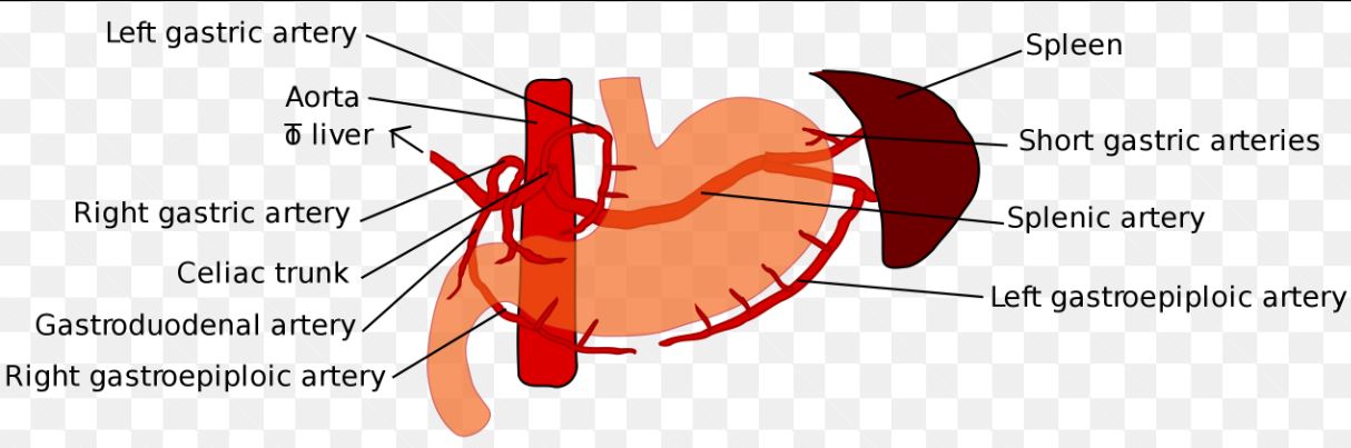
(1) Anterior part: Ventral branch of left gastric
(2) Proximal part of distal curvature: Dorsal branch of left gastric
(3) Distal part of lesser curvature (pyrlorus): Right gastric from common hepatic
(4) Greater curvature distal: Right gastroepiploic from gastroduodenal from common hepatic
(5) Greater curvature proximal: Left gastroepiploic from splenic
(6) Behind the stomach (fundus): Short gastric from splenic
99
Right gastroomental artery [ ] anastomoses with the left gastroomental artery [ ] midway along the of the stomach
Right gastroomental artery [common hepatic] anastomoses with the left gastroomental artery [splenic] midway along the greater curvature of the stomach 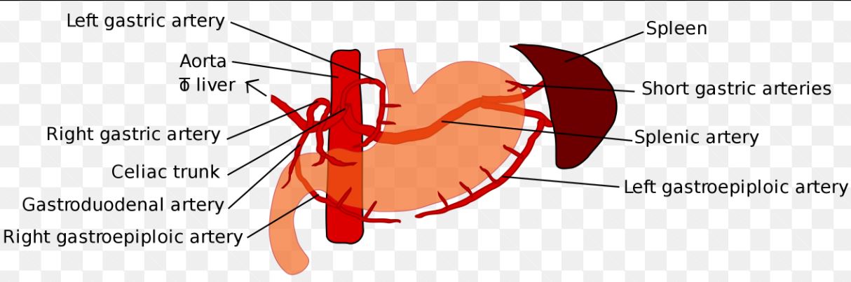
100
and anastomose at the and form the marginal arteries 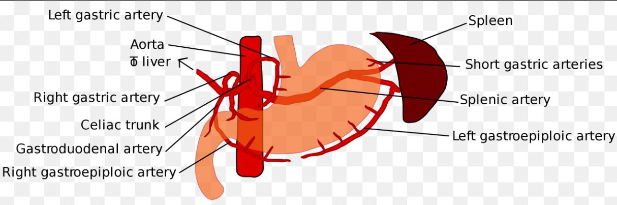
Right gastric artery [common hepatic] and ventral artery [left gastric] anastomose at the transverse colon and form the marginal arteries
101
What is the blood supply of the duodenum?
-
-
-
-
What is the blood supply of the duodenum?
-Superior pancreaticoduodenal (from the gastroduodenal from the common hepatic artery from the celiac trunk)
-Inferior pancreaticoduodenal artery (from the SMA)
-Superior pancreaticoduodenal (from the gastroduodenal from the common hepatic artery from the celiac trunk)
-Inferior pancreaticoduodenal artery (from the SMA)
102
Blood supply of the pancreas is the (from the of celiac trunk)
Blood supply of the pancreas is the pancreatic arteries (from the splenic artery of celiac trunk)
103
Blood supply of spleen is the and the
Blood supply of spleen is the Splenic artery and the inferior pancreatic artery
104
Blood supply of liver
(1)
(2)
(1)
(2)
Blood supply of liver
(1) Hepatic arteries
(2) Portal vein (brings deoxygenated blood for the liver to clean)
(1) Hepatic arteries
(2) Portal vein (brings deoxygenated blood for the liver to clean)
105
If patient has ulceration on the posterior wall of the stomach:
a.Between fundus and body of stomach that goes through the wall- the will be affected
b.At pyloric sphincter- the would be affected
a.Between fundus and body of stomach that goes through the wall- the will be affected
b.At pyloric sphincter- the would be affected
If patient has ulceration on the posterior wall of the stomach:
a.Between fundus and body of stomach that goes through the wall- the splenic arteries will be affected
b.At pyloric sphincter- the gastroduodenal artery would be affected
a.Between fundus and body of stomach that goes through the wall- the splenic arteries will be affected
b.At pyloric sphincter- the gastroduodenal artery would be affected
106
The SMA/SMV ride over the portion of the part of the .
The SMA/SMV ride over the anterior portion of the third part of the duodenum.
107
SMA Syndrome: Loss of fat between the and the leads to compression of the
SMA Syndrome: Loss of fat between the SMA and the abdominal aorta leads to compression of the third part of the duodenum
108
forms behind the head/neck of the pancreas from the union of the and the
Portal hepatic vein forms behind the head/neck of the pancreas from the union of the Superior Mesenteric Vein and the Splenic vein
109
The quadrate lobe is associated with the
The quadrate lobe is associated with the fossa of and the gallbladder.
110
The caudate lobe is related to the
The caudate lobe is related to the IVC.
111
Both the quadrate and caudate lobes are part of the of the liver.
Both the quadrate and caudate lobes are part of the right lobe of the liver.
112
What are the posterior, right, and left contents of the portal triad? **Be able to identify
(1) Portal vein = posterior
(2) Common bile duct = Right
(3) Hepatic artery = Left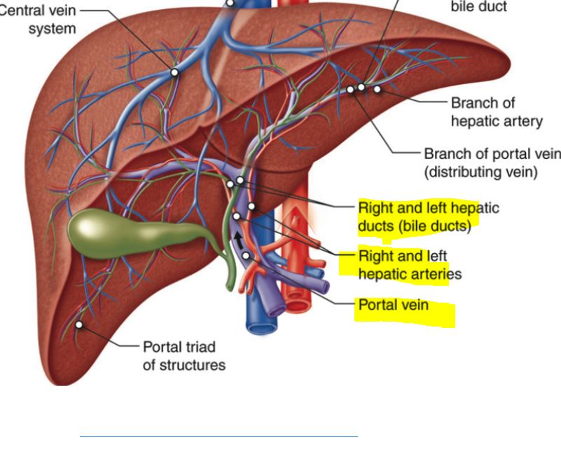
(2) Common bile duct = Right
(3) Hepatic artery = Left
113
If an ulcer perforates the , contents will empty into the
If an ulcer perforates the posterior wall of the stomach, contents will empty into the omental bursa/lesser sac.
114
What arteries supply the fundus of the stomach? Where do they come from?
1.
2.
1.
2.
What arteries supply the fundus of the stomach? Where do they come from?
1. Short gastric artery [splenic artery]
2. Posterior gastric artery [splenic artery]
1. Short gastric artery [splenic artery]
2. Posterior gastric artery [splenic artery]
115
What is the flow of bile throught the liver?
(Right + Left Hepatic Duct = ) to ( + = ) to ( + = ) to ( )
(Right + Left Hepatic Duct = ) to ( + = ) to ( + = ) to ( )
What is the flow of bile throught the liver?
(Right + Left Hepatic Duct = Common Hepatic) to (Common Hepatic + Cystic = Common Bile Duct) to (Common Bile Duct + Main Pancreatic = Ampulla of Vater) to (Sphincter of Oddi)
(Right + Left Hepatic Duct = Common Hepatic) to (Common Hepatic + Cystic = Common Bile Duct) to (Common Bile Duct + Main Pancreatic = Ampulla of Vater) to (Sphincter of Oddi)
116
What is the blood supply of the gall baldder?
Cystic Artery
(from Celiac trunk —> common hepatic artery —> right hepatic artery —> cystic artery)
(from Celiac trunk —> common hepatic artery —> right hepatic artery —> cystic artery)
117
Gallbladder is located in and between the and the at the
Gallbladder is located in and between the right costal margin and the right linea semilunaris at the Transpyloric plane (L1)
118
What are the anterior/superior, posterior, and inferior relationships of the gallbaldder?
Anterior and superior:
Posterior:
Inferior:
Anterior and superior:
Posterior:
Inferior:
What are the anterior/superior, posterior, and inferior relationships of the gallbaldder?
Anterior and superior: inferior border of liver, and anterior abdominal wall
Posterior: transverse colon and proximal duodenum
Inferior: biliary tree and remaining parts of duodenum
Anterior and superior: inferior border of liver, and anterior abdominal wall
Posterior: transverse colon and proximal duodenum
Inferior: biliary tree and remaining parts of duodenum
119
What are the 4 parts of the stomach?
1. Cardiac part (Cardia)
2. Fundus
3. Body
4. Pylorus
2. Fundus
3. Body
4. Pylorus
120
Test Question
The cardiac part of the stomach lies posterior to the costal cartlage, from the at level of .
The cardiac part of the stomach lies posterior to the costal cartlage, from the at level of .
Test Question
The cardiac part of the stomach lies posterior to the 7th left costal cartlage, 2-4 cm from the median plane at level of T11.
The cardiac part of the stomach lies posterior to the 7th left costal cartlage, 2-4 cm from the median plane at level of T11.
121
The fundus is to the cardiac orifice
The fundus is superior to the cardiac orifice
122
The fundus is related to the of the diaphram
The fundus is related to the left dome of the diaphram
123
The fundus lies posterior to the rib in the
The fundus lies posterior to the 5th rib in the mid-calvicular plane
124
The body of the stomach has a wide portion (pyloric ) and narrow portion (pyloric )
The body of the stomach has a wide portion (pyloric antrum) and narrow portion (pyloric canal)
125
The pyrlorus has the pyloric
The pyrlorus has the pyloric sphincter
126
In supine position, pylorus lies at level of costal cartilage at the level of , cm of . In erect position, it is at .
In supine position, pylorus lies at level of 9th costal cartilage at the level of L1, 1.25 cm right of midline . In erect position, it is at L2-L4.
127
Where is the accessory spleen present (if it is there at all)?
Gastrosplenic
128
What are the 2 ligaments of the spleen?
a.Gastrosplenic- short gastric and Left gastroepiploic vessels, accessory spleen when present
b. Splenorenal- splenic vessels and tail of pancreas
b. Splenorenal- splenic vessels and tail of pancreas
129
Main pancreatic duct drains the of pancreas and joins the to empty into the into the
Main pancreatic duct drains the body and tail of pancreas and joins the common bile duct to empty into the greater duodenal papilla into the second part of the duodenum
130
Accessory pancreatic duct drains the of the pancreas and empties into the into the (superior to the )
Accessory pancreatic duct drains the head of the pancreas and empties into the lesser duodenal papilla into the second part of the duodenum (superior to the greater duodenal papilla)
131
Where does the hepatiopancreatic duct drain?
Greater duodenal papilla- second part of the duodenum
132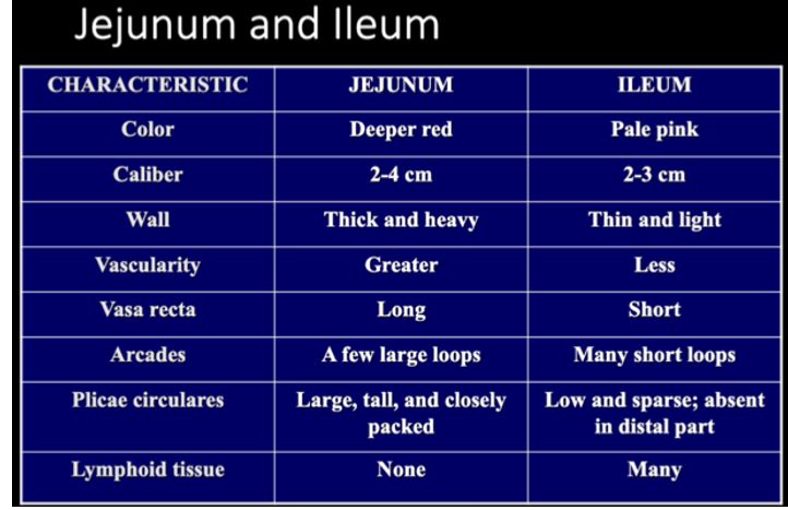
Differences between Jejunum and Ileum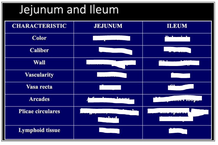
133
( + ) Anastamose→ Forms the Marginal Artery→ branches into many vasa recta
(Middle colic + Left Colic) Anastamose→ Forms the Marginal Artery→ branches into many vasa recta
134
Blood Supply of Colon
Distal ileum, Cecum, and Prox Asending Colon =
Mid-distal ascending colon =
Right Colic Flexure =
Transverse =
Desending colon =
Sigmoid =
Rectum =
Distal ileum, Cecum, and Prox Asending Colon =
Mid-distal ascending colon =
Right Colic Flexure =
Transverse =
Desending colon =
Sigmoid =
Rectum =
Blood Supply of Colon
Distal ileum, Cecum, and Prox Asending Colon = Ileocolic a.
Mid-distal ascending colon = Right Colic a.
Right Colic Flexure = Middle Colic artery
Transverse = Marginal Artery artery
Desending colon = Left colic artery
Sigmoid = Signmoid artery
Rectum = Superior rectal artery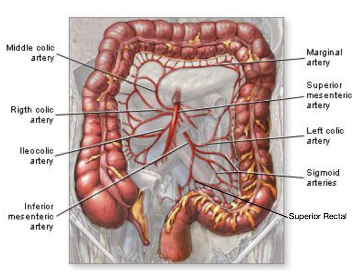
Distal ileum, Cecum, and Prox Asending Colon = Ileocolic a.
Mid-distal ascending colon = Right Colic a.
Right Colic Flexure = Middle Colic artery
Transverse = Marginal Artery artery
Desending colon = Left colic artery
Sigmoid = Signmoid artery
Rectum = Superior rectal artery
135
SMA anastamose with , ,
SMA anastamose with Middle Colic, Right Colic, Ileocolic
136
IMA anastamose with , ,
IMA anastamose with left colic, sigmoid, superior rectal
137
Marginal arteries anastomoses the and
Marginal arteries anastomoses the middle colic and left colic
138
Vasa recta are small arteries from to colon
Vasa recta are small arteries from marginal arteries to colon
139
TEST QUESTION BE CAREFUL OF WORDING
The appendix is located 1/3 of the way between the and
The appendix is located 1/3 of the way between the and
TEST QUESTION BE CAREFUL OF WORDING
The appendix is located 1/3 of the way between the ASIS and umbilicus
The appendix is located 1/3 of the way between the ASIS and umbilicus
140
The on the cecum converge to point to the location of the appendix
The teniae coli on the cecum converge to point to the location of the appendix
141
The appendix is . It lacks and
The appendix is intraperitoneal. It lacks taenia coli and haustra
142
What are the signs and symptoms of portal hypertension?
i. - distended superficial epigastric veins
ii.
iii.
iv. - backup drainage of the left gastric vein into the esophagus due to the anastomosis
v.
vi. - CNS dysfunction due to failure of liver to detox CNS toxic agents (can be caused by alcoholism, o structure, liver disease)
i. - distended superficial epigastric veins
ii.
iii.
iv. - backup drainage of the left gastric vein into the esophagus due to the anastomosis
v.
vi. - CNS dysfunction due to failure of liver to detox CNS toxic agents (can be caused by alcoholism, o structure, liver disease)
What are the signs and symptoms of portal hypertension?
i.Caput medusae- distended superficial epigastric veins
ii.Ascites
iii.Splenomegaly
iv.Esophogeal varices- backup drainage of the left gastric vein into the esophagus due to the anastomosis
v.Hemorrhoids
vi.Hepatic encephalopathy- CNS dysfunction due to failure of liver to detox CNS toxic agents (can be caused by alcoholism, o structure, liver disease)
i.Caput medusae- distended superficial epigastric veins
ii.Ascites
iii.Splenomegaly
iv.Esophogeal varices- backup drainage of the left gastric vein into the esophagus due to the anastomosis
v.Hemorrhoids
vi.Hepatic encephalopathy- CNS dysfunction due to failure of liver to detox CNS toxic agents (can be caused by alcoholism, o structure, liver disease)
143
Esophageal vein- via (systemic) anastomose with (portal)
Esophageal vein- via azygous vein (systemic) anastomose with left gastric vein (portal)
144
Superior rectal vein (portal) anastamose with (into pudendal vein-Systemic) and which drain into (systemic)
Superior rectal vein (portal) anastamose with inferior rectal veins (into pudendal vein-Systemic) and middle rectal veins which drain into internal ileal vein (systemic) 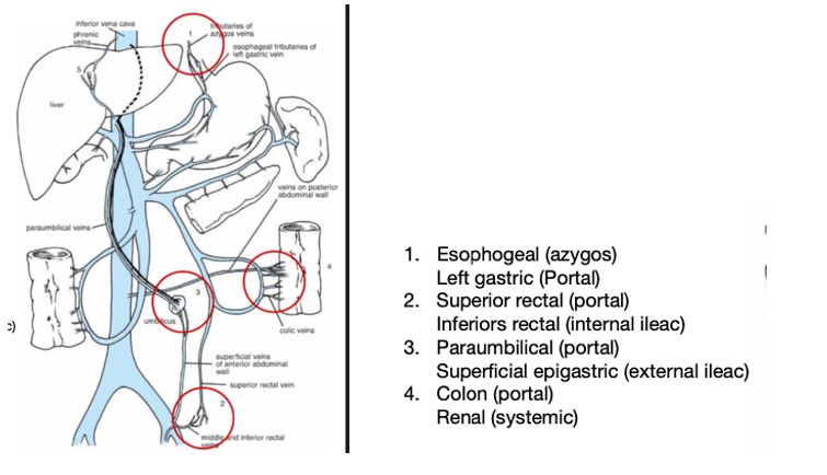
145
Paraumbilical veins (portal) anastamose with veins connected to which drain into the
Paraumbilical veins (portal) anastamose with veins connected to umbilicus (superficial Epigastric vein) which drain into the external ileal vein (systemic) 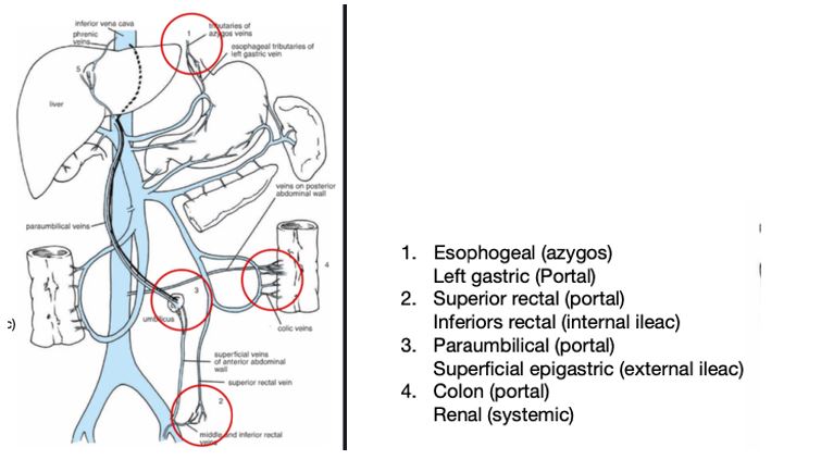
146
Veins of colon, pancreas, liver (drain into portal) anastamose with , and (systemic)
Veins of colon, pancreas, liver (drain into portal) anastamose with renal, lumbar and Phrenic veins (systemic) 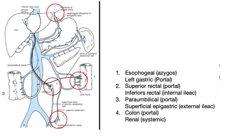
147
Portal vein is formed by the union of the and
Portal vein is formed by the union of the splenic vein and SMV
148
-Short gastric
-Left gastroepiploic
-Pancreatic (dorsal, great, caudal)
Tributaries of the portal vein: Splenic Vein
-Short gastric
-Left gastroepiploic
-Pancreatic (dorsal, great, caudal)
149
Tributaries of the portal vein: IMV
1. Sigmoid
2. Left colic
2. Left colic
150
Trubutaries of the Portal Vein: SMV
1. Illeal and jejunal
2. Ileocolic
3. RIght colic
4. Middle colic
5. Right gastroepiploic
6. Pancreaticoduodenal
2. Ileocolic
3. RIght colic
4. Middle colic
5. Right gastroepiploic
6. Pancreaticoduodenal
151
Tributaries of the portal vein: Pyloric vein (runs along the of the stomach)
Tributaries of the portal vein: Pyloric vein (runs along the lesser curvature of the stomach)
152
Tributaries of the portal vein: Cystic vein (drains the and accompanying )
Tributaries of the portal vein: Cystic vein (drains the gallbladder and accompanying cystic duct)
153
The 6 trubutaries of the portal vein are
1. Splenic
2. IMV
3. SMV
4. Pyloric
5. Cystic
6. Paraumbilical
2. IMV
3. SMV
4. Pyloric
5. Cystic
6. Paraumbilical
154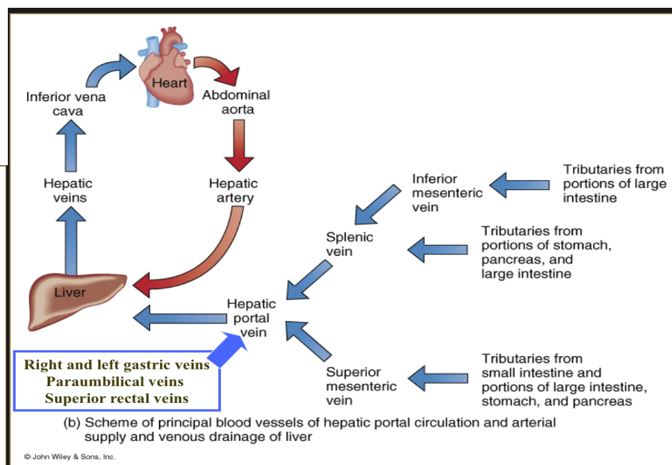
Draw through blood supply diaphram. Note which are oxygenated and which are deoxygenated.
155
What is Nuckcracker Syndrome?
Compression of the left renal vein between the SMA and abdominal aorta due to loss of fat
156
The kidneys are
The kidneys are retroperitoneal
157
The is longer and higher than the
The left kidney is longer and higher than the right kidney
158
In the erect position, the kidneys are from . In the supine position, they are from
In the erect position, the kidneys are from L1-L4. In the supine position, they are from T12-L3.
159
The kidneys are supported by peritioneal fat and renal vessels and when this support it lost, there will be kidneys will be lower)
The kidneys are supported by peritioneal fat and renal vessels and when this support it lost, there will be nephrotosis (kidneys will be lower)
160
What are the anterior, middle, and posterior boundaires of the renal hiatus? What else is present?
1. Renal vein (anterior)
2. Renal artery (middle)
3. Ureter (posterior)
4. Sympathetic and parasympathetic n.
2. Renal artery (middle)
3. Ureter (posterior)
4. Sympathetic and parasympathetic n.
161
What is the flow of urine through the kidney and to the urethra?
R enal papilla of Renal pyramid -> -> -> -> -> ->
R enal papilla of Renal pyramid -> -> -> -> -> ->
What is the flow of urine through the kidney and to the urethra?
R enal papilla of Renal pyramid -> minor calyx -> major calyx -> renal pelvis -> ureter -> urinary bladder -> urethra
R enal papilla of Renal pyramid -> minor calyx -> major calyx -> renal pelvis -> ureter -> urinary bladder -> urethra
162
What is the most posterior structure of the renal hiatus?
Ureter
163
What is superior to the kidneys?
Adrenal glands
164
What 5 things are posterior to the kidneys?
1. Psoas major
2. Quadratus lumbourm
3. Diaphragm
4. 12th rib
5. Lateral erector spinae muscles
2. Quadratus lumbourm
3. Diaphragm
4. 12th rib
5. Lateral erector spinae muscles
165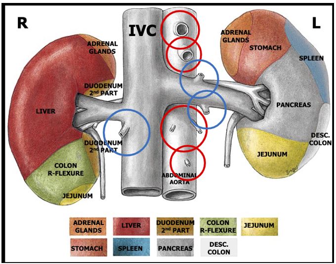
Review image of kidneys and which organs would need to be retracted? 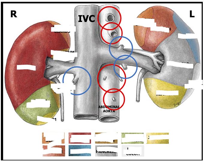
166
What are the 3 arterial supplies of the adrenal glands?
1. Superior adrenal (from inferior phrenic)
2. Middle adrenal (abdominak aorta)
3. Inferior adrenal (renal arteries)
2. Middle adrenal (abdominak aorta)
3. Inferior adrenal (renal arteries)
167
What are the 2 venous supplies of the adrenal gland? Where do they drain?
1. Right adrenal gland drains into IVC
2. Left adrenal gland drains into left renal vein
2. Left adrenal gland drains into left renal vein
168
What are teh 4 blood supplies of the ureter?
1. Gonadal
2. Renal
3. Aortic
4. Vesicular (from umbilical from iliac)
5. Common iliac
2. Renal
3. Aortic
4. Vesicular (from umbilical from iliac)
5. Common iliac
169
What are the 3 constricted points?
1. Pelvic brim
2. Ureteric pelvic (renal pelvis)
3. Bladder- as they enter obliquely into posterior wall
2. Ureteric pelvic (renal pelvis)
3. Bladder- as they enter obliquely into posterior wall
170
A kidneyreter stone (nephrolithiasis) can occur at any of these points and is treated with (extracorporeal shock wave lithotripsy)
A kidneyreter stone (nephrolithiasis) can occur at any of these points and is treated with ECSW (extracorporeal shock wave lithotripsy)
171
Recall that the jets right behind the ; that is, it passes to the
Recall that the right renal artery jets right behind the IVC; that is, it passes posterior to the IVC 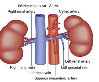
172
The abdominal cavity extends superiority into the osteocartilaginous thoracic cage to about the intercostal space in the supine position
The abdominal cavity extends superiority into the osteocartilaginous thoracic cage to about the 5th intercostal space in the supine position
173
(skin over upper inguinal region (groin), skin over iliac crest)
Iliohypogastric nerve (skin over upper inguinal region (groin), skin over iliac crest)
174
(skin over the superomedial aspect of the thigh, skin of the lower inguinal region, mons pubis, anterior scrotum, labia majora)
Ilioinguinal (skin over the superomedial aspect of the thigh, skin of the lower inguinal region, mons pubis, anterior scrotum, labia majora)
175
Superior to the medial part of the inguinal ligament, there is a triangular opening in the aponuerosis called the
Superior to the medial part of the inguinal ligament, there is a triangular opening in the aponuerosis called the superficial inguinal ring
176
Superficial inguinal ring has two crura on its side:
- inserts on the pubic tubercle
- inserts on the pubic symphysis
- inserts on the pubic tubercle
- inserts on the pubic symphysis
Superficial inguinal ring has two crura on its side:
Lateral crus- inserts on the pubic tubercle
Medial crus- inserts on the pubic symphysis
Lateral crus- inserts on the pubic tubercle
Medial crus- inserts on the pubic symphysis
177
What is the acronym for the boundaries of the inguinal ligament? What are the boundaries?
2 Malt
Superior wall (roof): 2 Muscles
internal oblique Muscle
transversus abdominis Muscle
Anterior wall: 2 Aponeuroses
Aponeurosis of external oblique
Aponeurosis of internal oblique
Lower wall (floor): 2 Ligaments
inguinal Ligament·
lacunar Ligament
Posterior wall: 2 Ts
Transversalis fascia
conjoint Tendon
Superior wall (roof): 2 Muscles
internal oblique Muscle
transversus abdominis Muscle
Anterior wall: 2 Aponeuroses
Aponeurosis of external oblique
Aponeurosis of internal oblique
Lower wall (floor): 2 Ligaments
inguinal Ligament·
lacunar Ligament
Posterior wall: 2 Ts
Transversalis fascia
conjoint Tendon
178
Infracolic compartment contains gutters that slope and , so when fluid accumulates in the peritoneal cavity, it will follow the gutters to the part of the abdomen when the patient is
Infracolic compartment contains gutters that slope superiority and posteriorly, so when fluid accumulates in the peritoneal cavity, it will follow the gutters to the superior part of the abdomen when the patient is supine
179
The abdominal esophagus measure cm from the groove in the left lobe of the liver and enters the stomach at the cardiac orifice posterior the the costal cartilage
The abdominal esophagus measure 1-2 cm from the groove in the left lobe of the liver and enters the stomach at the cardiac orifice posterior the the 7th costal cartilage
180
is suspended by the ligament of Treitz (extends from the upper aspect of the to the and the tissue around the celiac trunk)
Ascending duodenum is suspended by the ligament of Treitz (extends from the upper aspect of the ascending part of the duodenum to the right crus of the diaphragm and the tissue around the celiac trunk)
181
Right colic flexure-more than left side. lie deep to ribs and are overlapped by the
Right colic flexure-more posterior than left side. lie deep to ribs 9-10 and are overlapped by the liver
182
Left colic flexure- more , acute and less than the right colic flexure. Anterior to the left
Left colic flexure- more superior, acute and less mobile than the right colic flexure. Anterior to the left kidney
183
The and are enclosed by the head of the pancreas
The SMA and SMV are enclosed by the head of the pancreas
184
The tail of the pancreas ends near the
The tail of the pancreas ends near the spleen
185
The splenic artery runs on the surface of the pancreas and the splenic veins runs to the pancreas
The splenic artery runs on the superior surface of the pancreas and the splenic veins runs posterior to the pancreas
186
The liver is covered by the peritoneum except at the area. It is on the visceral surface on the aspect of the lobe. The is associated with it
The liver is covered by the peritoneum except at the bare area. It is on the visceral surface on the posterior aspect of the right lobe. The IVC is associated with it
187
When the falciform ligament gets to the visceral surface of the liver, it changes names:
ligament- anterior/posterior
ligament- right/left
ligament- anterior/posterior
ligament- right/left
When the falciform ligament gets to the visceral surface of the liver, it changes names:
Coronary ligament- anterior/posterior
Triangular ligament- right/left
Coronary ligament- anterior/posterior
Triangular ligament- right/left
188
Arterial supply of Kidney: arteries (branch off the abdominal aorta just below SMA)
-Left: may be
-Right: renal artery splits into , and posterior branches
-Left: may be
-Right: renal artery splits into , and posterior branches
Arterial supply of Kidney: renal arteries (branch off the abdominal aorta just below SMA)
-Left: may be more than one- supernumeral arteries
-Right: renal artery splits into upper, middle and lower posterior branches
-Left: may be more than one- supernumeral arteries
-Right: renal artery splits into upper, middle and lower posterior branches
189
Venous supply of Kindey: veins (empty into the IVC)
Left: receives tributaries from , , and
Right: There may be
Left: receives tributaries from , , and
Right: There may be
Venous supply of Kindey: renal veins (empty into the IVC)
Left: receives tributaries from left gonadal, left adrenal, diaphragm and body wall
Right: There may be more than one- supranumerary or accessory veins
Left: receives tributaries from left gonadal, left adrenal, diaphragm and body wall
Right: There may be more than one- supranumerary or accessory veins
190
Nerve supply of the kidney: aorticorenal plexus, inter mesenteric plexus, thoracic and lumbar splanchnic nerves
-Pain fibers form the refer pain to the testis, labia majora, and to the lumbar region of the back
-Pain fibers form the refer pain to the testis, labia majora, and to the lumbar region of the back
Nerve supply of the kidney: aorticorenal plexus, inter mesenteric plexus, thoracic and lumbar splanchnic nerves
-Pain fibers form the splanchnic nerves refer pain to the testis, labia majora, and to the lumbar region of the back
-Pain fibers form the splanchnic nerves refer pain to the testis, labia majora, and to the lumbar region of the back
191
Ureters: in females, near the ischial spine, they turn , and below the uterine vessels and they are about 1.5-2 cm from the cervix
Ureters: in females, near the ischial spine, they turn downward, forward and medially below the uterine vessels and they are about 1.5-2 cm from the cervix
192
Right Cruz of diaphragm arises from the sides of the bodies of and intervertebral discs and it splits to enclose the
Right Cruz of diaphragm arises from the sides of the bodies of L1-3 and intervertebral discs and it splits to enclose the esophogous
193
Left crus of diaphragm arises from the sides of the bodies of and the intervertebral disk
Left crus of diaphragm arises from the sides of the bodies of L1-2 and the intervertebral disk
194
Lateral to the crura, the diaphragm arises from the and
Lateral to the crura, the diaphragm arises from the medial and lateral arcuate ligaments
195
Medial arcuate ligament extends from the side of the to the
Medial arcuate ligament extends from the side of the body of L2 to the transverse process of L1
196
Lateral arcuate ligament extends from the to
Lateral arcuate ligament extends from the transverse process of L1 to rib 12