cardiovascular
Study these flashcardsFree · spaced repetition · iPhone, Android, Mac & web
All cards
1
distribute blood to the tissues
distribute blood to the tissues
arterial vessels
distribute blood to the tissues
distribute blood to the tissues
2
perfuse the tissue and exchange gases, metabolites, and waste products
capillaries perfuse the tissue and exchange gases, metabolites, and waste products
3
to return blood to the heart
venous vessels to return blood to the heart
4
All vessels, including the heart, are lined by endothelium, a (type of epithelium?)
All vessels, including the heart, are lined by endothelium, a simple squamous epithelium (type of epithelium?)
5
Capillaries, the tiniest vessels, are basically tubes of
Capillaries, the tiniest vessels, are basically tubes of endothelial cells
6
larger vessels have
additional layers or “tunics” of and .
additional layers or “tunics” of and .
larger vessels have
additional layers or “tunics” of muscle and connective tissue (CT).
additional layers or “tunics” of muscle and connective tissue (CT).
7
What are the 3 layers (inner to outer) in large vessels?
1. Tunica intima
2. Tunica Media
3. Tunic adventitia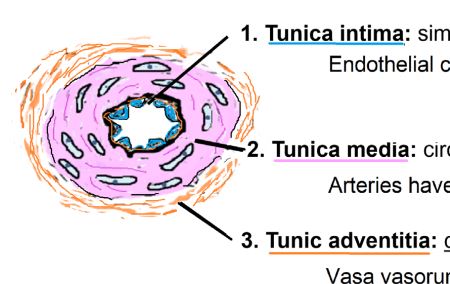
2. Tunica Media
3. Tunic adventitia
8
What is the tunica intima made of?
Simple squamous endothelial lining, basal lamina, and CT
9
How are endothelial cells lined in the tunica intima?
Longitudinally in direction of blood flow
10
What is the tunica media made out of?
Circularly arranged smooth muscle
11
In the tunica media, arteries have abundant
In the tunica media, arteries have abundant elastic fibers
12
What is the tunic adventitia made out of?
Connective tissue layer
13
In the tunica adventitia, what provides blood to walls of larger vessels?
Vasa vasorum
14
What is the vaso nervosa?
Nerves in the adventitia
15
In the tunic adventitia, what do large veins contain?
Smooth muscle
16
The heart is a muscular pump with two and two
The heart is a four-chambered muscular pump with two atria and two ventricles
17
What is the cardiac skeleton?
A connective tissue framework
18
What are the 3 layers of the wall of the heart?
1. Endocardium
2. Myocardium
3. Epicardiium
2. Myocardium
3. Epicardiium
19
What does the endocardium contain?
1. Inner lining of endothelium
2. Basal lamina
3. CT
2. Basal lamina
3. CT
20
Purkinje fibers are in the connective tissue
Purkinje fibers are in the subendocardial connective tissue
21
What is present in the myocardium?
Cardiac muscle
22
Thickness and fiber orientation in the myocardium varies, thickest in
Thickness and fiber orientation in the myocardium varies, thickest in ventricles
23
What does the epicardium contain?
1. Loose CT with adipose
2. Vessels
3. Nerves
4. Mesothelium (simple squamous)
2. Vessels
3. Nerves
4. Mesothelium (simple squamous)
24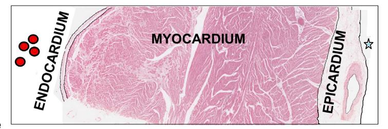
Image occlusion: name the hidden labels
25
The coronary artery is formed from the
The coronary artery is formed from the epicardium
26
, a fluid filled sac that cushions and lubricates the outer
wall of the heart, reducing friction during beating
wall of the heart, reducing friction during beating
Pericardium, a fluid filled sac that cushions and lubricates the outer
wall of the heart, reducing friction during beating
wall of the heart, reducing friction during beating
27
Inner lining of the pericardial sac is formed by
Epicardium (viseral pericardium) lined my mesothelium
28
The outer lining of the pericardial sac is formed by
Parietal pericardium lined by mesothelium
29
The produces the pericardial fluid.
The simple squamous mesothelium produces the pericardial fluid.
30
What is the cardiac skeleton?
Dense connective tissue
31
What are the 3 functions of the cardiac skeleton?
1. Separates the atria from ventricles
2. Anchors cardiac fibers
3. Forms supportive rings at base of valves
2. Anchors cardiac fibers
3. Forms supportive rings at base of valves
32
Heart valves are flaps of covered by .
Heart valves are flaps of connective tissue (collagen and elastic) covered by endothelium.
33
initiates and coordinates the rhythmic contraction of the heart
Cardiac conducting system initiates and coordinates the rhythmic contraction of the heart
34
What are the 4 components of the cardiac conducting system?
1. Sinoatrial (SA)
2. Atrioventicular (AV)
3. Atrioventricular bundle (of HIS)
4. Purkinje fibers
2. Atrioventicular (AV)
3. Atrioventricular bundle (of HIS)
4. Purkinje fibers
35
Purkinje fibers, located in the subendocardial CT, can be distinguished from typical cardiac muscle
cells by their , , and
cells by their , , and
Purkinje fibers, located in the subendocardial CT, can be distinguished from typical cardiac muscle
cells by their greater diameter, large clear spaces, and sparse myofibrils
cells by their greater diameter, large clear spaces, and sparse myofibrils
36
Purkinje fibers contain empty space histologically beause they have
Purkinje fibers contain empty space histologically beause they have glycogen
37
Purkinje fibers spread their excitation via
Purkinje fibers spread their excitation via gap junctions.
38
ENDOTHELIUM, a simple squamous epithelium, lines
ENDOTHELIUM, a simple squamous epithelium, lines ALL vessels
39
What are 5 important roles of endothelium?
1. Selective permeability barrier, allowing solute exchange
2. Is an anti-thrombogenic surface
3. Modulates blood flow by secreting nitric oxide and endothelin
4. Helps move WBC to connective tissue
5. Secretes growth factor
2. Is an anti-thrombogenic surface
3. Modulates blood flow by secreting nitric oxide and endothelin
4. Helps move WBC to connective tissue
5. Secretes growth factor
40
The arterial system transports blood at from the heart and distribute blood to all the parts of the body and controls of the flow of blood into capillaries
The arterial system transports blood at high pressure away from the heart and distribute blood to all the parts of the body and controls of the flow of blood into capillaries
41
Arteries have thick walls with
the being the thickest of the 3 layers
the being the thickest of the 3 layers
Arteries have thick walls with
the tunica media being the thickest of the 3 layers
the tunica media being the thickest of the 3 layers
42
Elastic fibers, produced by ,
is prevalent in arteries
is prevalent in arteries
Elastic fibers, produced by smooth muscle cells,
is prevalent in arteries
is prevalent in arteries
43
What are the 3 divisions of the arterial system?
1. Elastic arteries
2. Muscular arteries
3. Arterioles
2. Muscular arteries
3. Arterioles
44
What is in the tunica intima of elastic arteries?
Endothelium and CT 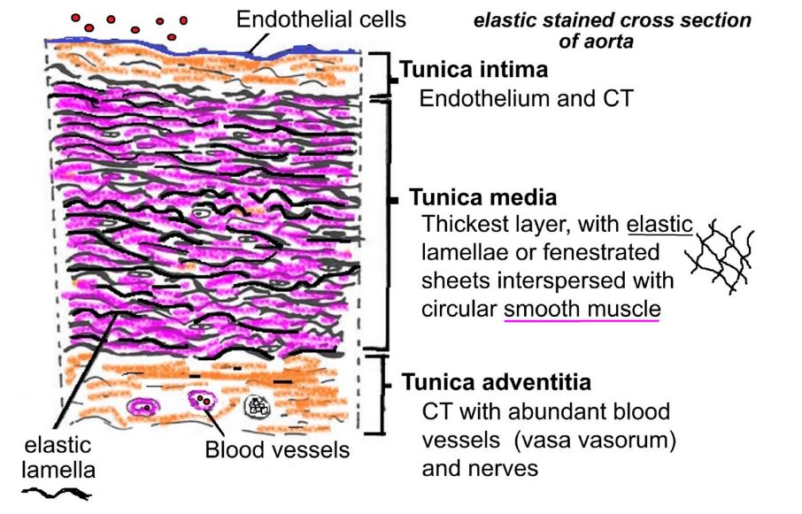
45
What is in the tunica media of elastic arteries?
Elastic lamellae or fenestrated sheets with circular smooth muscle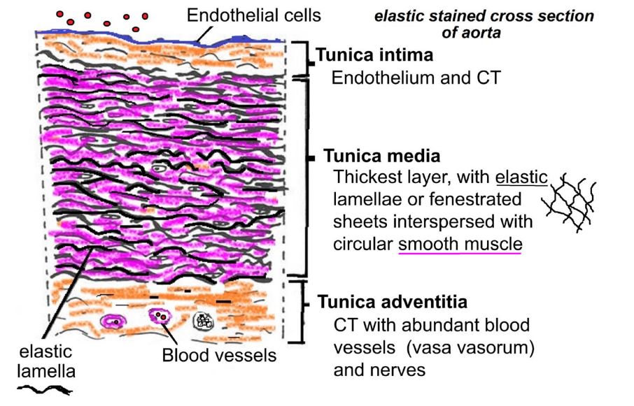
46
What is in the tunica adventitia of the elastic arteries?
CT with abundant blood vessels and nerves 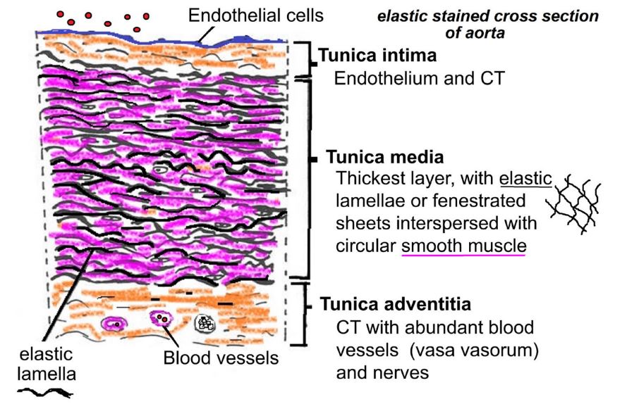
47
What is in the tunica intima of muscular arteries?
Endothelium, small amount of connective tissues, INTERNAL ELASTIC MEMBRANE 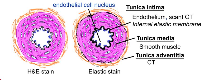
48
What is in the tunica intima of arterioles?
Endothelium 
49
What is in the tunica media of arterioles?
ONLY 1-2 layers of smooth muscle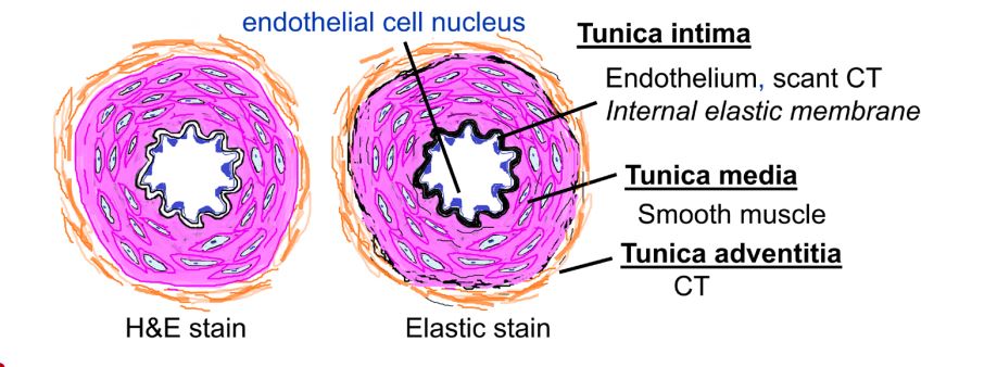
50
What is in the tunica adventitia of arteroles?
Connective tissue 
51
Changing lumin size of arterioles impacts what?
Blood pressure
52
The control the flow of blood into the capillary bed is done by by contracting or relaxing smooth muscle in the tunica media of
The control the flow of blood into the capillary bed is done by by contracting or relaxing smooth muscle in the tunica media of arterioles
53
What acts as the precapillary sphincter?
Arterioes
54
Capillaries consist of
1. Single layer of endothelial cells
2. Basal lamina
2. Basal lamina
55
Most capillaries
have a lumen around the size of a
have a lumen around the size of a
Most capillaries
have a lumen around the size of a single red blood cell
have a lumen around the size of a single red blood cell
56
What are pericytes?
Undifferentiated mesenchymal stem cells that wrap around capillaries and post postcapillary venules, sharing a basal lamina
57
What is the function of pericytes?
1. Provide vascular support
2. Can make endothelial, smooth muscle, and mesenchyme derived cells
3. They are contractile
2. Can make endothelial, smooth muscle, and mesenchyme derived cells
3. They are contractile
58
What are the 3 types of capillaries?
1. Continuous
2. Fenestrated
3. Discontinuous
2. Fenestrated
3. Discontinuous
59
What are continuous capillaries?
Endothelial cells are attached by tight junctions (zonula occludens) and have a continuous or
complete basal lamina
complete basal lamina
60
Where are continuous capillaries found?
muscle, connective tissue, skin, lungs, and CNS (blood brain barrier)
61
What are fenestrated capillaries?
Endothelial cells have small pores or fenestrations that allow passage of certain size
substances, like hormones. A continuous basal lamina is present.
substances, like hormones. A continuous basal lamina is present.
62
Where are fenestrated capillaries found?
Endocrine glands, kidney, and intestines
63
What are discontinuous (sinusoidal capillaries)?
Endothelial cells have large gaps between adjacent endothelial cells, fenestrations, and a
sparse, discontinuous, or incomplete basal lamina
sparse, discontinuous, or incomplete basal lamina
64
Discontinous capillaries have a (larger or smaller) diameter than other capillary types?
Larger
65
Discontinous capillaries have an _______ shape.
Irregular shape
66
Where are discontinous capillaries found?
Bone marrow, liver, spleen, anterior pituitary, adrenal cortex
67
Terminal arterioles control flow of blood into the capillary bed by contraction of its smooth muscle by
Terminal arterioles control flow of blood into the capillary bed by contraction of its smooth muscle by
changes the lumen size.
changes the lumen size.
68
The therefore acts as precapillary sphincter
The terminal arteriole therefore acts as precapillary sphincter
69
After exchange of gases and materials in the capillaries, the blood flows into
After exchange of gases and materials in the capillaries, the blood flows into postcapillary venules.
70
What is an arteriorvenous shunt/anatosomosis?
Ateriole to venule bypassing capillary bed 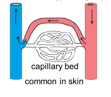
71
What is a portal system?
Two capillary beds with a connecting or portal vessel in between 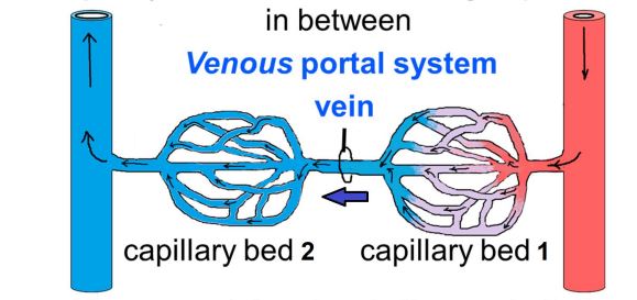
72
Where is the venous portal system found?
Liver and pituitary
73
Where is the arteriole portal system found?
Kidney
74
The venous system is a that collects blood from capillaries and returns it to
the heart.
the heart.
The venous system is a low-pressure system that collects blood from capillaries and returns it to
the heart.
the heart.
75
Compared to accompanying arteries, veins have a with loosely arranged
smooth muscle, larger and often collapsed lumen, and poorly defined tunics
smooth muscle, larger and often collapsed lumen, and poorly defined tunics
Compared to accompanying arteries, veins have a thinner wall with loosely arranged
smooth muscle, larger and often collapsed lumen, and poorly defined tunics
smooth muscle, larger and often collapsed lumen, and poorly defined tunics
76
Valves are present in
Valves are present in veins.
77
The is the
largest layer in veins while the is the largest in arteries.
largest layer in veins while the is the largest in arteries.
The adventitia is the
largest layer in veins while the tunica media is the largest in arteries.
largest layer in veins while the tunica media is the largest in arteries.
78
What are the three components of the venous system?
Venules, veins, and large veins
79
are the first and smallest venules, immediately adjacent to capillaries
Postcapillary venules are the first and smallest venules, immediately adjacent to capillaries
80
What are the components of a postcapillary venule
1. Endothelial lining
2. Basal lamina
3. Connective tissue
4. Pericytes
2. Basal lamina
3. Connective tissue
4. Pericytes
81
Comparied to a capillary, a vein has a
Comparied to a capillary, a vein has a larger lumen
82
Postcapillary venules are the primary site where leave the blood and enter
tissues at sites of infection and tissue damage.
tissues at sites of infection and tissue damage.
Postcapillary venules are the primary site where white blood cells leave the blood and enter
tissues at sites of infection and tissue damage.
tissues at sites of infection and tissue damage.
83
Lymphocytes exit blood and enter the lymphatic system through high
, which are a special type of
, which are a special type of
Lymphocytes exit blood and enter the lymphatic system through high
endothelial venules (HEV), which are a special type of postcapillary venule
endothelial venules (HEV), which are a special type of postcapillary venule
84
Blood enters veins under and moves towards the heart by contraction of smooth
muscle in the vessel wall and by external compressions from surrounding tissue.
muscle in the vessel wall and by external compressions from surrounding tissue.
Blood enters veins under low pressure and moves towards the heart by contraction of smooth
muscle in the vessel wall and by external compressions from surrounding tissue.
muscle in the vessel wall and by external compressions from surrounding tissue.
85
Veins have a thin layer of with small bundles of smooth muscle cells mixed with
connective tissue. The is thicker than the .
connective tissue. The is thicker than the .
Veins have a thin layer of tunica media with small bundles of smooth muscle cells mixed with
connective tissue. The adventitia is thicker than the media.
connective tissue. The adventitia is thicker than the media.
86
Veins, especially of the limbs, have valves to prevent
Veins, especially of the limbs, have valves to prevent backflow of blood.
87
Valves are folds of the projecting across the lumen. They are covered by
and have a core of connective tissue rich in
and have a core of connective tissue rich in
Valves are folds of the tunica intima projecting across the lumen. They are covered by
endothelium and have a core of connective tissue rich in elastic fibers.
endothelium and have a core of connective tissue rich in elastic fibers.
88
Large veins, like the , have a well-developed intima but only a thin media with
scattered circularly arranged smooth muscle.
scattered circularly arranged smooth muscle.
Large veins, like the vena cava, have a well-developed intima but only a thin media with
scattered circularly arranged smooth muscle.
scattered circularly arranged smooth muscle.
89
TEST QUESTION: What is the unique feasture of large veins?
Longitudinally arranged bundes of smooth muscle in the tunica adventitia
90
The wall of large veins is than that of elastic arteries
The wall of large veins is thinner than that of elastic arteries
91
In addition to blood vessels, there is a system of vessels, the lymphatic vessels, that
collects excess fluid from
collects excess fluid from
In addition to blood vessels, there is a system of thin-walled vessels, the lymphatic vessels, that
collects excess fluid from interstitial spaces
collects excess fluid from interstitial spaces
92
The lymph fluid is filtered and emptied into the venous
system near the
system near the
The lymph fluid is filtered and emptied into the venous
system near the heart
system near the heart
93
Lymphatic vessels are not present in the and
Lymphatic vessels are not present in the CNS and cartilage.
94
Describe lympathic capillaries
1. Blind-ended vessels
2. One laye of overlapping epithelium
3. Incomplete or absent basal lamina
2. One laye of overlapping epithelium
3. Incomplete or absent basal lamina
95
Interittial fluid flow in when pressure is
Interittial fluid flow in when pressure is high
96
Are pericytes involves with the lymph capillaries?
No
97
Compared to a vein, a lymphatic vessel has a
and a with few endothelial cell nuclei visible
and a with few endothelial cell nuclei visible
Compared to a vein, a lymphatic vessel has a
larger lumen and a thin wall with few endothelial cell nuclei visible
larger lumen and a thin wall with few endothelial cell nuclei visible
98
Lympathic vessels have to keep fluid moving in one direction.
Lympathic vessels have valves to keep fluid moving in one direction.
99
Only the l like those next to lymph nodes have
smooth muscle in its wall
smooth muscle in its wall
Only the largest lymphatic vessels like those next to lymph nodes have
smooth muscle in its wall
smooth muscle in its wall
100
are often present in lymphatic vessels because they utilize
the vessels to travel between lymphoid organ
the vessels to travel between lymphoid organ
Lymphocytes are often present in lymphatic vessels because they utilize
the vessels to travel between lymphoid organ
the vessels to travel between lymphoid organ
101
What will NOT be present in a lymathic vessel compared to a vein?
Will not contain RBC or WBC
102
What are the two processes of blood vessel development in the embryo?
vasculogenesis and angiogenesis
103
What is vasculogenesis?
Blood vessels form de novo, in blood islands
104
What are the steps of vasculogenesis?
1. Blood islands form from mesenchyeme cells in mesoderms
2. The cells in the center give rise to primitive blood cells
3. Cells differenitae into angioblasts that give rise to endothelial cells
4. Ajacent vessels fuse
2. The cells in the center give rise to primitive blood cells
3. Cells differenitae into angioblasts that give rise to endothelial cells
4. Ajacent vessels fuse
105
What is angiogenesis?
New blood vessels by growth from exisiting vessels
106
What process is responsible for most or all growth of new blood vessels for wound healing, development of collateral circulation, and tumor growth?
Angiogenesis
107
How does vasculogenesis occur in adults?
By recruitment of endothelial precursor cells from bone marrow
108
What is ateriorsclerosis?
Hardening of arteries due to thickening of vessel wall. Lumen is narrowed and elasciticty is lost
109
What are the two types of ateriosclerosis?
1. Atherosclerosis
2. Ateriolosclerosis
2. Ateriolosclerosis
110
Atherosclerosis – hardening of caused by growth of intimal lesions called
atherosclerotic or
atherosclerotic or
Atherosclerosis – hardening of large arteries caused by growth of intimal lesions called
atherosclerotic plaques or atheroma
atherosclerotic plaques or atheroma
111
What happens to the plaques formed in atherosclerosis?
The plaques can cause occulsions leading to ischemia or cause blot clots
112
Atherosclerosis is the underlying cause of most , , and disease. Common arteries that develop plaques include
and l
and l
Atherosclerosis is the underlying cause of most coronary, cerebral, and peripheral vascular disease. Common arteries that develop plaques include elastic arteries
and larger muscular arteries
and larger muscular arteries
113
The core difference between atheroscleosis and arteriolosclerosis?
Atherosclerosis is a large vessel disease, Arteriolosclerosis is a small vessel disease
114
Arteriolosclerosis- thickening of or
Arteriolosclerosis- thickening of wall of arterioles or small arteries
115
What are the two types of arterioloscleosis?
1. Hyaline anteriorlosclerosis
2. Hyperplastic anteriorlosclerosis
2. Hyperplastic anteriorlosclerosis
116
What is the special feature of hyaline arteriosclerosis? What diseases is it common in?
-Glassy, pink homogeneous staining due to plasma proteins leaking
-Benign hypertension and diabetes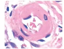
-Benign hypertension and diabetes
117
What is the special feature of hyperplastic ateriolosclerosis? What disease is it common in?
-Concentric lamellated smooth muscle in the wall resembles an “onion” skin.
-Malignant hypertension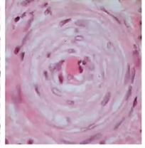
-Malignant hypertension
118
What are aneurysms?
Localized weakening of a segment of a vessel wall leading to a dilation or bulging
outward of the vessel.
outward of the vessel.
119
What conditions predispose someone to aneurysm?
Atherosclerosis and hypertension
120
What is an aortic dissection?
A tear in the intima allows blood (at high pressure) to enter the wall and
separate or “dissect” the media into layers.
separate or “dissect” the media into layers.
121
What disease predispose someone to aortic dissection?
MARFANS syndrome or aquired factors (hypertension, atherosclerosis, diabetes)
122
What are the two categories of low-pressure system diseases? What area of the body do these most often effect?
1. Thrombosis
2. Inadequate venous drainage
-The legs
2. Inadequate venous drainage
-The legs
123
– abnormally dilated torturous superficial veins produced by prolonged increase
in intraluminal pressure.
in intraluminal pressure.
Varicose veins – abnormally dilated torturous superficial veins produced by prolonged increase
in intraluminal pressure.
in intraluminal pressure.
124
– is a thrombus (blood clot) that forms in a major vein, usually in
the legs. The thrombus can restrict the return of blood to the heart and result in pain and
swelling as blood gathers behind the clot.
the legs. The thrombus can restrict the return of blood to the heart and result in pain and
swelling as blood gathers behind the clot.
Deep vein thrombosis (DVT) – is a thrombus (blood clot) that forms in a major vein, usually in
the legs. The thrombus can restrict the return of blood to the heart and result in pain and
swelling as blood gathers behind the clot.
the legs. The thrombus can restrict the return of blood to the heart and result in pain and
swelling as blood gathers behind the clot.
125
Deep vein thrombosis is especially dangerous because it can result in a .
This occurs when the clot detaches from the vein, travels through the heart to the lungs, and
wedges in a pulmonary vessel
This occurs when the clot detaches from the vein, travels through the heart to the lungs, and
wedges in a pulmonary vessel
Deep vein thrombosis is especially dangerous because it can result in a pulmonary embolism.
This occurs when the clot detaches from the vein, travels through the heart to the lungs, and
wedges in a pulmonary vessel
This occurs when the clot detaches from the vein, travels through the heart to the lungs, and
wedges in a pulmonary vessel
126
increases the probability of thrombus formation
Slow moving blood increases the probability of thrombus formation
127
can be placed in the inferior vena cava to “catch” any clots and prevent them from
causing a pulmonary
causing a pulmonary
IVC Filters can be placed in the inferior vena cava to “catch” any clots and prevent them from
causing a pulmonary
causing a pulmonary